. 2009 Jul;41(3):337-47.
doi: 10.1016/j.mcn.2009.04.007. Epub 2009 May 3.

Circumventricular organs: a novel site of neural stem cells in the adult brain

Affiliations

Circumventricular organs: a novel site of neural stem cells in the adult brain

Lori Bennett et al. Mol Cell Neurosci. 2009 Jul.

Abstract

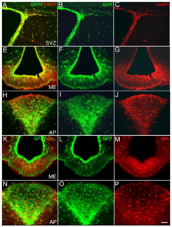
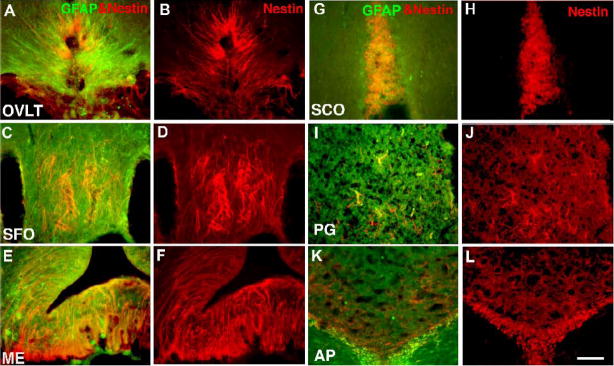
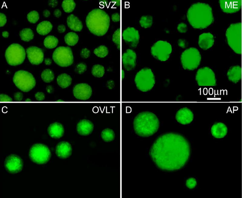
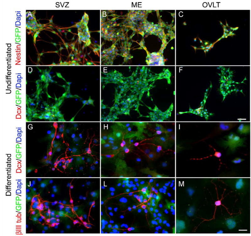
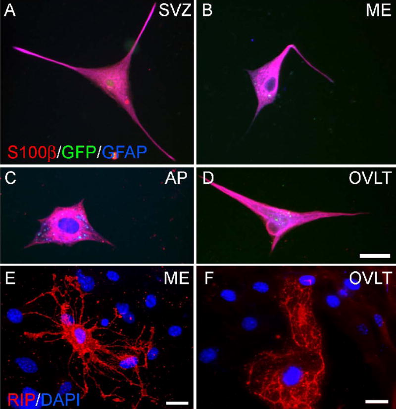
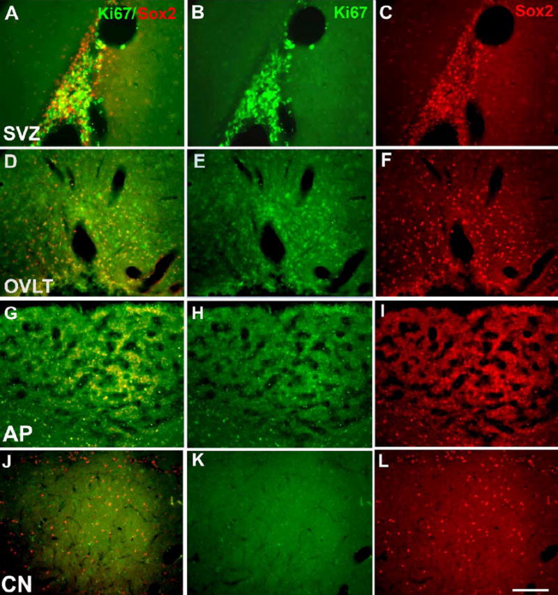
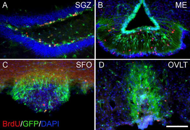
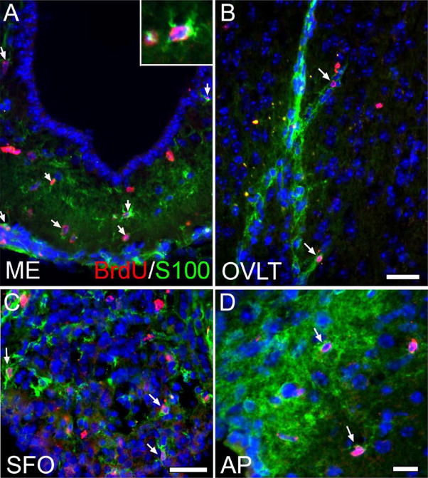
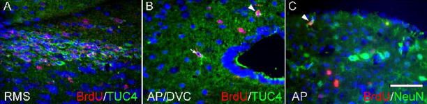
Neurogenesis in the adult mammalian nervous system is now well established in the subventricular zone of the anterolateral ventricle and subgranular zone of the hippocampus. In these regions, neurons are thought to arise from neural stem cells, identified by their expression of specific intermediate filament proteins (nestin, vimentin, GFAP) and transcription factors (Sox2). In the present study, we show that in adult rat and mouse, the circumventricular organs (CVOs) are rich in nestin+, GFAP+, vimentin+ cells which express Sox2 and the cell cycle-regulating protein Ki67. In culture, these cells proliferate as neurospheres and express neuronal (doublecortin+, beta-tubulin III+) and glial (S100beta+, GFAP+, RIP+) phenotypic traits. Further, our in vivo studies using bromodeoxyuridine show that CVO cells proliferate and undergo constitutive neurogenesis and gliogenesis. These findings suggest that CVOs may constitute a heretofore unknown source of stem/progenitor cells, capable of giving rise to new neurons and/or glia in the adult brain.


Figures

Figure 1
Figure 1
Expression of intermediate filaments in CVOs of the adult transgenic nestin-GFP mouse. Using immunocytochemistry for tissue sections, cells in the SVZ (A-C), ME (E-G, K-M) or AP (H-J, N-P) were both GFP fluorescent (B, F, I, L, O) and nestin+ (C, G, J) or vimentin+ (M, P) in merged images (A, E, H, K, N). Calibration bar = 100μm.
Figure 2
Figure 2
Immunocytochemical localization of intermediate filament proteins in the adult rat CVOs. Shown are nestin staining or merged images of nestin and GFAP staining in the OVLT (A, B), SFO (C, D), ME (E, F), SCO (G, H), PG (I, J) and AP (K, L). Calibration bar = 200μm.
Figure 3
Figure 3
Neurosphere formation from adult transgenic nestin-GFP mouse cells. GFP+ neurospheres from control region SVZ (A) and CVO regions ME (B), OVLT (C), and AP (D) are shown 6–14 days following microdissection and plating of single cells in suspension culture.
Figure 4
Figure 4
Clonal proliferation of adult rat CVO cells in vitro. Cells from the CVO region ME were plated at low density and single attached cells were marked and observed for colony formation. Here ME cells are shown in the act of cytokinesis (A) and developing expanding colonies (B, C). Immunocytochemical analysis demonstrated that like control SVZ cells (D), ME cells expressed nestin and the nuclear proliferation marker Ki67 (E).
Figure 5
Figure 5
Neuronal differentiation of nestin-GFP mouse CVO cells in vitro. Undifferentiated cells from the SVZ (A, D), ME (B, E), and OVLT (C, F) expressed nestin and GFP (A-C), but few expressed the neuroblast marker doublecortin (DCX, D-F). Following differentiation for 14 days, process-bearing cells expressed neuronal markers DCX (G-I) and β-tubulin III (βIIItub, J-M). Calibration bar = 50 μm for Panels A-F and 25 μm for Panels G-M.
Figure 6
Figure 6
Glial differentiation of nestin-GFP mouse cells in vitro. Undifferentiated nestin-GFP cells plated as adherent culture and differentiated for 9–14 days were analyzed by immunocytochemistry for expression of astrocyte markers S100β and GFAP and oligodendrocyte marker RIP. Shown are cells from the SVZ (A), ME (B), AP (C), and OVLT (D) co-expressing S100β and GFAP. Note that cells have lost expression of GFP, which would appear white if triple-labeled. ME (E) and OVLT (F) cells with characteristic oligodendrocyte morphology are shown expressing RIP. Calibration bar = 25 μm.
Figure 7
Figure 7
Immunocytochemical localization of transcription and proliferation factors in adult rat CVOs. Shown are merged images (A, D, G, J) for Ki67 (B, E, H, K) and Sox2 (C, F, I, L) staining in the SVZ (A-C), OVLT (D-F), AP (G-I) and in presumptive parenchymal progenitors of the caudate nucleus (J-L). Calibration bar = 200μm.
Figure 8
Figure 8
Constitutive proliferation of CVO cells in the nestin-GFP mouse. BrdU-labeled nuclei are seen in control region SGZ (A) and CVO regions ME (B), SFO (C), and OVLT (D) following administration of BrdU for one week. Calibration bar = 100μm.
Figure 9
Figure 9
Constitutive gliogenesis in CVOs of the nestin-GFP mouse. To track the differentiative fate of proliferating (BrdU+) CVO cells, intact Nestin-GFP animals were administered BrdU for four weeks. As indicated by the arrows, many BrdU+ cells co-labeled with the astrocyte marker S100β in the ME (A, see inset for enlargement of BrdU+, S100β+ cell), OVLT (B), SFO (C), and AP (D), indicating active gliogenesis at these sites. Scale bars = 25 μm.
Figure 10
Figure 10
Neurogenesis in the area postrema of the intact nestin-GFP mouse. Shown are BrdU-labeled cells co-expressing the early neuronal marker TUC-4 within the boundary of the AP (B, arrowhead), in the dorsal vagal complex (DVC, B, arrow), and in the rostral migratory stream, a known region of migrating neuroblasts (RMS, A). BrdU-labeled cells in the AP also express the neuronal marker NeuN (C, arrow). Scale bar = 50 μm.

Similar articles

Cited by

References

    1. Alvarez-Buylla A, Lim DA. For the long run: Maintaining germinal niches in the adult brain. Neuron. 2004;41:683–686. - PubMed
    1. Arochena M, Anadon R, Diaz-Regueira SM. Development of vimentin and glial fibrillary acidic protein immunoreactivities in the brain of gray mullet (chelon labrosus), an advanced teleost. J Comp Neurol. 2004;469:413–436. - PubMed
    1. Barkho BZ, Song H, Aimone JB, Smrt RD, Kuwabara T, Nakashima K, Gage FH, Zhao X. Identification of astrocyte-expressed factors that modulate neural stem/progenitor cell differentiation. Stem Cells Dev. 2006;15:407–421. - PMC - PubMed
    1. Barry D, McDermott K. Differentiation of radial glia from radial precursor cells and transformation into astrocytes in the developing rat spinal cord. Glia. 2005;50:187–197. - PubMed
    1. Bauer S, Hay M, Amilhon B, Jean A, Moyse E. In vivo neurogenesis in the dorsal vagal complex of the adult rat brainstem. Neuroscience. 2005;130:75–90. - PubMed

Publication types

MeSH terms