

Abnormalities of intrinsic functional connectivity in autism spectrum disorders
- PMID: 19409498
- PMCID: PMC2731579
- DOI: 10.1016/j.neuroimage.2009.04.069
Abnormalities of intrinsic functional connectivity in autism spectrum disorders
Abstract
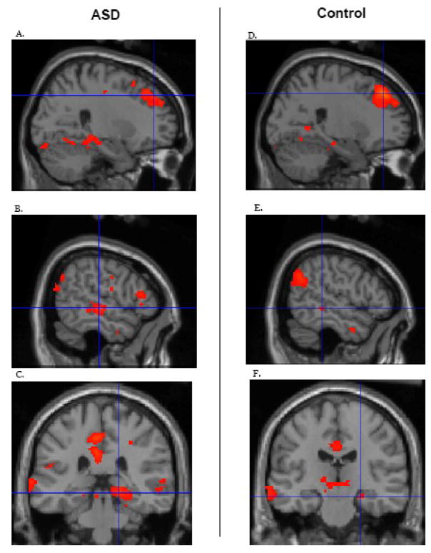
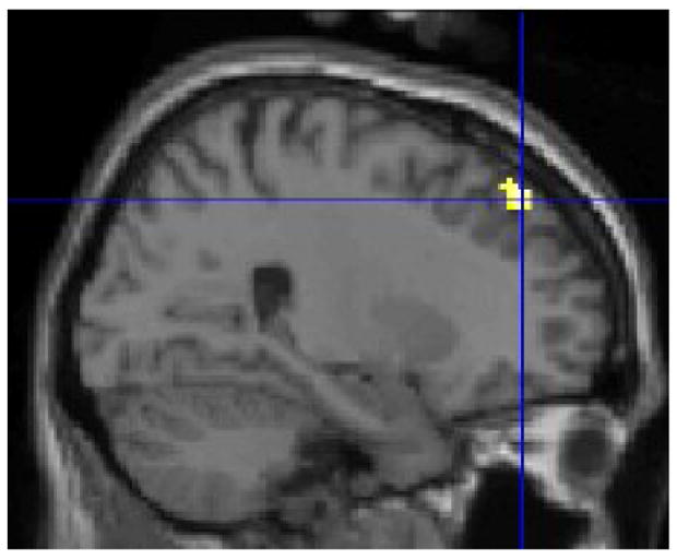
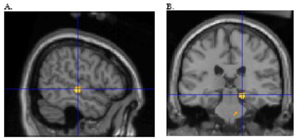
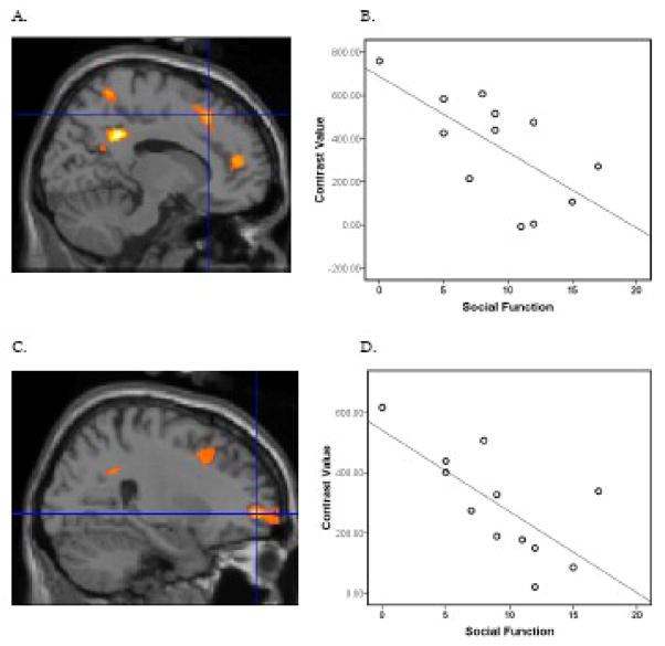
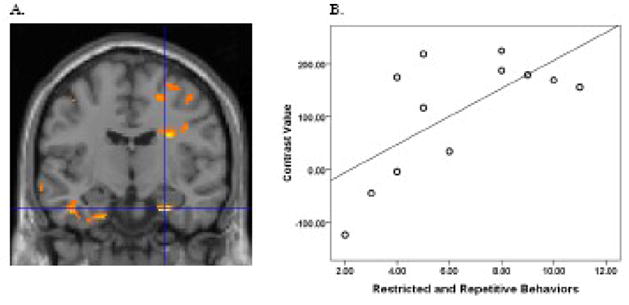
Autism spectrum disorders (ASD) impact social functioning and communication, and individuals with these disorders often have restrictive and repetitive behaviors. Accumulating data indicate that ASD is associated with alterations of neural circuitry. Functional MRI (FMRI) studies have focused on connectivity in the context of psychological tasks. However, even in the absence of a task, the brain exhibits a high degree of functional connectivity, known as intrinsic or resting connectivity. Notably, the default network, which includes the posterior cingulate cortex, retro-splenial, lateral parietal cortex/angular gyrus, medial prefrontal cortex, superior frontal gyrus, temporal lobe, and parahippocampal gyrus, is strongly active when there is no task. Altered intrinsic connectivity within the default network may underlie offline processing that may actuate ASD impairments. Using FMRI, we sought to evaluate intrinsic connectivity within the default network in ASD. Relative to controls, the ASD group showed weaker connectivity between the posterior cingulate cortex and superior frontal gyrus and stronger connectivity between the posterior cingulate cortex and both the right temporal lobe and right parahippocampal gyrus. Moreover, poorer social functioning in the ASD group was correlated with weaker connectivity between the posterior cingulate cortex and the superior frontal gyrus. In addition, more severe restricted and repetitive behaviors in ASD were correlated with stronger connectivity between the posterior cingulate cortex and right parahippocampal gyrus. These findings indicate that ASD subjects show altered intrinsic connectivity within the default network, and connectivity between these structures is associated with specific ASD symptoms.
Figures
Similar articles
-
Alterations of resting state functional connectivity in the default network in adolescents with autism spectrum disorders.Brain Res. 2010 Feb 8;1313:202-14. doi: 10.1016/j.brainres.2009.11.057. Epub 2009 Dec 11. Brain Res. 2010. PMID: 20004180 Free PMC article.
-
Dynamic functional connectivity analysis reveals decreased variability of the default-mode network in developing autistic brain.Autism Res. 2018 Nov;11(11):1479-1493. doi: 10.1002/aur.2020. Epub 2018 Oct 1. Autism Res. 2018. PMID: 30270547
-
Decreased interhemispheric functional connectivity rather than corpus callosum volume as a potential biomarker for autism spectrum disorder.Cortex. 2019 Oct;119:258-266. doi: 10.1016/j.cortex.2019.05.003. Epub 2019 May 15. Cortex. 2019. PMID: 31167156
-
The nature of brain dysfunction in autism: functional brain imaging studies.Curr Opin Neurol. 2010 Apr;23(2):124-30. doi: 10.1097/WCO.0b013e32833782d4. Curr Opin Neurol. 2010. PMID: 20154614 Free PMC article. Review.
-
The role of the posterior cingulate cortex in cognition and disease.Brain. 2014 Jan;137(Pt 1):12-32. doi: 10.1093/brain/awt162. Epub 2013 Jul 18. Brain. 2014. PMID: 23869106 Free PMC article. Review.
Cited by
-
A model of functional brain connectivity and background noise as a biomarker for cognitive phenotypes: application to autism.PLoS One. 2013 Apr 17;8(4):e61493. doi: 10.1371/journal.pone.0061493. Print 2013. PLoS One. 2013. PMID: 23613864 Free PMC article.
-
EEG resting-state functional connectivity: evidence for an imbalance of external/internal information integration in autism.J Neurodev Disord. 2022 Aug 27;14(1):47. doi: 10.1186/s11689-022-09456-8. J Neurodev Disord. 2022. PMID: 36030210 Free PMC article.
-
Abnormal brain activity in social reward learning in children with autism spectrum disorder: an fMRI study.Yonsei Med J. 2015 May;56(3):705-11. doi: 10.3349/ymj.2015.56.3.705. Yonsei Med J. 2015. PMID: 25837176 Free PMC article.
-
Atypical lexicosemantic function of extrastriate cortex in autism spectrum disorder: evidence from functional and effective connectivity.Neuroimage. 2012 Sep;62(3):1780-91. doi: 10.1016/j.neuroimage.2012.06.008. Epub 2012 Jun 12. Neuroimage. 2012. PMID: 22699044 Free PMC article.
-
Hypoconnectivity of insular resting-state networks in adolescents with Autism Spectrum Disorder.Psychiatry Res Neuroimaging. 2019 Jan 30;283:104-112. doi: 10.1016/j.pscychresns.2018.12.003. Epub 2018 Dec 7. Psychiatry Res Neuroimaging. 2019. PMID: 30594068 Free PMC article.
References
-
- Alexander AL, Lee JE, Lazar M, Boudos R, DuBray MB, Oakes TR, Miller JN, Lu J, Jeong EK, McMahon WM, Bigler ED, Lainhart JE. Diffusion tensor imaging of the corpus callosum in Autism. Neuroimage. 2007;34:61–73. - PubMed
-
- APA. Diagnostic and Statistical Manual of Mental Disorders. Washington, DC: American Psychiatric Association; 1994.
-
- Barnea-Goraly N, Kwon H, Menon V, Eliez S, Lotspeich L, Reiss AL. White matter structure in autism: preliminary evidence from diffusion tensor imaging. Biol Psychiatry. 2004;55:323–6. - PubMed
Publication types
MeSH terms
Grants and funding
LinkOut - more resources
Full Text Sources
Other Literature Sources
Medical
