

Abnormalities of intrinsic functional connectivity in autism spectrum disorders
- PMID: 19409498
- PMCID: PMC2731579
- DOI: 10.1016/j.neuroimage.2009.04.069
Abnormalities of intrinsic functional connectivity in autism spectrum disorders
Abstract
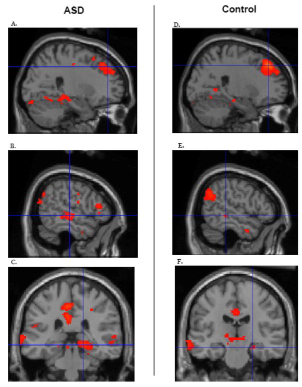
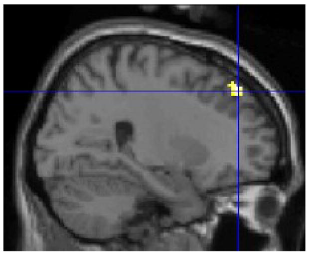
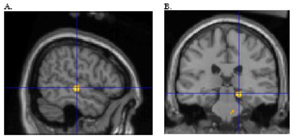
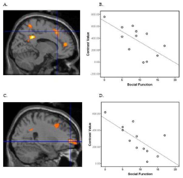
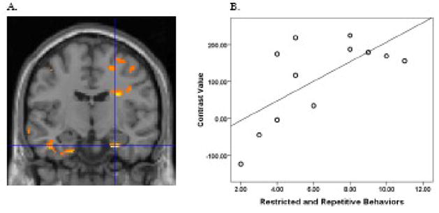
Autism spectrum disorders (ASD) impact social functioning and communication, and individuals with these disorders often have restrictive and repetitive behaviors. Accumulating data indicate that ASD is associated with alterations of neural circuitry. Functional MRI (FMRI) studies have focused on connectivity in the context of psychological tasks. However, even in the absence of a task, the brain exhibits a high degree of functional connectivity, known as intrinsic or resting connectivity. Notably, the default network, which includes the posterior cingulate cortex, retro-splenial, lateral parietal cortex/angular gyrus, medial prefrontal cortex, superior frontal gyrus, temporal lobe, and parahippocampal gyrus, is strongly active when there is no task. Altered intrinsic connectivity within the default network may underlie offline processing that may actuate ASD impairments. Using FMRI, we sought to evaluate intrinsic connectivity within the default network in ASD. Relative to controls, the ASD group showed weaker connectivity between the posterior cingulate cortex and superior frontal gyrus and stronger connectivity between the posterior cingulate cortex and both the right temporal lobe and right parahippocampal gyrus. Moreover, poorer social functioning in the ASD group was correlated with weaker connectivity between the posterior cingulate cortex and the superior frontal gyrus. In addition, more severe restricted and repetitive behaviors in ASD were correlated with stronger connectivity between the posterior cingulate cortex and right parahippocampal gyrus. These findings indicate that ASD subjects show altered intrinsic connectivity within the default network, and connectivity between these structures is associated with specific ASD symptoms.
Figures
References
-
- Alexander AL, Lee JE, Lazar M, Boudos R, DuBray MB, Oakes TR, Miller JN, Lu J, Jeong EK, McMahon WM, Bigler ED, Lainhart JE. Diffusion tensor imaging of the corpus callosum in Autism. Neuroimage. 2007;34:61–73. - PubMed
-
- APA. Diagnostic and Statistical Manual of Mental Disorders. Washington, DC: American Psychiatric Association; 1994.
-
- Barnea-Goraly N, Kwon H, Menon V, Eliez S, Lotspeich L, Reiss AL. White matter structure in autism: preliminary evidence from diffusion tensor imaging. Biol Psychiatry. 2004;55:323–6. - PubMed
Publication types
MeSH terms
Grants and funding
LinkOut - more resources
Full Text Sources
Other Literature Sources
Medical
