Case Reports
. 2009 May;84(5):594-604.
doi: 10.1016/j.ajhg.2009.04.004. Epub 2009 Apr 30.

The mitochondrial disulfide relay system protein GFER is mutated in autosomal-recessive myopathy with cataract and combined respiratory-chain deficiency

Affiliations
Case Reports

The mitochondrial disulfide relay system protein GFER is mutated in autosomal-recessive myopathy with cataract and combined respiratory-chain deficiency

Alessio Di Fonzo et al. Am J Hum Genet. 2009 May.

Abstract

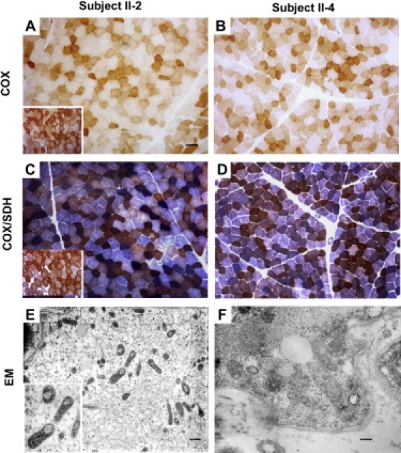
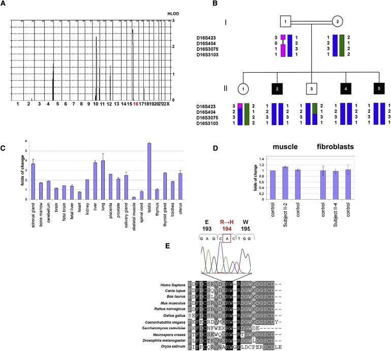
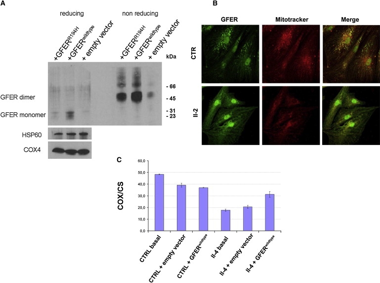
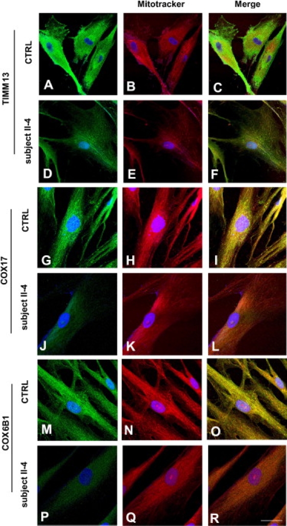
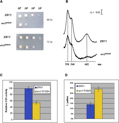
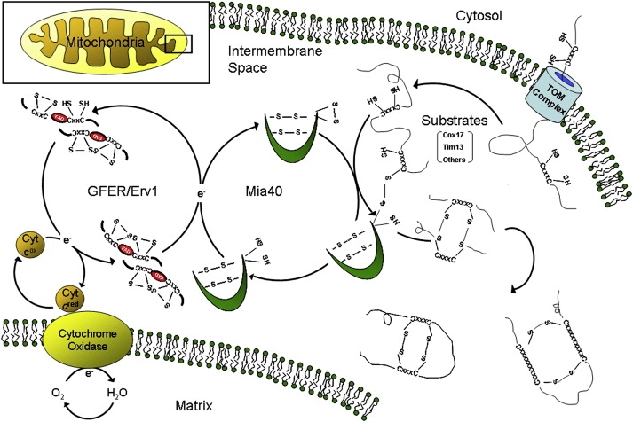
A disulfide relay system (DRS) was recently identified in the yeast mitochondrial intermembrane space (IMS) that consists of two essential components: the sulfhydryl oxidase Erv1 and the redox-regulated import receptor Mia40. The DRS drives the import of cysteine-rich proteins into the IMS via an oxidative folding mechanism. Erv1p is reoxidized within this system, transferring its electrons to molecular oxygen through interactions with cytochrome c and cytochrome c oxidase (COX), thereby linking the DRS to the respiratory chain. The role of the human Erv1 ortholog, GFER, in the DRS has been poorly explored. Using homozygosity mapping, we discovered that a mutation in the GFER gene causes an infantile mitochondrial disorder. Three children born to healthy consanguineous parents presented with progressive myopathy and partial combined respiratory-chain deficiency, congenital cataract, sensorineural hearing loss, and developmental delay. The consequences of the mutation at the level of the patient's muscle tissue and fibroblasts were 1) a reduction in complex I, II, and IV activity; 2) a lower cysteine-rich protein content; 3) abnormal ultrastructural morphology of the mitochondria, with enlargement of the IMS space; and 4) accelerated time-dependent accumulation of multiple mtDNA deletions. Moreover, the Saccharomyces cerevisiae erv1(R182H) mutant strain reproduced the complex IV activity defect and exhibited genetic instability of the mtDNA and mitochondrial morphological defects. These findings shed light on the mechanisms of mitochondrial biogenesis, establish the role of GFER in the human DRS, and promote an understanding of the pathogenesis of a new mitochondrial disease.


Figures

Figure 1
Figure 1
Histochemical and Ultrastructural Analysis of Probands' Skeletal Muscle (A–D) COX histochemistry reveals the presence of several COX-deficient fibers in probands II-2 and II-4 (A and B). Double staining for COX/SDH. Scattered fibers with increased SDH staining, indicative of mitochondrial proliferation, are present in patient II-2. A COX deficiency is confirmed in both patient II-2 and patient II-4. (C and D) Insets within panels (A) and (C) represent the normal controls. (E and F) Electron microscopy shows several mitochondria with thickened cristae; many mitochondria are also present with large vacuolization in (E). Scale bars represent 25 μm (A–D), 700 nm (E), or 250 nm (F).
Figure 2
Figure 2
Linkage Analysis, Expression Studies, and Mutation Analysis (A) Multipoint linkage analysis of the genome-wide scan with the Affymetrix GeneChip Human Mapping 50K Array Xba240. (B) Family pedigree and haplotype analysis of the GFER locus. Affected individuals are indicated by black symbols. (C) Quantitative RT-PCR of GFER expression in 20 human control tissues. GAPDH was used as the control housekeeping gene. The thymus mRNA level was used for normalization of the expression data. All determinations have been performed in replicates (n = 6). (D) Quantitative RT-PCR of GFER expression in muscle and fibroblasts from patients and controls. Results are presented as mean ± SD. (E) CLUSTALW multiple-alignment sequence of the GFER region containing the mutated residue in our family. The electropherogram of the c.581 G→A mutation in exon 3 of GFER, resulting in a p.R194H substitution, is shown above.
Figure 3
Figure 3
Western Blot Analysis of Overexpressed GFER, Immunocytochemical Analysis of Endogenous GFER in Patient Myoblasts, and Functional Rescue in Patient Fibroblasts (A) HEK293 cells were stably transfected with vector overexpressing GFERwild-type and GFERR194H cDNA. Immunoblot analysis showed a reduction of GFERR194H in mitochondrial fractions under both reducing (DTT 15 mM) and nonreducing conditions. COX4 and HSP60 were used for normalization. (B) Antibodies against human GFER showed specific GFER colocalization with MitoTracker and DAPI in control myoblasts, whereas immunofluorescence was almost absent in the cytoplasm. Instead, primary myoblasts from patient II-2 showed a reduced punctuate mitochondrial fluorescence compared to control cells with more diffuse cytoplasmic staining. Scale bar represents 40 μm. (C) Biochemical functional rescue in proband II-4 fibroblasts that were transfected with GFERwild-type cDNA. The analysis of COX activity (normalized to citrate synthase [CS]) shows a restoration of mitochondrial cytochrome c oxidation (complex IV). Results are presented as mean of triplicate determinations ± SD.
Figure 4
Figure 4
Confocal Immunocytochemical Analysis of IMS Proteins in Patient Fibroblasts Immunocytochemistry of patient and control fibroblasts, using antibodies against TIMM13, COX17, and COX6B1. Confocal analysis revealed reduced colocalization of COX17, TIMM13, and COX6B1 immunostaining with MitoTracker compared to the colocalization signal observed in control cells from healthy donors. Scale bars represent 40 μm (A–F) or 20 μm (G–R).
Figure 5
Figure 5
Yeast Model (A) Phenotypic analysis of the DMT1-4C strain (Δerv1) carrying either ERV1 or the erv1R182H allele. The growth phenotype was that observed at 37°C. Equal amounts of serial dilutions of cells from exponentially grown cultures were spotted onto YNB medium supplemented with 2% glucose. (B) Cytochrome spectra of cells grown on YNB medium supplemented with 2% glucose at 28°C and then shifted to 37°C overnight (A denotes absorbance). (C) COX activity measured in the ERV1 and erv1R182H strains. (D) Petite frequency of Δerv1 strain transformed with wild-type ERV1 and the erv1R182H mutant allele. COX activity and petite frequency determinations were performed in triplicate. The error bars represent SD.
Figure 6
Figure 6
The Mitochondrial Disulfide Relay System Schematic representation of the mitochondrial DRS, which mediates the import of cysteine-rich substrates into the IMS by an oxidative trapping mechanism. Mia40 performs the oxidation of these proteins and is reoxidized by Erv1/GFER in a disulfide-transfer reaction. For Erv1 to be recycled into its oxidized form, electrons are transferred to cytochrome c, connecting the DRS to the electron-transport chain of the mitochondria.

References

    1. DiMauro S., Schon E.A. Mitochondrial disorders in the nervous system. Annu. Rev. Neurosci. 2008;31:91–123. - PubMed
    1. Zeviani M., Carelli V. Mitochondrial disorders. Curr. Opin. Neurol. 2007;20:564–571. - PubMed
    1. Mesecke N., Terziyska N., Kozany C., Baumann F., Neupert W., Hell K., Herrmann J.M. A disulfide relay system in the intermembrane space of mitochondria that mediates protein import. Cell. 2005;121:1059–1069. - PubMed
    1. Hell K. The Erv1-Mia40 disulfide relay system in the intermembrane space of mitochondria. Biochim. Biophys. Acta. 2008;1783:601–609. - PubMed
    1. Heckmatt J.Z., Dubowitz V. Needle biopsy of skeletal muscle. Muscle Nerve. 1984;7:594. - PubMed

Publication types

MeSH terms

Substances