. 2009 Aug;30(23-24):4021-8.
doi: 10.1016/j.biomaterials.2009.04.005. Epub 2009 May 1.

Tissue-specific extracellular matrix coatings for the promotion of cell proliferation and maintenance of cell phenotype

Affiliations

Tissue-specific extracellular matrix coatings for the promotion of cell proliferation and maintenance of cell phenotype

Yuanyuan Zhang et al. Biomaterials. 2009 Aug.

Abstract

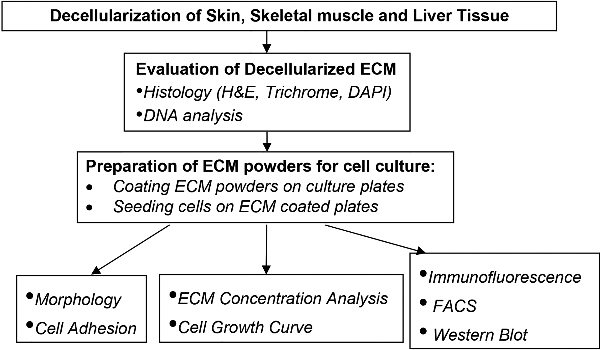
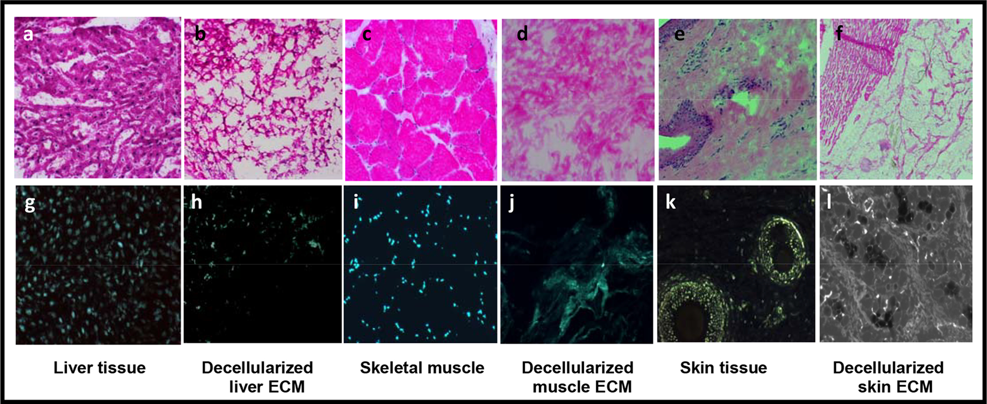
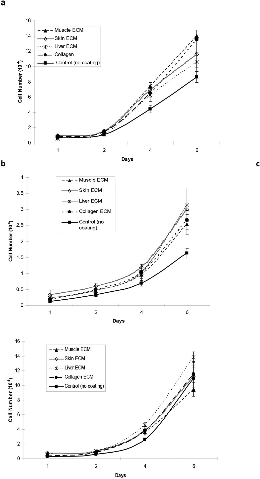
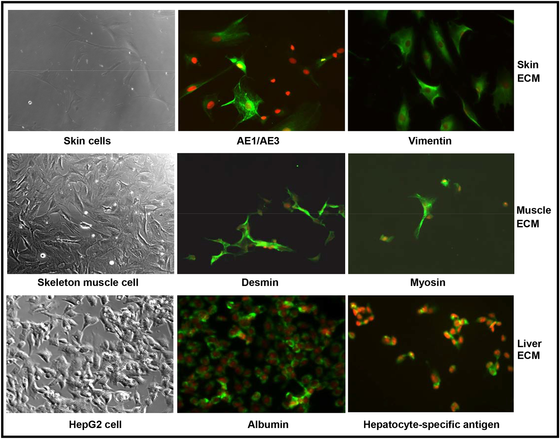
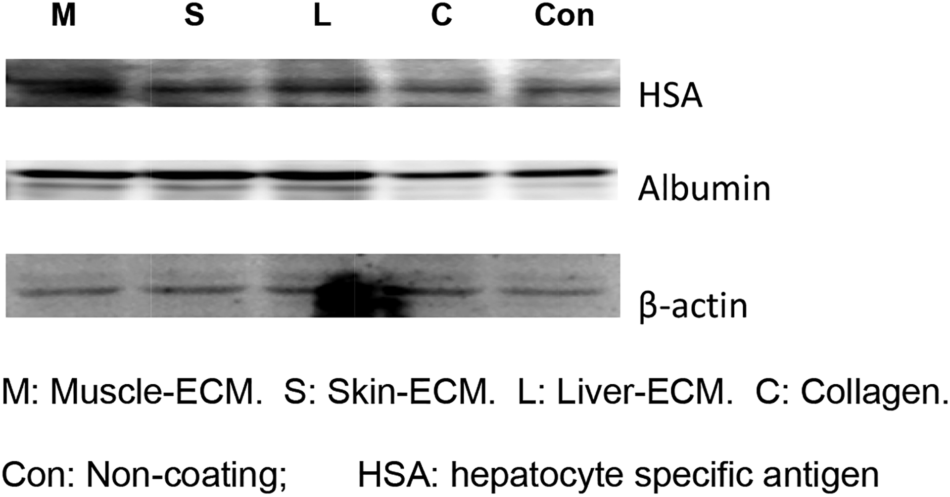
Recent studies have shown that extracellular matrix (ECM) substitutes can have a dramatic impact on cell growth, differentiation and function. However, these ECMs are often applied generically and have yet to be developed for specific cell types. In this study, we developed tissue-specific ECM-based coating substrates for skin, skeletal muscle and liver cell cultures. Cellular components were removed from adult skin, skeletal muscle, and liver tissues, and the resulting acellular matrices were homogenized and dissolved. The ECM solutions were used to coat culture dishes. Tissue matched and non-tissue matched cell types were grown on these coatings to assess adhesion, proliferation, maintenance of phenotype and cell function at several time points. Each cell type showed better proliferation and differentiation in cultures containing ECM from their tissue of origin. Although subtle compositional differences in the three ECM types were not investigated in this study, these results suggest that tissue-specific ECMs provide a culture microenvironment that is similar to the in vivo environment when used as coating substrates, and this new culture technique has the potential for use in drug development and the development of cell-based therapies.


Figures

Fig. 1
Fig. 1
Schematic representation of the experimental design
Fig. 2
Fig. 2. Histology of fresh liver, muscle and skin tissue and corresponding decellularized ECM using H&E and DAPI-staining.
Histology of fresh liver (a,b) and decellularized liver (g,h) fresh muscle (c,d) and decellularized muscle (I,j) and fresh skin (e,f) and decellularized skin (k,l). The top panels show H&E (a-f) and the bottom panels, DAPI staining (g-l). Magnification, 100x. Cellular components are clearly visible in the fresh-frozen tissues (a, c, e) and rare or absent in the decellularized ECM tissues (b, d, f). Well-organized, punctate nuclei were clearly visible in fresh-frozen tissues (g,i,k), whereas efficient cellular removal was noted in the decellularized tissues. Only strands of disrupted DNA and RNA remnants can be seen, indicating that cells were no longer intact (h, j, l).
Fig. 3
Fig. 3. Growth curve of skeletal muscle, skin and liver cells on tissue-specific ECM coatings.
(a) Skeletal muscle cells, (b) skin cells and (c) liver cells were cultured on different tissue-specific ECM coatings for 6 days in 24 well plates. Media was replaced every other day and cells were counted using a Coulter counter on day 1, 2, 4 and 6. Growth and proliferation of each cell type was maximum on ECM derived from its tissue of origin.
Fig. 4
Fig. 4. Morphology and immunofluorescence analysis of cells cultured on tissue-specifc ECM coatings.
Cells were cultured on their respective tissue-specific ECM for 5 days. The cells were stained for specific protein markers by immunofluorescence. The top row ( AE1/AE3 and vimentin) for skin cells, middle row (desmin and myosin) for skelton muscle cells, and low row (albumin and hepatocyte-specific antigen) for Hep G2 cells depict fluorescent images. Magnification, X100
Fig. 5
Fig. 5. Immunoblot cell extracts from HepG2 cells cultured on tissue-specific ECM coatings.
HepG2 cells were cultured on different tissue-specific ECM coatings for 14 days before protein lysates were prepared. Proteins were electrophoresed and transferred to a membrane, which was probed with antibodies to HAS, albumin and β-actin (loading control). Higher expression of liver-specific proteins are visible when cells are cultured on ECM versus control (no ECM coating) plates.

References

    1. Sadoshima J, Izumo S. The cellular and molecular response of cardiac myocytes to mechanical stress. Annu Rev Physiol 1997;59:551–571. - PubMed
    1. Syedain ZH, Weinberg JS, Tranquillo RT. Cyclic distension of fibrin-based tissue constructs: evidence of adaptation during growth of engineered connective tissue. Proc Natl Acad Sci USA 2008; 105:6537–6542. - PMC - PubMed
    1. Funderburgh ML, Mann MM, Funderburgh JL. Keratocyte phenotype is enhanced in the absence of attachment to the substratum. Mol Vis 2008;14:308–317. - PMC - PubMed
    1. Duffy DM, Garrett SM, Ellis SE, Scott TR. Influence of supramammary lymph node extract on in vitro cell proliferation. Cell Prolif 2008;41:299–309. - PMC - PubMed
    1. Yoshida A, Kanno H, Watabe D, Akasaka T, Sawai T. The role of heparin-binding EGF-like growth factor and amphiregulin in the epidermal proliferation of psoriasis in cooperation with TNFalpha. Arch Dermatol Res 2008;300:37–45. - PubMed

Publication types

Substances