. 2009 May 1;137(3):433-44.
doi: 10.1016/j.cell.2009.02.046.

HIV enters cells via endocytosis and dynamin-dependent fusion with endosomes

Affiliations

HIV enters cells via endocytosis and dynamin-dependent fusion with endosomes

Kosuke Miyauchi et al. Cell. .

Abstract

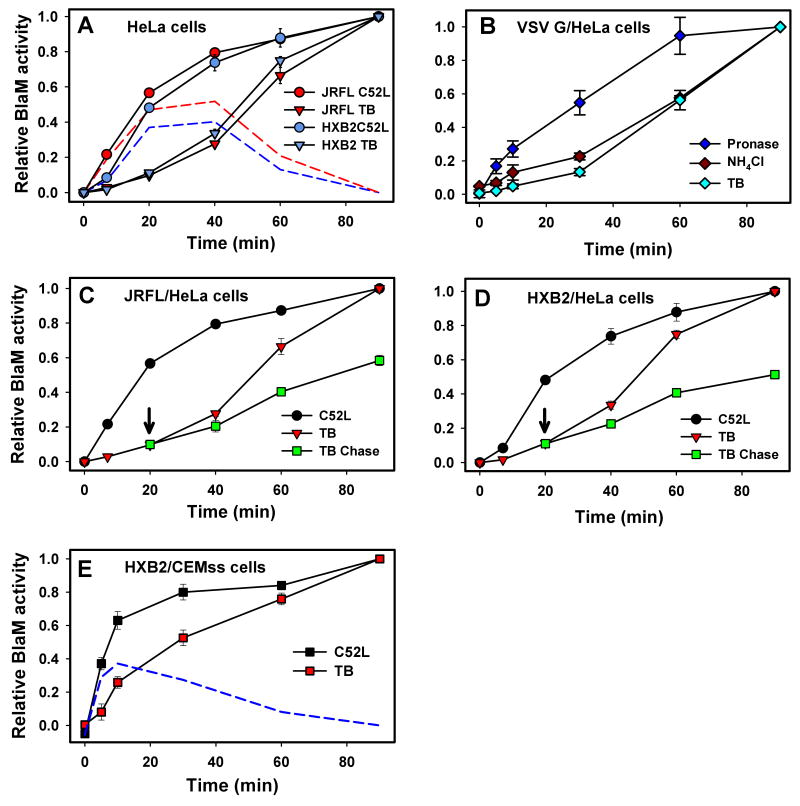
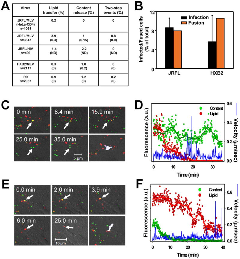
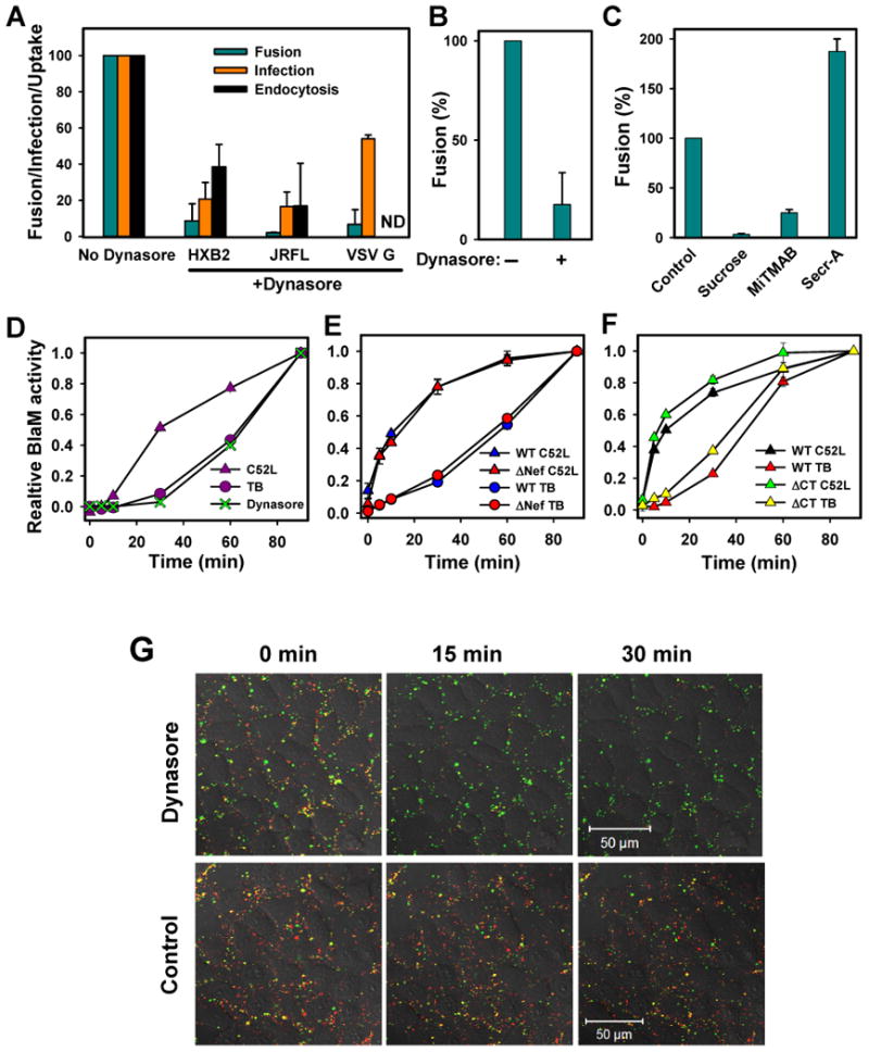
Enveloped viruses that rely on a low pH-dependent step for entry initiate infection by fusing with acidic endosomes, whereas the entry sites for pH-independent viruses, such as HIV-1, have not been defined. These viruses have long been assumed to fuse directly with the plasma membrane. Here we used population-based measurements of the viral content delivery into the cytosol and time-resolved imaging of single viruses to demonstrate that complete HIV-1 fusion occurred in endosomes. In contrast, viral fusion with the plasma membrane did not progress beyond the lipid mixing step. HIV-1 underwent receptor-mediated internalization long before endosomal fusion, thus minimizing the surface exposure of conserved viral epitopes during fusion and reducing the efficacy of inhibitors targeting these epitopes. We also show that, strikingly, endosomal fusion is sensitive to a dynamin inhibitor, dynasore. These findings imply that HIV-1 infects cells via endocytosis and envelope glycoprotein- and dynamin-dependent fusion with intracellular compartments.


Conflict of interest statement

The authors have no conflict of interest to declare.

Figures

Figure 1
Figure 1
Dissection of surface and endosomal HIV-1 fusion. (A) Virus fusion with TZM-bl cells was stopped by adding C52L after indicated times of incubation at 37°C, and incubation was continued up to 90 min, at which point the cells were briefly placed on ice and loaded with the BlaM substrate. Alternatively, fusion was stopped by placing cells on ice after varied times of incubation at 37°C (TB). After loading the substrate, cells were incubated overnight at 13.5°C regardless of the fusion protocol to allow the substrate cleavage. The red and blue dashed lines were obtained by subtracting the TB plot from the C52L escape plot for JRFL and HXB2, respectively. Unless stated otherwise, data points are means +/-SEM from triplicate measurements. (B) Fusion of VSV G pseudotypes with TZM-bl cells was blocked at indicated times either by treating cells with 2 mg/ml pronase on ice (10 min), adding 50 mM NH4Cl, or chilling the samples (TB). Cells were then loaded with CCF2 and incubated overnight at 12°C. (C, D) After 20 min at 37°C, viruses remaining at the surface of TZM-bl cells were rendered non-fusogenic by adding C52L (arrow), and the extent of fusion over time at 37°C was determined by chilling cells either immediately or at indicated time points (red triangles). (E) HXB2 virus escape from C52L and from the TB in CEMss cells was measured as described above. The dashed blue line represents the difference between the C52L and TB curves. Error bars are SEM (n=4).
Figure 2
Figure 2
Identification of HIV-1 fusion sites by single virus imaging. (A) Schematic presentation of redistribution of viral lipid and content markers upon fusion with a plasma membrane (left) and with an endosome (right). Viruses co-labeled with membrane (red) and content (green) markers are pseudocolored yellow. (B, C) Partial fusion of JRFL with the plasma membrane of TZM-bl cells. The time from the beginning of imaging is shown. The two-dimensional projection of the particle's trajectory (cyan) is overlaid on the last image. Changes in fluorescence intensities (in arbitrary units) of membrane (red) and content (green) markers, as well as the instantaneous velocity (blue trace) of the particle, are shown. (D-G) Complete fusion of JRFL (D, E) and HXB2 (F, G) viruses following the fast retrograde movement from the cell periphery (cyan traces on last images). Fusion is evident from the disappearance of GFP signal. Graphs (E and G) show changes in fluorescence of membrane and content markers (smoothed for visual clarity) and 3D trajectories for the viruses marked by arrows in panels D and F. See supplemental movies 3-5.
Figure 3
Figure 3
Fusion of HIV core-based pseudoviruses and of infectious HIV-1 with TZM-bl cells. (A) The efficiency of lipid mixing with the plasma membrane, the viral content release from endosomes, and the sequential 2-step fusion events mediated by JRFL Env (pseudotyped with MLV or HIV core), HXB2 Env, and by infectious R9 viruses. The first row is the negative control for JRFL fusion using HeLa-CD4 cells lacking CCR5. The number of respective fusion-related events was normalized to the total number of cell-associated double-labeled virions at the beginning of experiment. The extents of viral lipid and content mixing in the presence of 4 μM C52L are shown in parentheses. The rare false-positive events in the presence of C52L were usually delayed relative to those in the absence of the inhibitor (data not shown) and were minimized by limiting the duration of imaging experiments. ND, not determined. (B) The fraction of cells supporting viral content release was measured by imaging (orange bars), and the fraction of infected cells in the same sample was determined by a β-Gal assay (black bars) after an additional 48 hr-cultivation in the presence of C52L. The somewhat larger fraction of cells supporting HXB2 fusion was due to a more efficient binding of HXB2 to target cells compared to JRFL viruses (data not shown). (C, D) Lipid transfer initiated by the infectious R9 HIV-1 labeled with DiD and MA-GFP-CA at the surface of the TZM-bl cell. (E, F) Complete endosomal fusion of the infectious R9 particle. See the supplementary movie 6.
Figure 4
Figure 4
Lipid mixing at the cell surface precedes the content release from endosomes. (A) The kinetics of JRFL (circles) and HXB2 (triangles) fusion with TZM-bl cells. Waiting times from the shift to 37°C to the point of lipid (red symbols) or content (green symbols) transfer were measured, rank ordered, and plotted as cumulative distributions of the fraction of fused viruses over time. (B) Comparative kinetics of partial fusion at the cell surface (red circles), endosomal fusion (green circles), and of sequential lipid and content transfer exhibited during the 2-step events (red and green triangles, respectively). The time intervals between sequential content (TC) and lipid (TL) transfer were ranked and plotted as cumulative distribution (crosses). (C) The kinetics of lipid and content mixing during fusion of infectious R9 HIV-1 viruses with TZM-bl cells.
Figure 5
Figure 5
Sequential lipid and content transfer events exhibited by HIV-1. (A, B) A 2-step fusion event exhibiting a short delay between lipid and content transfer. (C, D) A rare 2-step event characterized by stepwise release of viral content. Complete content discharge (double arrow) occurs after the onset of quick movement (see the movie 7). The the predicted dynamics of a fusion pore (panel D) is shown by a thick line above the panel. The initial and final coordinates of particles on 3D-plots are marked by pink crosses and stars, respectively. (E, F) The 2-step fusion of infectious R9 HIV-1 co-labeled with DiD and MA-GFP-CA. Changes in fluorescence intensities of viral lipid and content markers upon incubation with TZM-bl cells at 37°C are smoothed for visual clarity.
Figure 6
Figure 6
Blocking the dynamin function inhibits HIV-1 uptake, fusion and infection. (A) TZM-bl cells were pre-treated with 80 μM dynasore and allowed to bind viruses in the cold. Virus uptake after a 1 hr-incubation at 37°C was measured by the accumulation of intracellular p24 (black bars, n=6), as described in Supplemental Experimental Procedures. The extent of fusion was quantified by the BlaM assay (dark cyan bars, n=4), and single-cycle infection was measured by a β-Gal assay (orange bars, n=6); (B) Inhibition of HXB2 fusion with CEMss cells pretreated with dynasore. (C) Inhibition of HXB2 fusion in TZM-bl cells pretreated with 0.45 M sucrose, 80 μM MiTMAB, and 15 μM secramine A. (D) The kinetics of HXB2 escape from 80 μM dynasore added at indicated times of incubation at 37°C. The background fusion (∼20% of total) in the presence of dynasore was subtracted from the dynasore curve to ease the comparison with the C52L and TB curves. (E) Fusion of wild-type (WT) and Nef-deficient (ΔNef) viruses bearing HXB2 Env with TZM-bl cells. (F) Kinetics of fusion mediated by the wild-type and cytoplasmic tail-deleted (ΔCT) JRFL Env. (G) Pre-treatment of TZM-bl cells with dynasore blocks the HXB2 content (NC-GFP) transfer while permitting lipid mixing (disappearance of DiD) with the plasma membrane. The reduction in the number of GFP-labeled particles at a later time point is mainly due to virus detachment.

Comment in

  • HIV Entry Revisited.
    Uchil PD, Mothes W. Uchil PD, et al. Cell. 2009 May 1;137(3):402-4. doi: 10.1016/j.cell.2009.04.033. Cell. 2009. PMID: 19410537 Free PMC article.

References

    1. Aiken C. Pseudotyping human immunodeficiency virus type 1 (HIV-1) by the glycoprotein of vesicular stomatitis virus targets HIV-1 entry to an endocytic pathway and suppresses both the requirement for Nef and the sensitivity to cyclosporin A. J Virol. 1997;71:5871–5877. - PMC - PubMed
    1. Bayer N, Schober D, Prchla E, Murphy RF, Blaas D, Fuchs R. Effect of bafilomycin A1 and nocodazole on endocytic transport in HeLa cells: implications for viral uncoating and infection. J Virol. 1998;72:9645–9655. - PMC - PubMed
    1. Beer C, Andersen DS, Rojek A, Pedersen L. Caveola-dependent endocytic entry of amphotropic murine leukemia virus. J Virol. 2005;79:10776–10787. - PMC - PubMed
    1. Brandt SM, Mariani R, Holland AU, Hope TJ, Landau NR. Association of chemokine-mediated block to HIV entry with coreceptor internalization. J Biol Chem. 2002;277:17291–17299. - PubMed
    1. Campbell EM, Nunez R, Hope TJ. Disruption of the actin cytoskeleton can complement the ability of Nef to enhance human immunodeficiency virus type 1 infectivity. J Virol. 2004;78:5745–5755. - PMC - PubMed

Publication types

MeSH terms