. 2009 May 1;137(3):472-84.
doi: 10.1016/j.cell.2009.02.039.

Centromere-specific assembly of CENP-a nucleosomes is mediated by HJURP

Affiliations

Centromere-specific assembly of CENP-a nucleosomes is mediated by HJURP

Daniel R Foltz et al. Cell. .

Abstract

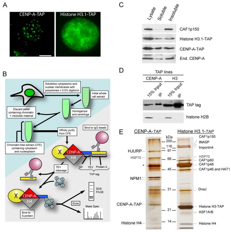
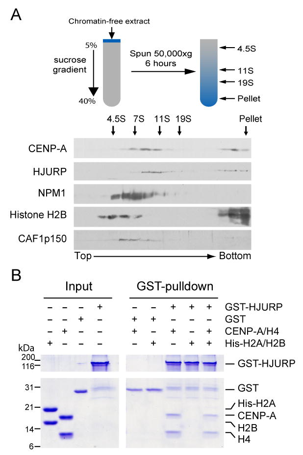
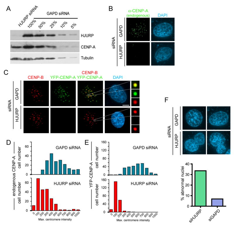
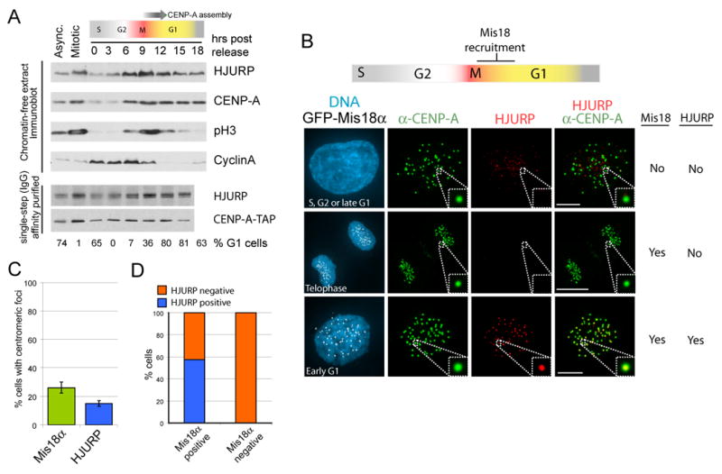
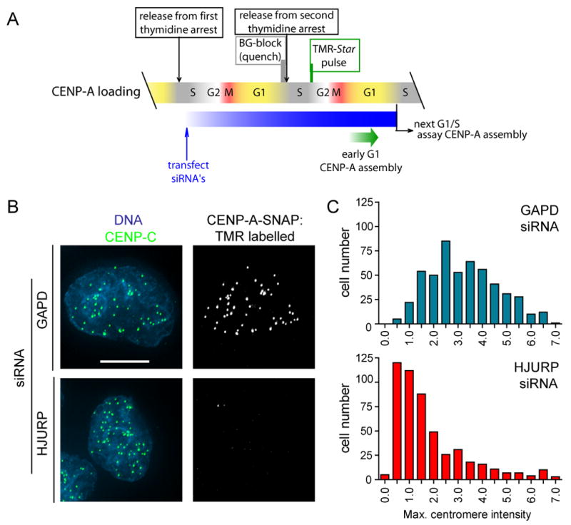
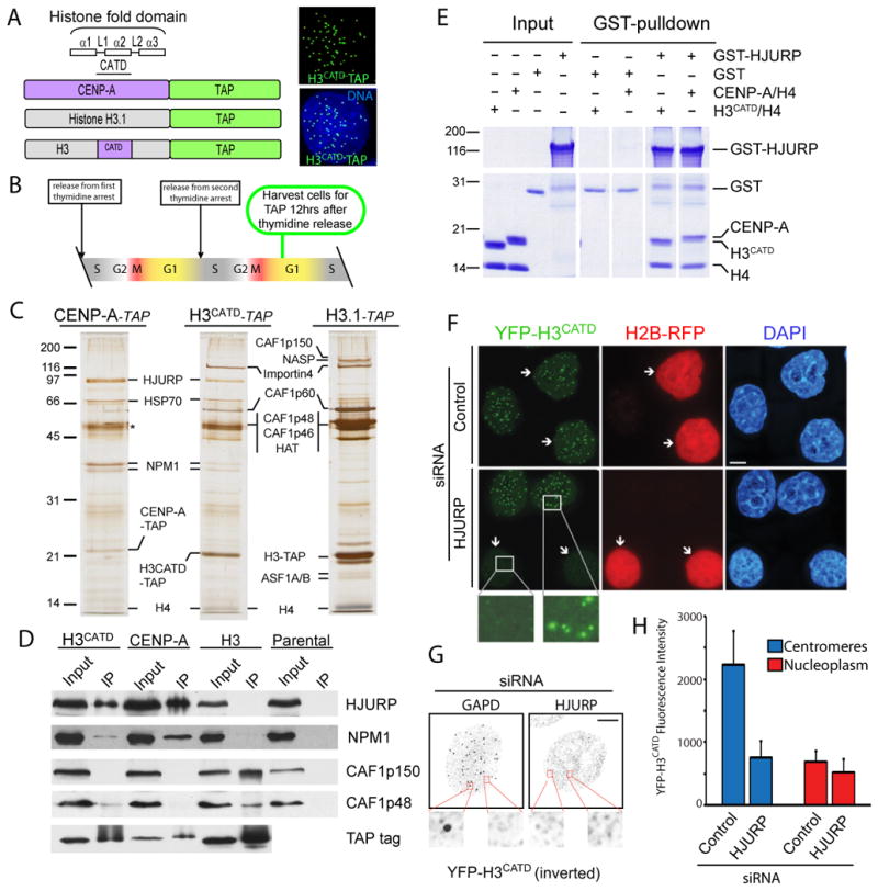
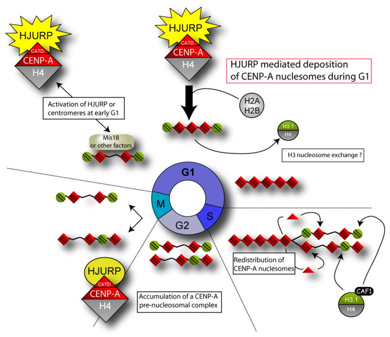
The centromere is responsible for accurate chromosome segregation. Mammalian centromeres are specified epigenetically, with all active centromeres containing centromere-specific chromatin in which CENP-A replaces histone H3 within the nucleosome. The proteins responsible for assembly of human CENP-A into centromeric nucleosomes during the G1 phase of the cell cycle are shown here to be distinct from the chromatin assembly factors previously shown to load other histone H3 variants. Here we demonstrate that prenucleosomal CENP-A is complexed with histone H4, nucleophosmin 1, and HJURP. Recruitment of new CENP-A into nucleosomes at replicated centromeres is dependent on HJURP. Recognition by HJURP is mediated through the centromere targeting domain (CATD) of CENP-A, a region that we demonstrated previously to induce a unique conformational rigidity to both the subnucleosomal CENP-A heterotetramer and the corresponding assembled nucleosome. We propose HJURP to be a cell-cycle-regulated CENP-A-specific histone chaperone required for centromeric chromatin assembly.


Figures

Figure 1
Figure 1. Affinity purification of the CENP-A prenucleosomal complex
(A) Localization of CENP-A-TAP and histone H3.1-TAP in stable cell lines to centromeres and chromatin, respectively. The scale bar equals 5μm. (B) Purification scheme for identification of a soluble CENP-A prenucleosomal complex by production of a chromatin-free extract followed by tandem affinity purification. (C) Immunoblots of chromatin-free extract extracts derived from parental HeLa cells and stable cell lines expressing TAP-tagged CENP-A or histone H3.1 demonstrate the presence of tagged and endogenous histones as well as chromatin assembly factors. (D) Single-step affinity purifications of TAP-tagged CENP-A and histone H3.1 immunoblotted for the affinity tag as well as histone H2A. (E) Tandem affinity purified CENP-A and histone H3.1 and the associated complexes from chromatin free extracts were visualized by silver stain. Asterisk indicates contaminant present in both preparations. Proteins associated with the soluble complexes were identified in solution by MudPIT mass spectrometry.
Figure 2
Figure 2. Identification of an endogenous CENP-A prenucleosomal complex
(A) Chromatin-free extract derived from HeLa cells was subjected to sucrose gradient sedimentation. The bottom of the gradient appears on the right. The migration of sedimentation coefficient standards bovine serum albumin, aldolase, catalase and thyroglobulin are indicated by arrows at the top. (B) A direct interaction was observed between recombinant GST-HJURP and recombinant untagged CENP-A:H4 but not recombinant His-H2A:H2B dimer. Following purification on glutathione agarose, proteins associated with GST-HJURP were separated by SDS-PAGE and stained with Coomassie blue.
Figure 3
Figure 3. Loss of CENP-A recruitment in HJURP depleted cells
(A) Extracts from HeLa cells depleted of endogenous HJURP by treatment with siRNA pools for 72 hours were subjected to immunoblot. Serial dilution of GAPD control treated cell extracts was used to determine the degree of HJURP knockdown. Parental HeLa cells (B,D) or HeLa cells stably expressing YFP-CENP-A (C and E) treated with HJURP siRNA pools. Cells were pre-extracted and fixed 72 hours after the initiation of siRNA treatment and CENP-A and YFP-CENP-A were detected by immunofluoresence. Centromeres were identified by using anti-CENP-B monoclonal antibody in (C). Reduction of endogenous (B, n=200) and YFP-CENP-A (C, n=250) in response to HJURP siRNA was assessed by measuring the maximum pixel intensity per nucleus. All values are background corrected. Scale bar equals 5μm. (F) Abnormally shaped nuclei were observed by DAPI stain in HJURP siRNA treated cells at a greater frequency than controls. Abnormal nuclei included those that were multi lobed or contained micronuclei.
Figure 4
Figure 4. Cell cycle regulation of HJURP accumulation and recruitment
(A) The levels of soluble CENP-A and HJURP across the cell cycle were assessed in chromatin-free extracts from synchronized cells by immunoblot. Each lane contains extract from 1 X 10⁵ cells. The percent of cells in G1 was determined by FACS analysis. (B) Cells stably expressing GFP-Mis18α were pre-extracted and fixed prior to immunostaining for HJURP and CENP-A. (C,D) Cells were scored for the accumulation of HJURP and GFP-Mis18α at CENP-A foci (n=280 cells). Data are represented as the mean (+/- S.D.), Scale bar equals 5μm.
Figure 5
Figure 5. HJURP is required for recruitment of new CENP-A nucleosomes
(A) Scheme for siRNA treatment and SNAP labeling to visualize loading of newly synthesized CENP-A-SNAP in the absence of HJURP. (B) Loading of newly synthesized CENP-A was assessed by the ability of cells to recruit TMR-Star labeled (i.e. newly synthesized) CENP-A to centromeres in cells treated with HJURP or GAPD siRNA and SNAP labeled as described in (A). Centromeres are shown by immunostaining for CENP-C. Scale bar equals 5μm. (C) The degree of TMR labeled CENP-A-SNAP loading into centromeres in siRNA treated cells was quantified by measuring the maximum pixel intensity per nucleus in >500 cells per condition.
Figure 6
Figure 6. Histone H3CATD chimeric protein recruits HJURP
(A) Stable cell lines expressing a chimeric H3CATD in which the loop1 and α2 helix of CENP-A were swapped into histone H3 and fused to the TAP tag. H3CATD-TAP localizes to discreet centromeric foci. (B) Cells were harvested for tandem affinity purification (TAP) 12 hours after release from double thymidine arrest when the majority of cells are in the G1 phase. (C) Tandem affinity purifications conducted from stable cell lines during the G1 phase of the cell cycle. Proteins were identified by in-gel trypsin digestion and mass spectrometry, (D) Immunoblots of single-step affinity purifications. (E) Recombinant co-expressed chimeric H3CATD and histone H4 directly interacts with GST-HJURP in vitro. (F) A cell line that stably expresses YFP-H3CATD was treated with control or HJURP-directed siRNA and processed for immunofluorescence 72 hours following transfection. Arrowheads indicate cells co-transfected with the indicated siRNA plasmids and a plasmid encoding H2B-RFP. (G) Inverted grayscale images of YFP-H3CATD in which soluble protein was pre-extracted prior to fixation. Scale bars equal 5μm. (H) Quantitation of the effect of HJURP siRNA or control treatment on YFP-H3CATD fluorescent intensity. Centromeres and nucleoplasm were distinguished based on ACA immunostaining (n=12).
Figure 7
Figure 7. Model of CENP- A nucleosome deposition at centromeres
Replication of sister chromatids requires that new CENP-A be deposited specifically at centromeric loci in order to maintain the epigenetic mark of the centromere. Existing CENP-A nucleosomes are distributed to sister chromosomes during DNA replication. Cells progress through G2 and mitosis with half the maximal centromeric complement of CENP-A nucleosomes. CENP-A nucleosomes are assembled in a HJURP dependent manner during the G1 phase of the cell cycle. HJURP and CENP-A levels accumulate during G2 and peak during M even though active assembly of CENP-A is restricted to G1. Restriction of CENP-A assembly may be achieved by modification of the CENP-A prenucleosomal complex or through the recruitment of other proteins recruited to the centromere during G1, such as the Mis18 complex, that may serve to prime the centromere and restrict CENP-A nucleosomes assembly.

References

    1. Ahmad K, Henikoff S. The histone variant H3.3 marks active chromatin by replication-independent nucleosome assembly. Mol Cell. 2002;9:1191–1200. - PubMed
    1. Black BE, Brock MA, Bedard S, Woods VL, Jr, Cleveland DW. An epigenetic mark generated by the incorporation of CENP-A into centromeric nucleosomes. Proc Natl Acad Sci U S A. 2007;104:5008–5013. - PMC - PubMed
    1. Black BE, Foltz DR, Chakravarthy S, Luger K, Woods VL, Jr, Cleveland DW. Structural determinants for generating centromeric chromatin. Nature. 2004;430:578–582. - PubMed
    1. Black BE, Jansen LE, Maddox PS, Foltz DR, Desai AB, Shah JV, Cleveland DW. Centromere identity maintained by nucleosomes assembled with histone H3 containing the CENP-A targeting domain. Mol Cell. 2007;25:309–322. - PubMed
    1. Blower MD, Sullivan BA, Karpen GH. Conserved organization of centromeric chromatin in flies and humans. Dev Cell. 2002;2:319–330. - PMC - PubMed

Publication types