. 2009 Jul;46(1):30-8.
doi: 10.1016/j.ceca.2009.03.018. Epub 2009 May 2.

Homeostatic and stimulus-induced coupling of the L-type Ca2+ channel to the ryanodine receptor in the hippocampal neuron in slices

Affiliations

Homeostatic and stimulus-induced coupling of the L-type Ca2+ channel to the ryanodine receptor in the hippocampal neuron in slices

Jonathan Berrout et al. Cell Calcium. 2009 Jul.

Abstract

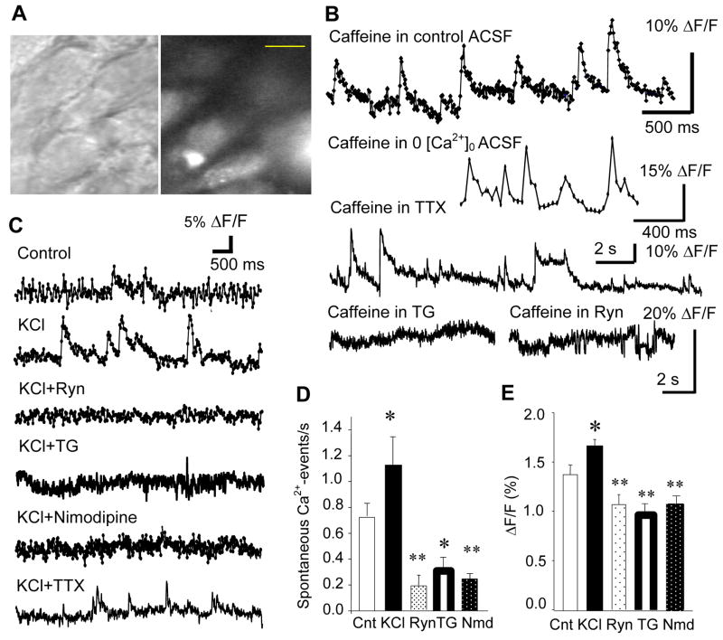
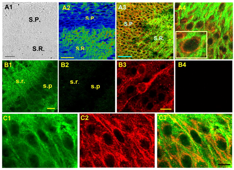
Activity-dependent increase in cytosolic calcium ([Ca(2+)](i)) is a prerequisite for many neuronal functions. We previously reported a strong direct depolarization, independent of glutamate receptors, effectively caused a release of Ca(2+) from ryanodine-sensitive stores and induced the synthesis of endogenous cannabinoids (eCBs) and eCB-mediated responses. However, the cellular mechanism that initiated the depolarization-induced Ca(2+)-release is not completely understood. In the present study, we optically recorded [Ca(2+)](i) from CA1 pyramidal neurons in the hippocampal slice and directly monitored miniature Ca(2+) activities and depolarization-induced Ca(2+) signals in order to determine the source(s) and properties of [Ca(2+)](i)-dynamics that could lead to a release of Ca(2+) from the ryanodine receptor. In the absence of depolarizing stimuli, spontaneously occurring miniature Ca(2+) events were detected from a group of hippocampal neurons. This miniature Ca(2+) event persisted in the nominal Ca(2+)-containing artificial cerebrospinal fluid (ACSF), and increased in frequency in response to the bath-application of caffeine and KCl. In contrast, nimodipine, the antagonist of the L-type Ca(2+) channel (LTCC), a high concentration of ryanodine, the antagonist of the ryanodine receptor (RyR), and thapsigargin (TG) reduced the occurrence of the miniature Ca(2+) events. When a brief puff-application of KCl was given locally to the soma of individual neurons in the presence of glutamate receptor antagonists, these neurons generated a transient increase in the [Ca(2+)](i) in the dendrosomal region. This [Ca(2+)](i)-transient was sensitive to nimodipine, TG, and ryanodine suggesting that the [Ca(2+)](i)-transient was caused primarily by the LTCC-mediated Ca(2+)-influx and a release of Ca(2+) from RyR. We observed little contribution from N- or P/Q-type Ca(2+) channels. The coupling between LTCC and RyR was direct and independent of synaptic activities. Immunohistochemical study revealed a cellular localization of LTCC and RyR in a juxtaposed configuration in the proximal dendrites and soma. We conclude in the hippocampal CA1 neuron that: (1) homeostatic fluctuation of the resting membrane potential may be sufficient to initiate functional coupling between LTCC and RyR; (2) the juxtaposed localization of LTCC and RyR has anatomical advantage of synchronizing a Ca(2+)-release from RyR upon the opening of LTCC; and (3) the synchronized Ca(2+)-release from RyR occurs immediately after the activation of LTCC and determines the peak amplitude of depolarization-induced global increase in dendrosomal [Ca(2+)](i).


Figures

Figure 1
Figure 1
Miniature [Ca2+]i events in the CA1 pyramidal cell. A. Visually-identified CA1 pyramidal cells with DIC (left) were loaded with fluo-3AM (right). Calibration bar: 10 μm. B. Caffeine enhanced the probability of generating miniature [Ca2+]i-events, which persisted in the Ca2+-free ACSF and in the tetrodotoxin (TTX, 1 μM)-containing ACSF. However, caffeine was not effective of generating any [Ca2+]i-event in the presence of thapsigargin (TG, 4 μM) and ryanodine (Ryn, 10 μM). C. Miniature [Ca2+]i-events in the control ACSF, 80 mM K+-containing ACSF, 80 mM K+ and ryanodine-containing ACSF, 80 mM K+ and TG-containing ACSF, 80 mM K+ and nimodipine-containing ACSF, and 80 mM K+ and TTX-containing ACSF. D. Mean frequency of miniature [Ca2+]i-events in the above 5 experimental conditions (excluding TTX experiment, see text for explanations). E. Mean amplitude (measured as %ΔF/F0) of miniature [Ca2+]i events in the 5 conditions. Cnt: Control, KCl: 80mM K+-containing ACSF, Ryn: ryanodine and 80 mM K+-containing ACSF, TG: thapsigargin and 80 mM K+-containing ACSF, Nmd: nimodipine and 80 mM K+-containing ACSF.
Figure 2
Figure 2
Cellular expression of RyR and caffeine-induced [Ca2+]i-responses. A. A confocal image of RyR immunoreactivity in the CA1 pyramidal cell. B. Fluo-3AM-loaded pyramidal cells increased [Ca2+]i-signals in response to a puff-application of caffeine (B1: before, B2: after). Calibration bar: 10 μm in A and B. C. Caffeine-induced [Ca2+]i-increase in control ACSF (C1:Mean ± SEM in 5 cells). Inset in C1 shows a single caffeine response in single neuron. Repeated caffeine puffs (5s-puff, indicated by black arrows) depleted caffeine-sensitive Ca2+ stores (two representative patterns of depletion in C2 and C3). Caffeine puffs were not effective of generating any [Ca2+]i-increase in the presence of thapsigargin (TG: 4 μM, C4). D. Time-dependent changes in the caffeine-induced [Ca2+]i-response in 0[Ca2+]o ACSF in two neurons, each with a representative release property. Inset shows a Ca2+ response to a single caffeine puff in a single neuron in 0[Ca2+]o ACSF. E. KCl-induced [Ca2+]i-increase in 0[Ca2+]o ACSF. The plot shows the average and SEM of peak [Ca2+]i-increase caused by KCl in 7 neurons. Inset shows a [Ca2+ ]i-increase in response to a single KCl application in a single neuron in 0[Ca2+]o ACSF.
Figure 3
Figure 3
Juxtaposed localization of RyR and LTCC (CaV1.2). A1. A bright field image of the hippocampal slice culture that was used for dual labeling of RyR and CaV1.2. A2. CaV1.2 immunoreactivity in CA1 (warmer color represents stronger immunoreactivity). A3. Dual labeling of RyR (red) and CaV1.2 (green). SP: Stratum Pyramidale, SR: Stratum Radiatum. A4. Dual labeling in the pyramidal cell layer with higher magnification. B1. CaV1.2. B2. CaV1.2 with a blocking peptide. B3. RyR, B4. RyR with a blocking peptide. C13. Dual labeling of CaV1.2 and RyR with an image of CaV1.2 only (C1), RyR only (C2), and a merged image of both CaV1.3 and RyR (C3). These three images were taken sequentially from the identical population of the neurons in the same slice. Calibration bar: 50 μm in A1 and A2, 20 μm in B1 and B3, 10 μm in C3.
Figure 4
Figure 4
A. Depolarization-induced [Ca2+]i-elevation and its modulation by ryanodine (traces are shown as mean ± SEM). Inset shows Fluo-3 images before (1) and after (2) KCl puff. Calibration bar: 15 μm. B1. Depolarization-induced [Ca2+]i-elevation in the presence of kynurenic acid, nimodipine, and ryanodine. B2. Responses to caffeine in the control ACSF and the thapsigargin (TG)-containing ACSF in neurons. B3. The same neurons in B2 generated a [Ca2+]i-increase in response to KCl-induced depolarization. A caffeine-puff of 5s immediately before the depolarization (indicated by a horizontal bar) amplified a peak amplitude of the [Ca2+]i-increase. On the other hand, TG reduced the [Ca2+]i-peak to less than 1/3 of the control. C1. Depolarization-induced [Ca2+]i-elevation in the control ACSF and its reduction by nimodipine and ryanodine. C2. Effect of ω-conotoxin on depolarization-induced [Ca2+]i-elevation. C3. Effect of ω-agatoxin on depolarization-induced [Ca2+]i-elevation. D. Time-dependent changes in depolarization-induced [Ca2+]i-elevation in control, nimodipine, and ryanodine. E1. Changes in peak amplitude and peak latency of depolarization-induced [Ca2+]i-elevation by ryanodine and nimodipine. Inset shows differences in peak [Ca2+]i-elevation and the peak latency between control and nimodipine (green) and between nimodipine and nimodipine plus ryanodine (black). E2. Changes in the identical parameters shown in E1 in the presence of kynurenic acid in ACSF.

References

    1. Sharp AH, McPherson PS, Dawson TM, Aoki C, Campbell KP, Snyder SH. Differential immunohistochemical localization of inositol 1,4,5-trisphosphate- and ryanodine-sensitive Ca2+ release channels in rat brain. J Neurosci. 1993;13:3051–3063. - PMC - PubMed
    1. Chavis P, Fagni L, Lansman JB, Bockaert J. Functional coupling between ryanodine receptors and L-type calcium channels in neurons. Nature. 1996;382:719–722. - PubMed
    1. Bito H, Deisseroth K, Tsien RW. CREB phosphorylation and dephosphorylation: a Ca and stimulus duration-dependent switch for hippocampal gene expression. Cell. 1996;87:1203–1214. - PubMed
    1. Bouchard R, Pattarini R, Geiger JD. Presence and functional significance of presynaptic ryanodine receptors. Prog Neurobiol. 2003;69:391–418. - PubMed
    1. Melis M, Perra S, Muntoni AL, Pillolla G, Lutz B, Marsicano G, Di Marzo V, Gessa GL, Pistis M. Prefrontal cortex stimulation induces 2-arachidonoylglycerol-mediated suppression of excitation in dopamine neurons. J Neurosci. 2004;24:10707–10715. - PMC - PubMed

Publication types