. 2009 Jul;297(1):H304-12.
doi: 10.1152/ajpheart.00077.2008. Epub 2009 May 1.

Role of cGMP-dependent protein kinase in regulation of pulmonary vascular smooth muscle cell adhesion and migration: effect of hypoxia

Affiliations

Role of cGMP-dependent protein kinase in regulation of pulmonary vascular smooth muscle cell adhesion and migration: effect of hypoxia

S Negash et al. Am J Physiol Heart Circ Physiol. 2009 Jul.

Abstract

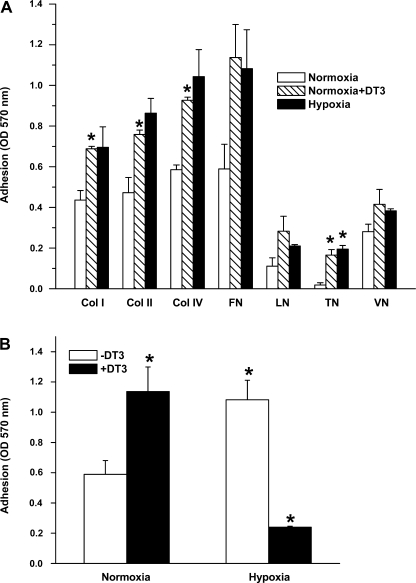
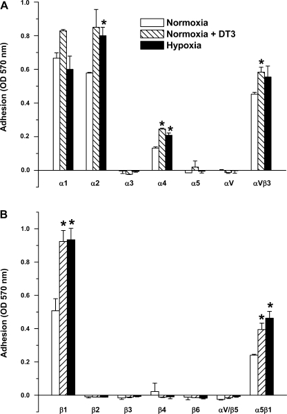
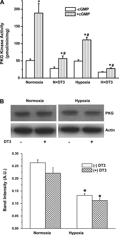
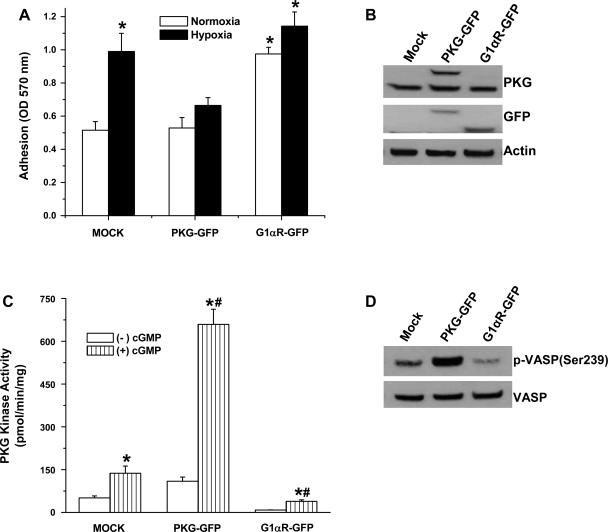
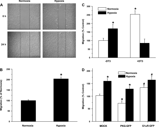
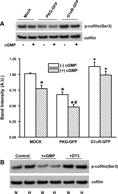
Exposure to prolonged hypoxia can result in pulmonary vascular remodeling and pulmonary hypertension. Hypoxia induces pulmonary vascular smooth muscle cell (PVSMC) proliferation and vascular remodeling by affecting cell adhesion and migration and secretion of extracellular matrix proteins. We previously showed that acute hypoxia decreases cGMP-dependent protein kinase (PKG) activity in PVSMC and that PKG plays a role in maintaining the differentiated contractile phenotype in normoxia. In this study, we investigated the effect of hypoxia on PVSMC adhesion and migration and the role of PKG in these functions. Ovine fetal pulmonary artery SMC were incubated in normoxia (Po(2) approximately 100 Torr) or hypoxia (Po(2) approximately 30-40 Torr) or treated with the PKG inhibitor DT-3 for 24 h in normoxia. To further study the role of PKG in the modulation of adhesion and migration, PVSMC were transiently transfected with a full-length PKG1alpha [PKG-green fluorescent protein (GFP)] or a dominant-negative construct (G1alphaR-GFP). Cell adhesion to extracellular matrix proteins was determined, and integrin-mediated adhesion was assessed by alpha/beta-integrin-mediated cell adhesion array. Exposure to hypoxia (24 h) and pharmacological inhibition of PKG1 by DT-3 significantly promoted adhesion mediated by alpha(4)-, beta(1)-, and alpha(5)beta(1)-integrins to fibronectin, laminin, and tenacin and also resulted in increased cell migration. Likewise, inhibition of PKG by expression of a dominant-negative PKG1alpha construct increased cell adhesion and migration, comparable to that induced by hypoxia. Dynamic actin reorganization associated with integrin-mediated cell adhesion is partly regulated by the actin-binding protein cofilin, the (Ser3) phosphorylation of which inhibits its actin-severing activity. We found that increased PKG expression and activity is associated with decreased cofilin (Ser3) phosphorylation, implying a role for PKG in the modulation of cofilin activity and actin dynamics. Together, these findings identify cGMP/PKG1 signaling as central to the functional differences between PVSMC exposed to normoxia versus hypoxia.


Figures

Fig. 1.
Fig. 1.
A: hypoxia-induced increased adhesion of fetal pulmonary artery smooth muscle cells (FPASMC) to extracellular matrix proteins and effect of pharmacological inhibition of cGMP-dependent protein kinase (PKG) by DT-3. col I, col II, col IV, FN, LN, TN, and VN correspond to collagen I-, collagen II-, collagen IV-, fibronectin-, laminin-, and vitronectin-coated wells, respectively. FPASMC were exposed to normoxia, hypoxia, or normoxia in the presence of DT-3 (3 μM) for 24 h, harvested, and incubated for 1 h at 37°C in wells coated with extracellular matrix proteins. Values are means ± SE (n = 4). *Significantly different from respective values observed in control (P < 0.05). OD, optical density. B: effect of DT-3 on adhesion of FPASMC exposed to hypoxia to fibronectin. Cells were incubated in normoxia or hypoxia in the presence or absence of DT-3 for 24 h. Values are means ± SE (n = 4). *Significantly different from normoxia (−DT-3) (P < 0.05).
Fig. 2.
Fig. 2.
α-Integrin (A)- and β-integrin (B)-mediated binding profile of FPASMC exposed to normoxia, hypoxia, or normoxia in the presence of PKG inhibitor DT-3 (3 μM) for 24 h, harvested, and incubated for 1 h at 37°C in wells coated with anti-α- or anti-β-integrin monoclonal antibodies. Values are means ± SE (n = 3). *Significantly different from respective values observed in control (P < 0.05).
Fig. 3.
Fig. 3.
Downregulation of PKG kinase activity (A) and protein expression (B) in FPASMC after exposure to normoxia or hypoxia for 24 h in the presence or absence of 3 μM DT-3. (−)cGMP, without cGMP; (+)cGMP, with 3 × 10⁻⁶ M cGMP. Values are means ± SE (n = 4). *Significantly different from respective (−)cGMP values (P < 0.05); #significantly different from normoxia (+)cGMP value (P < 0.05). B: PKG and actin expression in cell extracts of FPASMC exposed to 24-h normoxia or hypoxia in the presence or absence of DT-3. Top: representative blots of PKG and actin expression in homogenates exposed to normoxia (control), normoxia + DT-3, hypoxia, or hypoxia + DT-3. Bottom: densitometric quantification of PKG protein normalized to actin. AU, arbitrary units. Values are means ± SE (n = 4). *Significantly different from normoxia (−DT-3) (P < 0.05).
Fig. 4.
Fig. 4.
Increased cell adhesion to fibronectin by PKG inhibition with transient transfection of FPASMC with a green fluorescent protein (GFP)-tagged dominant-negative PKG construct. A: FPASMC transfected with pEGFP-N1 vector (mock), wild-type PKG (PKG-GFP), or PKG 1αR-GFP (dominant negative) were exposed to 24-h normoxia or hypoxia, harvested, and allowed to adhere to fibronectin-coated wells for 1 h at 37°C. Values are means ± SE (n = 3). *Significantly different from mock, normoxia value (P < 0.05). B and C: immunoblot detection of PKG expression (B) using anti-PKG antibody and measurement of kinase activity (C) in the presence or absence of 3 μM cGMP in FPASMC transiently transfected with vector alone (mock), wild-type PKG (PKG-GFP), and a dominant-negative PKG construct (G1αR-GFP). Because of the similarity in size of GFP (∼30 kDa) to the deleted portion of PKG, the molecular mass of the fusion protein is 85 kDa. *Significantly different from respective (−)cGMP values (P < 0.05); #significantly different from normoxia (+)cGMP value (P < 0.05). D: expression of endogenous PKG substrate vasodilator-stimulated phosphoprotein (VASP) and immunodetection of phospho (p)-VASP (Ser239) in transfected cells.
Fig. 5.
Fig. 5.
Downregulation of PKG in hypoxia or inhibition of PKG in normoxia with a dominant- negative PKG construct (G1αR-GFP) or PKG1 inhibitor DT-3 (3 μM) leads to increased FPASMC migration. A: hypoxia-induced increase in migration of FPASMC by wound healing model. Scratch on cell monolayer (0 h) and migration at 24-h normoxia and hypoxia. B: migration was assessed at 24 h by counting the number of cells that migrated across the scratch, normalized to scratch area and presented as %, with normoxia set at 100%. Values are means ± SE (n = 4). C: Boyden chamber migration assay of FPASMC treated with normoxia, hypoxia, or normoxia in the presence or absence of PKG1 inhibitor (DT3, 3 μM). Migration is presented as %, with normoxia (−DT3) set at 100%. Values are means ± SE (n = 3). *Significantly different from normoxia (−DT3) value (P < 0.05). D: Boyden chamber migration assay of FPASMC transfected with vector alone (pEGFP-N1, mock), PKG-GFP, or PKG 1αR-GFP (dominant negative) constructs. Migration is presented as %, with vector set at 100%. Values are means ± SE (n = 3). *Significantly different from respective normoxia values (P < 0.05); #significantly different from mock, normoxia value (P < 0.05).
Fig. 6.
Fig. 6.
Modulation of cofilin phosphorylation by PKG. A: cofilin expression and phospho-cofilin(Ser3) levels were determined in cell extracts prepared from FPASMC transfected with vector alone (pEGFP-N1, mock), PKG-GFP, or PKG1αR-GFP (dominant negative) constructs and incubated in the presence or absence of cGMP (3 μM, 10 min, 37°C). Top: representative blots of phospho-cofilin(Ser3) and cofilin expression. Bottom: densitometric quantification of phospho-cofilin levels normalized to cofilin expression in each sample. Values are means ± SE (n = 4). *Significantly different from respective −cGMP values (P < 0.05); #significantly different from PKG-GFP, −cGMP value (P < 0.05); +significantly different from PKG-GFP −cGMP and +cGMP values (P < 0.05). B: cofilin expression and phospho-cofilin(Ser3) levels were determined in FPASMC exposed to normoxia (N) or hypoxia (H) in the presence of cGMP or DT-3. FPASMC were exposed to normoxia, hypoxia, or normoxia in the presence or absence of DT-3 (3 μM) for 24 h. cGMP (3 μM, 10 min, 37°C) was added to cells 10 min before the end of a 24-h incubation period.

References

    1. Abedi H, Zachary I. Signaling mechanisms in the regulation of vascular cell migration. Cardiovasc Res 30: 544–556, 1995. - PubMed
    1. Blaschke F, Stawowy P, Goetze S, Hintz O, Grafe M, Kintscher U, Fleck E, Graf K. Hypoxia activates beta1-integrin via ERK 1/2 and p38 MAP kinase in human vascular smooth muscle cells. Biochem Biophys Res Commun 296: 890–896, 2002. - PubMed
    1. Browning DD, McShane M, Marty C, Ye RD. Functional analysis of type 1alpha cGMP-dependent protein kinase using green fluorescent fusion proteins. J Biol Chem 276: 13039–13048, 2001. - PubMed
    1. Chen H, Bernstein BW, Bamburg JR. Regulating actin-filament dynamics in vivo. Trends Biochem Sci 25: 19–23, 2000. - PubMed
    1. Corley KM, Taylor CJ, Lilly B. Hypoxia-inducible factor 1alpha modulates adhesion, migration, and FAK phosphorylation in vascular smooth muscle cells. J Cell Biochem 96: 971–985, 2005. - PubMed

Publication types

MeSH terms

LinkOut - more resources