

CT metrics of airway disease and emphysema in severe COPD
- PMID: 19411295
- PMCID: PMC2733947
- DOI: 10.1378/chest.08-2858
CT metrics of airway disease and emphysema in severe COPD
Abstract
Background: CT scan measures of emphysema and airway disease have been correlated with lung function in cohorts of subjects with a range of COPD severity. The contribution of CT scan-assessed airway disease to objective measures of lung function and respiratory symptoms such as dyspnea in severe emphysema is less clear.
Methods: Using data from 338 subjects in the National Emphysema Treatment Trial (NETT) Genetics Ancillary Study, densitometric measures of emphysema using a threshold of -950 Hounsfield units (%LAA-950) and airway wall phenotypes of the wall thickness (WT) and the square root of wall area (SRWA) of a 10-mm luminal perimeter airway were calculated for each subject. Linear regression analysis was performed for outcome variables FEV(1) and percent predicted value of FEV(1) with CT scan measures of emphysema and airway disease.
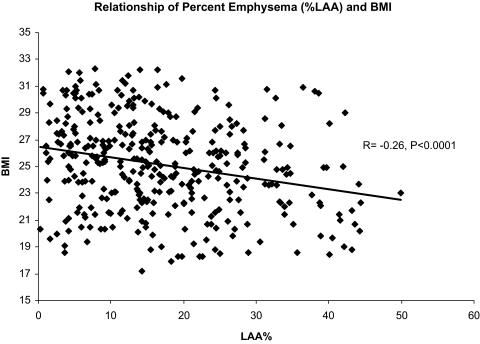
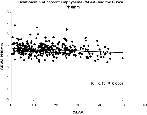
Results: In univariate analysis, there were significant negative correlations between %LAA-950 and both the WT (r = -0.28, p = 0.0001) and SRWA (r = -0.19, p = 0.0008). Airway wall thickness was weakly but significantly correlated with postbronchodilator FEV(1)% predicted (R = -0.12, p = 0.02). Multivariate analysis showed significant associations between either WT or SRWA (beta = -5.2, p = 0.009; beta = -2.6, p = 0.008, respectively) and %LAA-950 (beta = -10.6, p = 0.03) with the postbronchodilator FEV(1)% predicted. Male subjects exhibited significantly thicker airway wall phenotypes (p = 0.007 for WT and p = 0.0006 for SRWA).
Conclusions: Airway disease and emphysema detected by CT scanning are inversely related in patients with severe COPD. Airway wall phenotypes were influenced by gender and associated with lung function in subjects with severe emphysema.
Figures
References
-
- Rabe KF, Hurd S, Anzueto A, et al. Global strategy for the diagnosis, management, and prevention of chronic obstructive pulmonary disease: GOLD executive summary. Am J Respir Crit Care Med. 2007;176:532–555. - PubMed
-
- Hogg JC. Pathophysiology of airflow limitation in chronic obstructive pulmonary disease. Lancet. 2004;364:709. - PubMed
-
- de Jong PA, Muller NL, Pare PD, et al. Computed tomographic imaging of the airways: relationship to structure and function. Eur Respir J. 2005;26:140–152. - PubMed
-
- Nakano Y, Muro S, Sakai H, et al. Computed tomographic measurements of airway dimensions and emphysema in smokers: correlation with lung function. Am J Respir Crit Care Med. 2000;162:1102–1108. - PubMed
-
- Hasegawa M, Nasuhara Y, Onodera Y, et al. Airflow limitation and airway dimensions in chronic obstructive pulmonary disease. Am J Respir Crit Care Med. 2006;173:1309–1315. - PubMed
Publication types
MeSH terms
Grants and funding
- N01 HR076103/HL/NHLBI NIH HHS/United States
- N01 HR076102/HL/NHLBI NIH HHS/United States
- N01 HR076114/HL/NHLBI NIH HHS/United States
- N01 HR076113/HL/NHLBI NIH HHS/United States
- N01 HR076104/HL/NHLBI NIH HHS/United States
- N01 HR076101/HL/NHLBI NIH HHS/United States
- N01 HR076110/HL/NHLBI NIH HHS/United States
- N01 HR076112/HL/NHLBI NIH HHS/United States
- N01 HR076109/HL/NHLBI NIH HHS/United States
- N01 HR076105/HL/NHLBI NIH HHS/United States
- 1K23HL089353-01A1/HL/NHLBI NIH HHS/United States
- N01 HR076118/HL/NHLBI NIH HHS/United States
- N01 HR076115/HR/NHLBI NIH HHS/United States
- N01 HR076107/HL/NHLBI NIH HHS/United States
- K25 HL104085/HL/NHLBI NIH HHS/United States
- N01 HR076106/HL/NHLBI NIH HHS/United States
- N01 HR076119/HL/NHLBI NIH HHS/United States
- N01 HR076108/HL/NHLBI NIH HHS/United States
- N01 HR076116/HL/NHLBI NIH HHS/United States
- K23 HL089353/HL/NHLBI NIH HHS/United States
- N01 HR076111/HL/NHLBI NIH HHS/United States
LinkOut - more resources
Full Text Sources
Other Literature Sources
Medical
