. 2009 Jul;297(1):L143-52.
doi: 10.1152/ajplung.90618.2008. Epub 2009 May 1.

Aberrant cell adhesion molecule expression in human bronchopulmonary sequestration and congenital cystic adenomatoid malformation

Affiliations

Aberrant cell adhesion molecule expression in human bronchopulmonary sequestration and congenital cystic adenomatoid malformation

Maryann V Volpe et al. Am J Physiol Lung Cell Mol Physiol. 2009 Jul.

Abstract

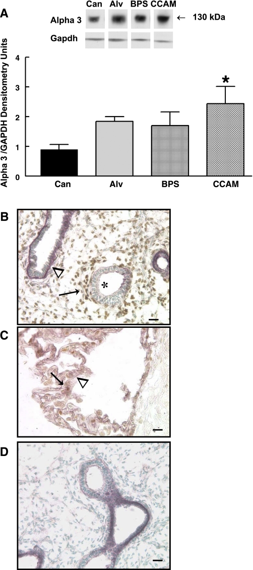
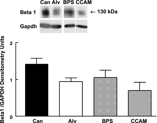
In many organs, integrins and cadherins are partly regulated by Hox genes, but their interactions in airway morphogenesis and congenital lung diseases are unknown. We previously showed that the Hox protein HoxB5 is abnormally increased in bronchopulmonary sequestration (BPS) and congenital cystic adenomatoid malformation (CCAM), congenital lung lesions with abnormal airway branching. We now report on alpha(2)-, alpha(3)-, and beta(1)-integrin and E-cadherin expression in normal human lung and in BPS and CCAM tissue previously shown to have abnormal HoxB5 expression and on the relationship of cell adhesion molecule expression to Hoxb5 regulation. alpha(2)-, alpha(3)-, and beta(1)-integrins and E-cadherin expression in normal human lung and BPS and CCAM were evaluated using Western blot and immunohistochemistry. Fetal mouse lung fibroblasts with Hoxb5-specific siRNA downregulation were evaluated for alpha(2)-integrin protein levels by Western blot. Compared with normal human lung, a previously undetected alpha(2)-integrin isoform potentially lacking essential cytoplasmic sequences was significantly increased in BPS and CCAM, and alpha(2)-integrin spatial and cellular expression was more intense. E-cadherin protein levels were also significantly increased, whereas alpha(3) increased in CCAM compared with canalicular, but not with alveolar, stage lung. beta(1)-integrin levels were unchanged. We conclude that in BPS and CCAM, altered alpha(2)-integrin cytoplasmic signaling contributes to abnormal cellular behavior in these lung lesions. Aberrant cell adhesion molecule and Hox protein regulation are likely part of the mechanism involved in the development of BPS and CCAM.


Figures

Fig. 1.
Fig. 1.
Schematic of primary antibody binding sites to α2-integrin transmembrane protein. Antibody 1 (Chemicon, Temecula, CA) detects essential regulatory sequences in the cytoplasmic domain of α2-integrin of mouse and human origin. Antibody 2 (Santa Cruz Biotechnology, Santa Cruz, CA) detects sequences in the extracellular domain of α2-integrin of human origin.
Fig. 2.
Fig. 2.
α2-integrin representative Western blots and summary densitometry. A: Western blots performed with antibody 1 (detects cytoplasmic sequences of α2-integrin) did not demonstrate any difference in protein levels of the 150-kDa protein isoform of α2-integrin between normal human lung from the canalicular (Can) and alveolar (Alv) stage of lung development and bronchopulmonary sequestration (BPS) and congenital cystic adenomatoid malformation (CCAM) tissue. B: Western blots using antibody 2 (detects extracellular sequences of α2-integrin) detected a significantly upregulated 130-kDa protein isoform of α2-integrin in BPS and CCAM compared with normal alveolar or canalicular stage lung tissue, but there were no differences in the 150-kDa α2-integrin isoform (*P = 0.005, means ± SE; BPS and CCAM vs. Alv or Can). A and B: n = 6, Can; n = 3, Alv; n = 4, BPS; n = 7, CCAM. C: representative Western blot of one CCAM sample in absence (−) or presence (+) of blocking peptide for antibody 2 showed that when antibody 2 activity was blocked with α2-specific blocking peptide against antibody 2, neither the 150- nor the 130-kDa protein isoform was detected. D: mouse fetal lung fibroblasts treated with Hoxb5-specific siRNA to downregulate Hoxb5 protein had significantly decreased α2-integrin protein levels compared with scramble-treated cells or control (no treatment) (*P = 0.04, means ± SE, n = 5).
Fig. 3.
Fig. 3.
α2-integrin immunostaining in CCAM and BPS. A–F: representative CCAM tissue sections stained for α2-integrin with blue alkaline phosphatase immunohistochemistry and nuclear Fast Red counterstain. G–I: α2-integrin stained with purple VIP-HRP immunolocalization and Hoxb5 protein (H, I) immunolocalized with brown DAB-HRP immunolocalization and counterstained with methyl green. Bar = 100 μm in D, 50 μm in A and E, and 25 μm in B, C, F, G, H, and I. A: α2-integrin antibody 1, immunolocalized α2-integrin protein to mesenchymal and epithelial cells of CCAM cysts (* in A and B) and adjacent compressed lung (arrow in A and C) with similar intensity. D, E, F: in contrast, α2-integrin antibody 2 immunostaining of adjacent section of CCAM shown in A–C revealed different regional localization of α2-integrin. Compared with adjacent compressed lung regions (# in D, E, F), increased α2-integrin staining in mesenchyme and epithelial cells was seen around CCAM cyst (* in D, E, F). Using antibody 2, BPS tissue (G) showed intense purple epithelial (arrow in G) and mesenchymal localization (* in G) of α2-integrin compared with human fetal lung (17-wk gestation) (H), which had strong but less intense purple α2-integrin localization in epithelial cells of branching airway tips (long arrows in H). These α2-positive purple epithelial cells were surrounded by closely adjacent subepithelial fibroblasts with intense brown nuclear staining for HoxB5 protein (arrowheads in H). Regions of airways with less purple α2-integrin staining (< in H) are not surrounded by closely adjacent HoxB5-positive brown cells. α2-integrin was also localized diffusely in mesenchyme, especially in fibroblasts with brown nuclear Hoxb5 staining (short arrow in H). In absence of α2-integrin antibody, there is no purple α2-integrin immunostaining (I) but continued Hoxb5 brown nuclear staining in presence of Hoxb5 antibody.
Fig. 4.
Fig. 4.
α3-integrin Western blots and immunohistochemistry. A: representative α3-integrin Western blot and summary densitometry showed similar levels of α3-integrin in BPS and CCAM tissue compared with alveolar stage lung tissue. CCAM tissue, however, had significantly increased α3-integrin protein levels compared with canalicular period lung tissue samples (*P = 0.03, CCAM vs. Can, means ± SE; n = 6, Can; n = 3, Alv; n = 4, BPS; n = 7, CCAM). B: human fetal lung tissue (17-wk human gestation) showed α3-integrin purple VIP staining at basal surface of columnar epithelial cells that had less intense subepithelial brown nuclear staining for Hoxb5 protein in mesenchymal cells (arrowhead). In contrast, airways (*) with less intense purple epithelial α2-integrin staining had more intense brown Hoxb5 staining in adjacent subepithelial fibroblasts (arrow). α3-integrin (purple) was also seen diffusely in mesenchyme but to a lesser degree than in airway epithelial cells and subepithelial fibroblasts. C: CCAM tissue had diffuse purple staining for α3-integrin (arrowhead) and scattered brown nuclear staining for Hoxb5 protein (arrow). D: immunostaining control showed that in absence of Hoxb5 primary antibody, no brown DAB staining was seen with only purple VIP staining for α3-integrin in presence of α3-integrin primary antibody.
Fig. 5.
Fig. 5.
β1-integrin representative Western blot and summary densitometry. β1-integrin protein levels were not altered in BPS and CCAM compared with alveolar or canalicular stage human lung tissue. N = 6, Can; n = 3, Alv; n = 4, BPS; n = 7, CCAM.
Fig. 6.
Fig. 6.
E-cadherin representative Western Blot and summary densitometry. Compared with alveolar stage human lung, E-cadherin protein levels were increased significantly in CCAM and showed a trend towards increased levels in BPS. Levels of E-cadherin in CCAM and BPS were more similar to canalicular stage lung tissue. *P = 0.04, CCAM vs. Alv, means ± SE, n = 6, Can; n = 3, Alv; n = 4, BPS; n = 7, CCAM.

References

    1. Achiron R, Hegesh J, Yagel S. Fetal lung lesions: a spectrum of disease. New classification based on pathogenesis, two dimensional and color doppler ultrasound. Ultrasound Obstet Gynecol 24: 107–114, 2004. - PubMed
    1. Achiron R, Zalel Y, Lipitz S, Hegesh J, Mazkereth R, Kuint J, Jacobson J, Yagel S. Fetal lung dysplasia: clinical outcome based on a new classification system. Ultrasound Obstet Gynecol 24: 127–133, 2004. - PubMed
    1. Aulicino MR, Reis ED, Dolgin SE, Unger PD, Shah KD. Intra-abdominal pulmonary sequestration exhibiting congenital cystic adenomatoid malformation: report of a case and review of literature. Arch Pathol Lab Med 118: 1034–1037, 1994. - PubMed
    1. Barr LF, Cambell SE, Bochner BS, Dang CV. Association of the decreased expression of alpha3 beta 1 integrin with the altered cell: environmental interactions and enhanced soft agar cloning ability of c-myc overexpressing small cell cancer cells. Cancer Res 58: 5537–5545, 1998. - PubMed
    1. Blystone SD Integrating an integrin: a direct route to actin. Biochim Biophys Acta 1692: 47–54, 2004. - PubMed

Publication types

MeSH terms

LinkOut - more resources