. 2009 Jul;297(1):L115-24.
doi: 10.1152/ajplung.90587.2008. Epub 2009 May 1.

Pleural mesothelial cell transformation into myofibroblasts and haptotactic migration in response to TGF-beta1 in vitro

Affiliations

Pleural mesothelial cell transformation into myofibroblasts and haptotactic migration in response to TGF-beta1 in vitro

Najmunnisa Nasreen et al. Am J Physiol Lung Cell Mol Physiol. 2009 Jul.

Abstract

Idiopathic pulmonary fibrosis (IPF) is a disease of unknown etiology characterized by the development of subpleural foci of myofibroblasts that contribute to the exuberant fibrosis noted in the pulmonary parenchyma. Pleural mesothelial cells (PMC) are metabolically dynamic cells that cover the lung and chest wall as a monolayer and are in intimate proximity to the underlying lung parenchyma. The precise role of PMC in the pathogenesis of pulmonary parenchymal fibrosis remains to be identified. Transforming growth factor (TGF)-beta1, a cytokine known for its capacity to induce proliferative and transformative changes in lung cells, is found in significantly higher quantities in the lungs of patients with IPF. High levels of TGF-beta1 in the subpleural milieu may play a key role in the transition of normal PMC to myofibroblasts. Here we demonstrate that PMC activated by TGF-beta1 undergo epithelial-mesenchymal transition (EMT) and respond with haptotactic migration to a gradient of TGF-beta1 and that the transition of PMC to myofibroblasts is dependent on smad-2 signaling. The EMT of PMC was marked by upregulation of alpha-smooth muscle actin (alpha-SMA), fibroblast specific protein-1 (FSP-1), and collagen type I expression. Cytokeratin-8 and E-cadherin expression decreased whereas vimentin remained unchanged over time in transforming PMC. Knockdown of smad-2 gene by silencing small interfering RNA significantly suppressed the transition of PMC to myofibroblasts and significantly inhibited the PMC haptotaxis. We conclude that PMC undergo EMT when exposed to TGF-beta1, involving smad-2 signaling, and PMC may be a possible source of myofibroblasts in IPF.


Figures

Fig. 1.
Fig. 1.
Transforming growth factor (TGF)-β1 induces a mesenchymal morphology in pleural mesothelial cells (PMC). Primary cultures of human PMC were cultured in the presence (5 ng/ml) or absence (control) of TGF-β1 for 96 h in serum-free medium. Phenotypic change (transition toward a myofibroblast-like phenotype) was evaluated by phase-contrast microscopy. In control culture, typical cobblestone morphology of PMC was clearly observed; at 24 h few PMC transdifferentiated to fibroblasts; at 48 h, 72 h, and 96 h TGF-β1-treated PMC showed decrease in cell-cell contacts and transition to a more elongated morphological shape of myofibroblasts. Scale bar, 50 μm.
Fig. 2.
Fig. 2.
Expression of epithelial-mesenchymal transition (EMT) markers in response to TGF-β1 in PMC. PMC were incubated in presence of TGF-β1 for indicated period of time or left untreated. Whole protein cell lysates were immunoblotted with specific antibodies. Expression of α-smooth muscle actin (α-SMA) was increased; at 72 h maximum expression was noted compared with control PMC. Cytokeratin decreased after 48 h of TGF-β1 activation. Vimentin remained unaffected. E-cadherin expression decreased significantly after 48 h of TGF-β1 activation. Smad-2 transcription factor expression was upregulated at 24 h. The blot was reprobed for β-actin to ensure equal loading of protein in each lane. Results are representative data of 3 separate experiments.
Fig. 3.
Fig. 3.
TGF-β1 induces EMT type of changes in primary PMC. Immunofluorescence staining for α-SMA, vimentin, and E-cadherin (green) was evaluated by confocal laser microscopy. A: TGF-β1-treated PMC exhibited a fibrillar pattern of α-SMA-specific proteins at 48 and 72 h. Also note that TGF-β1 caused spindle-shaped morphology, which was not observed in control. B: expression of vimentin remained unaffected. C: there was apparent loss of cell membrane-bound E-cadherin in TGF-β1-treated PMC in a time-dependent manner. 4′,6-Diamidino-2-phenylindole (DAPI, blue) was used for nuclear staining; FITC (green) was used for respective target proteins. Scale bars, 20 μm.
Fig. 4.
Fig. 4.
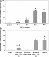
TGF-β1 induces fibroblast-specific protein-1 (FSP-1) expression in primary PMC. mRNA expression of FSP-1 was evaluated by quantitative RT-PCR. A: expression of FSP-1 over time. B: PMC pretreatment with antisense oligo for FSP-1 significantly blocked expression of FSP-1 mRNA compared with mismatch oligo. Relative amount of FSP-1 was normalized to β-actin and is expressed relative to control. Values are means ± SE of at least 3 independent experiments. *P < 0.05 vs. control; #P < 0.05 vs. mismatch oligo.
Fig. 5.
Fig. 5.
TGF-β1 induces collagen type I expression in PMC. A: level of collagen type I mRNA expression was evaluated by quantitative RT-PCR. Relative amount of collagen type I was normalized to β-actin and is expressed relative to control. Values are means ± SE of at least 3 independent experiments. *P < 0.05 vs. control. B: immunofluorescence staining for collagen type I (green) was evaluated by fluorescence microscopy. TGF-β1-treated PMC showed upregulated collagen expression over time; untreated PMC (0 h) did not show any collagen deposition. Scale bar, 20 μm.
Fig. 6.
Fig. 6.
TGF-β1 induces smad-2 signaling in PMC. Whole cell lysates were obtained and analyzed for total and phosphorylated forms of smad-2/4. Expression of smad-2 and smad-4 was upregulated in TGF-β1-treated PMC compared with control (resting PMC). Smad-2 transcription factor phosphorylation was noted up to 2 h, and it decreased at 24 h. β-Actin was used as a loading control to demonstrate equal protein loading. Results are representative of 3 separate experiments.
Fig. 7.
Fig. 7.
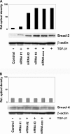
Smad-2 small interfering RNA (siRNA) suppresses the expression of smad-2 in TGF-β1-activated PMC. Whole protein cell lysates were immunoblotted with anti-smad-2 and smad-4 antibodies. PMC was transfected with siRNA smad-2 and activated with TGF-β1 for 4 h; transfection efficiency was confirmed. A: 3 specific siRNAs for smad-2 along with a control siRNA were used. siRNA-smad-2 no. 1 showed highly significant knockdown for smad-2 protein compared with the other 2 siRNAs or control siRNA. B: smad-2 siRNA did not effect smad-4 expression. Transfection of PMC with siRNA-smad-2 no. 1, siRNA-smad-2 no. 2, or siRNA-smad-2 no. 3 did not inhibit the expression of smad-4 compared with siRNA-control. Blot was reprobed with β-actin to ensure equal loading of protein for each lane. Relative optical densities were plotted, and blots presented are representative of 3 independent experiments.
Fig. 8.
Fig. 8.
smad-2 siRNA transfection preserves E-cadherin expression and suppresses α-SMA in TGF-β1-treated PMC. A: immunofluorescence staining for E-cadherin (green) in PMC transfected with smad-2 siRNA. PMC were cultured in the absence (control) and presence of TGF-β1 for 72 h and then were examined by confocal microscopy. DAPI stained blue for nuclei. smad-2 siRNA, but not control siRNA (sc-siRNA), preserved E-cadherin expression. B: α-SMA expression was decreased in smad-2 siRNA-treated PMC but not in control PMC. Smad-2 siRNA-treated cells showed a granular nonfibrillar distribution of α-SMA, while control siRNA-treated PMC preserved a polarized fibrillar α-SMA pattern after TGF-β1 treatment. C: phase-contrast microscopy of PMC exposed to TGF-β1 alone or siRNA-smad-2-transfected PMC; phenotypic changes (transition toward a myofibroblast-like phenotype) were clearly evident after 72 h. smad-2 siRNA, but not control siRNA, preserved the cobblestone morphology of PMC. FITC (green fluorescent staining) is respective target proteins. Scale bars, 20 μm. D: PMC were transfected with siRNA-smad-2 or control siRNA and treated with TGF-β1 for 72 h. Whole protein lysates were subjected to Western blot analysis for EMT markers E-cadherin and α-SMA. β-Actin was used as loading control to demonstrate equal protein loading in the lanes. E: FSP-1 mRNA expression was inhibited when smad-2 protein was knocked down in PMC treated with TGF-β1. FSP-1 expression was significantly decreased compared with TGF-β1-alone activated PMC or control siRNA. Values are means ± SE of triplicate wells from 3 independent experiments. **P < 0.05, smad-2 siRNA vs. control siRNA in presence of TGF-β1.
Fig. 9.
Fig. 9.
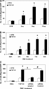
siRNA-smad-2 suppresses TGF-β1-induced haptotaxis in PMC. A: PMC were cultured in the presence of serum-free medium (SFM), and the haptotaxis of PMC against varied concentrations of TGF-β1 was evaluated. Bovine serum albumin (BSA) was used as a control. B: PMC were cultured in the presence or absence (SFM) of TGF-β1, and the haptotaxis against TGF-β1 was evaluated over time. C: siRNA smad-2 but not control siRNA transfection of PMC significantly decreased the haptotactic migration of PMC against TGF-β1. BSA was used as a control. Values are means ± SE of triplicate wells from 3 representative experiments. *P < 0.05 significantly increased compared with BSA; $P < 0.05 significantly decreased compared with TGF-β1-activated PMC; #P < 0.05 significantly decreased compared with smad-2 siRNA vs. control siRNA in the presence of TGF-β1. Values are mean ± SE number of cells migrated/10 high-power fields (HPF) of triplicate wells from 3 independent experiments.

References

    1. Abe R, Donnelly SC, Peng T, Bucala R, Metz CN. Peripheral blood fibrocytes: differentiation pathway and migration to wound sites. J Immunol 166: 7556–7562, 2001. - PubMed
    1. Antony VB Immunological mechanisms in pleural disease. Eur Respir J 21: 539–544, 2003. - PubMed
    1. Aroeira LS, Aguilera A, Selgas R, Ramirez-Huesca M, Perez-Lozano ML, Cirugeda A, Bajo MA, del Peso G, Sanchez-Tomero JA, Jimenez-Heffernan JA, Lopez-Cabrera M. Mesenchymal conversion of mesothelial cells as a mechanism responsible for high solute transport rate in peritoneal dialysis: role of vascular endothelial growth factor. Am J Kidney Dis 46: 938–948, 2005. - PubMed
    1. Bonniaud P, Kolb M, Galt T, Robertson J, Robbins C, Stampfli M, Lavery C, Margetts PJ, Roberts AB, Gauldie J. Smad3 null mice develop airspace enlargement and are resistant to TGF-beta-mediated pulmonary fibrosis. J Immunol 173: 2099–2108, 2004. - PubMed
    1. Broekelmann TJ, Limper AH, Colby TV, McDonald JA. Transforming growth factor beta 1 is present at sites of extracellular matrix gene expression in human pulmonary fibrosis. Proc Natl Acad Sci USA 88: 6642–6646, 1991. - PMC - PubMed

Publication types

MeSH terms