. 2009 May;19(5):757-69.
doi: 10.1101/gr.085738.108.

Meiotic recombination generates rich diversity in NK cell receptor genes, alleles, and haplotypes

Affiliations

Meiotic recombination generates rich diversity in NK cell receptor genes, alleles, and haplotypes

Paul J Norman et al. Genome Res. 2009 May.

Abstract

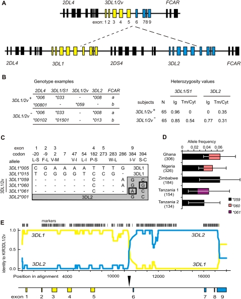
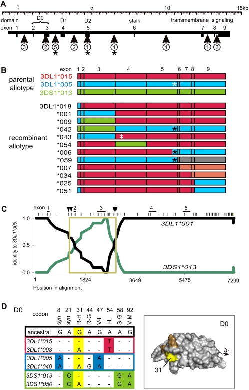
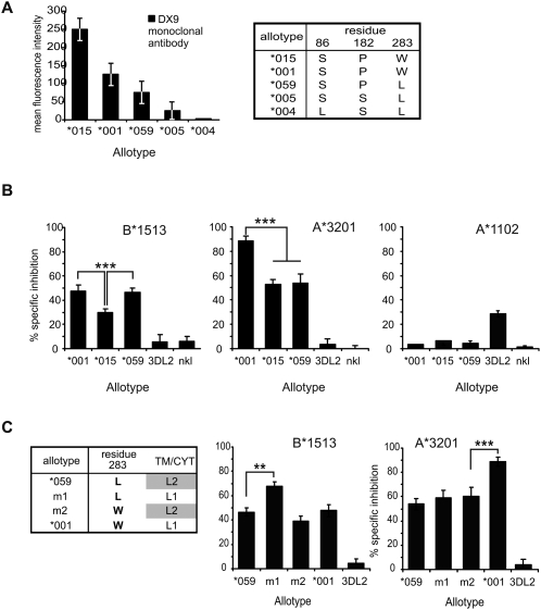
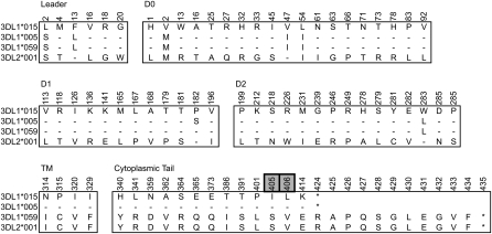
Natural killer (NK) cells contribute to the essential functions of innate immunity and reproduction. Various genes encode NK cell receptors that recognize the major histocompatibility complex (MHC) Class I molecules expressed by other cells. For primate NK cells, the killer-cell immunoglobulin-like receptors (KIR) are a variable and rapidly evolving family of MHC Class I receptors. Studied here is KIR3DL1/S1, which encodes receptors for highly polymorphic human HLA-A and -B and comprises three ancient allelic lineages that have been preserved by balancing selection throughout human evolution. While the 3DS1 lineage of activating receptors has been conserved, the two 3DL1 lineages of inhibitory receptors were diversified through inter-lineage recombination with each other and with 3DS1. Prominent targets for recombination were D0-domain polymorphisms, which modulate enhancer function, and dimorphism at position 283 in the D2 domain, which influences inhibitory function. In African populations, unequal crossing over between the 3DL1 and 3DL2 genes produced a deleted KIR haplotype in which the telomeric "half" was reduced to a single fusion gene with functional properties distinct from its 3DL1 and 3DL2 parents. Conversely, in Eurasian populations, duplication of the KIR3DL1/S1 locus by unequal crossing over has enabled individuals to carry and express alleles of all three KIR3DL1/S1 lineages. These results demonstrate how meiotic recombination combines with an ancient, preserved diversity to create new KIR phenotypes upon which natural selection acts. A consequence of such recombination is to blur the distinction between alleles and loci in the rapidly evolving human KIR gene family.


Figures

Figure 1.
Figure 1.
The 3DL1/2v fusion gene is allelic to 3DL1/S1 and 3DL2. (A, top) Schematic of the four 3DL1/2v haplotypes that were sequenced here (Supplemental Fig. S2). (Bottom) A KIR A haplotype; (dashed lines) the genomic segment absent from 3DL1/2v haplotypes; (yellow) 3DL1; (blue) 3DL2. Exons 1–5 encode the leader peptide and Ig domains (Ig), exon 6 the stalk, and exons 7–9 the transmembrane and cytoplasmic domains (Tm/Cyt). (B, left) Representative genotypes from the group of 65 subjects who carry 3DL1/2v (3DL1/2v+) and the group of 65 who do not (3DL1/2v). (Right) Comparisons of heterozygosity observed in the 3DL1/2v+ and 3DL1/2v groups from segments of 3DL1 and 3DL2. All of the 3DL1/2v+ subjects are hemizygous for exons 6–9 of 3DL1/S1 and exons 1–5 of 3DL2, which corresponds to the portion absent from the KIR A haplotype. This shows the 3DL1/2v+ subjects have 3DL1 and 3DL2 on one haplotype and 3DL1/2v on the other. (C) Shown are the nucleotide differences in exons 1–5 that distinguish 3DL1/2v (3DL1*059, 3DL1*060, and 3DL1*061) from (white) 3DL1 and in exons 7–9 that distinguish 3DL1/2v from (gray) 3DL2. The many nucleotide differences (N = 102) that distinguish 3DL1 and 3DL2 are not shown. Shown are 3DL1*00501 and 3DL1*01502, which represent the 005 and 015 lineages of inhibitory receptors; exons 1–5 of 3DL1/2v are related to 3DL1*00501. In exons 7–9, 3DL1*059 is identical to 3DL2*001; 3DL1*060 and 3DL1*061 being distinguished by the SNPs boxed. Codons are numbered according to the mature protein, and amino acid changes are indicated by single letter code. (D) Shown are the frequencies of three 3DL1/2v alleles (3DL1*059–61) in the five sub-Saharan African populations where they were detected. The number of haplotypes examined from each population is shown in parentheses; error bars show the 95% confidence interval of the allele frequency measurements. (E) Shown is a pairwise identity plot from alignment of genomic sequences. (Yellow line) Signifies identity of 3DL1/2v with 3DL1; (blue line) the identity of 3DL1/2v with 3DL2; (vertical bars) the SNP markers used in this analysis. (Shown below by the vertical arrow) The crossover occurred in intron 5 during the interval from 386 to 356 bp upstream from exon 6. A continuous sequence trace that spans the crossover is shown in Supplemental Figure S2.
Figure 2.
Figure 2.
Motif and domain shuffling between three lineages diversifies 3DL1/S1. (A) Shown to scale is the genomic organization of the 3DL1/S1 locus. (Boxes) Exons; exons 2–5 encode the three Ig domains, D0, D1, and D2. (Vertical arrows) The genomic regions and the number of recombination events detected from comparison of 3DL1/S1 alleles. (*) Recombination event that placed residue leucine 283 onto a different background allotype. (B) Schematic of 12 recombinant alleles of 3DL1/S1 that were identified using domain-by-domain phylogenetic analysis. The recombinant allotypes are represented by segments colored according to allelic lineage: (red) 015, (blue) 005, (green) 3DS1. The allotypes shown at the top of the panel are encoded by the most common modern alleles: 3DL1*01502, 3DL1*00501, and 3DS1*01301. For example, 3DL1*001 is identical to 3DL1*00501 in exons 1–3 and 3DL1*01501 in exons 4–9. (Gray) Recombination from another locus (3DL2); (pink) within-lineage recombinants. (White asterisk) Tryptophan–leucine substitution at residue 283; (black asterisk) recombination has replaced tryptophan at residue 283 with leucine; (‡) threonine 118 that distinguishes 3DL1*043 from 3DL1*001 and is shared with 3DL1*038. (C) The pairwise identity plot shows that 3DL1*009 formed by gene conversion. (Black line) Identity of 3DL1*009 with 3DL1*001; (green line) identity of 3DL1*009 with 3DS1*01301. The recombination included exons 2 and 3, which encode D0. (Across the top, vertical bars) SNP markers; (vertical arrows) the minimum and maximum limits of the gene conversion. Genomic sequences used for the RDP analysis include representatives of the three allelic lineages of 3DL1/S1 (listed in Methods). The 3DL1*009 cDNA sequence that was independently obtained (Middleton et al. 2007) corresponds precisely to the reading frame of the locus characterized here. (D) Shown for each of the 3DL1/S1 lineages is a pair of alleles that are dimorphic at (yellow) codon 31. Nucleotide differences in D0 are shown, and those that distinguish the three lineages are colored as in panel A. (Top) Amino acid substitutions with the ancestral residue first and the ancestral nucleotide immediately below. (Right panel) A homology model of D0; residue 31 (yellow) occurs in a patch of positively selected residues (orange) that were identified in Norman et al. (2007).
Figure 3.
Figure 3.
3DL1/2v mediates HLA-allotype specific inhibition of NK cells. (A) Binding of the 3DL1-specific monoclonal antibody DX9 to NK cells shows that 3DL1/2v is expressed at the cell surface at intermediate level. Shown are FACS analyses of peripheral-blood NK cells from donors who express single 3DL1 allotypes. (Right) The table shows amino acid differences that correspond to the 3DL1 expression level. 3DL1*004 is not expressed because of leucine-for-serine substitution at residue 86 (Pando et al. 2003). 3DL1*015 has further differences in D0 that are shown in Figure 1. Error bars indicate the standard deviation of DX9-PE fluorescence intensity, from all of the lymphocytes that stained positive with DX9. (B,C) Results from Cr51-release cytotoxicity assays. The basis for this assay is killing of HLA Class I–deficient cells (221) by an NK cell line (NKL). Inhibition of cytotoxicity occurs if NKL is transduced with inhibitory KIR and 721.221 is transfected with cognate HLA Class I ligand. Blocking the interaction with specific antibody restores target killing to that obtained using non-transduced NKL. The specificity of inhibition was determined in replicate assays using DX9, which specifically blocks 3DL1, or DX31, which specifically blocks 3DL2. The percent specific inhibition that is shown was calculated from killing in the presence of control/specific-blocking antibody. Results are mean (±SE) of five experiments at an effector:target ratio of 20:1. (***) P < 0.001; (**) P < 0.01 from Student's t-test for comparison of means. All of the effectors killed HLA-negative targets. Each of these assays shows different combinations of KIR and ligand. (B) The degree of inhibition mediated by natural KIR allotypes; these are 3DL1*001, 3DL1*015, 3DL1/2v (3DL1*059), and 3DL2*001 or no KIR (nkl). Target cells were 721.221 cells transfected with (left) HLA-B*1513, (center) A*3201, and (right) A*1102. B*1513 and –A*3201 are ligands for 3DL1, and A*1102 is a ligand for 3DL2. (C, left) The table shows the composition of the mutant KIR allotypes. Tail-swap mutant m1 (*001-L283) has the 3DL1*059 Ig-domain with a 3DL1*001 tail. m2 (*059-W283) has the 3DL1*001 Ig-domain with a 3DL2*001 tail. Results of the cytotoxicity analysis are shown for (center) HLA-A*3201 and (right) HLA-B*1513.
Figure 4.
Figure 4.
KIR3DL1 and KIR3DL2 differ at 68 amino acid residues spread throughout the polypeptide. Shown are the amino acid differences that distinguish 3DL1, 3DL2, and 3DL1/2v (3DL1*059). (Gray boxes) Differences that occur in the second ITIM motif of the cytoplasmic tail. Residue numbers are those of the mature protein. The 3DL1*059 sequence that was independently obtained from cDNA (Shilling et al. 2002) is identical to the amino acid sequence encoded by the 3DL1/2v gene characterized here (Fig. 1).
Figure 5.
Figure 5.
Simultaneous detection of KIR3DL1/S1 polymorphism and copy-number using pyrosequencing. (A, top) The diploid sequence of a 17-bp fragment of exon 3 from an individual heterozygous for 3DL1*01502 and 3DS1*01301. (Gray) SNP g336a. Underneath is a pyrogram obtained from the same individual. To generate the pyrogram, nucleotides were added to a single-strand template in the sequence shown, 1–11, and correspond to peaks of the same number. The peak height is proportional to the quantity of nucleotides that were incorporated, as shown by peak 1 (the sequence is gg), which is twice the height of peak 6 (g). Each peak is a sum of the haplotypes present, so that the monomorphic positions (relative peak height = 1) are used for calibration. The combined peak height at the heterozygous position shown (peaks 10 + 11) is equal to the peak from a single monomorphic position. Shown are four different pyrograms; (top left of each diagram) the derived genotype; (brackets) the peak-height ratio compared with the single monomorphic peak. (A) At SNP g336a, there is one peak for g and one for a (peaks 10 and 11), and each peak is half the height of a single peak (peak 6). This individual has one copy of 3DL1*01502 and one copy of 3DS1*01301 (0.5g: 0.5a). (B) At SNP g336a, there is one peak for g (peak 10) that is the same height as a single monomorphic position (peak 6) and no peak on addition of nucleotide a-11. This individual is homozygous, having two copies of 3DL1*01502 (1g: 0a). (C) There are two peaks as for A, but g is twice the height of a (0.67g: 0.33a), and their sum is the same as peak 6. This individual has two copies of 3DL1*01502 and one 3DS1*01301. (D) This individual has one copy of 3DL1*01502 and two of 3DS1*01301 (0.33g: 0.67a).
Figure 6.
Figure 6.
Duplicating KIR loci diversifies the NK cell repertoire in quantity and quality. (A) Schematic of the donor haplotypes and duplication haplotype. (Across the top) The KIR loci; (shaded boxes) indicate presence of the locus. The donor KIR haplotypes B (black) and A (orange); the composite duplication haplotype D is colored accordingly. (Dotted line) Non-allelic homologous recombination that was mediated by sequence similarity in the 5′-regions of 3DP1 and 2DL5A (Martin et al. 2003; Gomez-Lozano et al. 2005). (B) The seven different 2DL4, 3DL1, and 3DL2 haplotypes deduced from analysis of 80 individuals who have the duplication, and their haplotype frequencies in the populations where they were detected. The number of haplotypes analyzed from each population is indicated in parentheses. Georgia is the country. The Piramalai Kaller and the Yadheva are two distinct populations from Tamil Nadu in Southern India. (C) Shown are FACS analyses of freshly isolated 3DL1/S1-expressing NK cells from a European family with a duplicate haplotype. (At the top of each plot) The pyrosequencing genotype. Son 1 inherited 3DS1*013 and 3DL1*002 (haplotype 3, panel B) from the mother and 3DS1*013 from the father. Monoclonal antibody Z27 discriminates 3DL1/S1 allotypes, having high (3DL1*002 and 3DL1*015), low (3DL1*005 and 3DL1*007), and very low (3DS1*013) staining patterns. Showing that 3DL1/S1 expression depends on the number of copies of 3DS1 present, Son 1 has twice the number of 3DS1+ NK cells as the mother or father. Son 2 has inherited the opposite pair of haplotypes to Son 1, and expresses two distinct populations of high (*015) and low (*007) 3DL1 staining and no 3DS1. Further differences among allotypes are quantitative, with *015 being expressed by more cells than *002 (Chan et al. 2003), as observed here by comparing 3DL1 expression of the mother and Son 1. (D) Three unrelated individuals chosen for their 3DL1/S1 genotype. (Left) An individual heterozygous for 3DL1*015 and 3DL1*005 is shown as a negative control for the very low-staining 3DS1*013 peak. (Center and right) Two individuals who both express all three lineages of 3DL1/S1 receptors; these FACS analyses are from NK cells that had been stored frozen. A comparison of fresh and frozen NK cell staining is shown in Supplemental Figure S6.

References

    1. Alvarez-Arias D.A., Campbell K.S. Protein kinase C regulates expression and function of inhibitory killer cell Ig-like receptors in NK cells. J. Immunol. 2007;179:5281–5290. - PubMed
    1. Artavanis-Tsakonas K., Eleme K., McQueen K.L., Cheng N.W., Parham P., Davis D.M., Riley E.M. Activation of a subset of human NK cells upon contact with Plasmodium falciparum-infected erythrocytes. J. Immunol. 2003;171:5396–5405. - PubMed
    1. Bailey J.A., Eichler E.E. Primate segmental duplications: Crucibles of evolution, diversity and disease. Nat. Rev. Genet. 2006;7:552–564. - PubMed
    1. Bashirova A.A., Martin M.P., McVicar D.W., Carrington M. The killer immunoglobulin-like receptor gene cluster: Tuning the genome for defense. Annu. Rev. Genomics Hum. Genet. 2006;7:277–300. - PubMed
    1. Belle I., Hou L., Chen M., Steiner N.K., Ng J., Hurley C.K. Investigation of killer cell immunoglobulin-like receptor gene diversity in KIR3DL1 and KIR3DS1 in a transplant population. Tissue Antigens. 2008;71:434–439. - PubMed