. 2009 Jul 2;114(1):165-73.
doi: 10.1182/blood-2008-10-180489. Epub 2009 May 1.

GATA-1 associates with and inhibits p53

Affiliations

GATA-1 associates with and inhibits p53

Cecelia D Trainor et al. Blood. .

Abstract

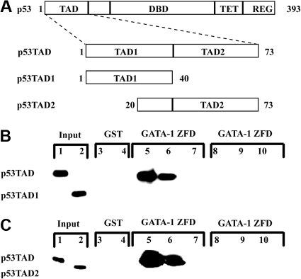
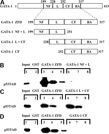
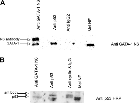
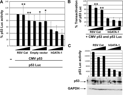
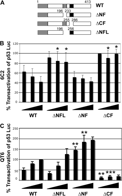
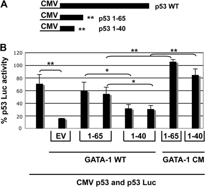
In addition to orchestrating the expression of all erythroid-specific genes, GATA-1 controls the growth, differentiation, and survival of the erythroid lineage through the regulation of genes that manipulate the cell cycle and apoptosis. The stages of mammalian erythropoiesis include global gene inactivation, nuclear condensation, and enucleation to yield circulating erythrocytes, and some of the genes whose expression are altered by GATA-1 during this process are members of the p53 pathway. In this study, we demonstrate a specific in vitro interaction between the transactivation domain of p53 (p53TAD) and a segment of the GATA-1 DNA-binding domain that includes the carboxyl-terminal zinc-finger domain. We also show by immunoprecipitation that the native GATA-1 and p53 interact in erythroid cells and that activation of p53-responsive promoters in an erythroid cell line can be inhibited by the overexpression of GATA-1. Mutational analysis reveals that GATA-1 inhibition of p53 minimally requires the segment of the GATA-1 DNA-binding domain that interacts with p53TAD. This inhibition is reciprocal, as the activation of a GATA-1-responsive promoter can be inhibited by p53. Based on these findings, we conclude that inhibition of the p53 pathway by GATA-1 may be essential for erythroid cell development and survival.


Figures

Figure 1
Figure 1
The full-length p53TAD binds to the zinc-finger domains of GATA-1 in an in vitro pulldown assay. (A) Schematic of the TAD of p53 demonstrating position of the full-length p53TAD and the location of the 2 independent subdomains: p53TAD1 and p53TAD2. (B) Binding of p53TAD and p53TAD1 to GST-GATA-1 ZFD (GATA-1199-317). Various concentrations (1.0, 0.1, and 0.01 μM) of p53TAD (lanes 5-7) and p53TAD1 (lanes 8-10) were incubated with 1 μM GST-GATA-1 ZFD. In the GST lanes, 1 μM purified p53TAD (lane 3) and p53TAD1 (lane 4) was incubated with 1 μM GST as a control. The input lanes are p53TAD (lane 1) and p53TAD1 (lane 2) at 0.5%. Bound protein was detected with an anti-p53 antibody DO-1. (C) Binding of p53TAD and p53TAD2 to GATA-1 ZFD. Various concentrations (1.0, 0.1, and 0.01 μM) of p53TAD (lanes 5-7) and p53TAD2 (lanes 8-10) were incubated with 1 μM GST-GATA-1 ZFD. In the GST lanes, 1 μM purified p53TAD (lane 3) and p53TAD2 (lane 4) was incubated with 1 μM GST as a control. The input lanes are p53TAD (lane 1) and p53TAD2 (lane 2) at 0.5%. Bound protein was detected with an anti-p53 antibody Pab 1801.
Figure 2
Figure 2
The linker region, the carboxyl-terminal zinc finger, and the basic arm of GATA-1 are sufficient for binding p53TAD in an in vitro pulldown assay. (A) Schematic of GATA-1 highlighting the zinc-finger domains (GATA-1 ZFD) and the various deletion mutants of GATA-1 ZFD used as GST fusion proteins. (B) Comparative binding of GST-GATA-1 ZFD and GST-GATA-1 NF plus L (GST-GATA-1200-251) to p53TAD. Various concentrations (1.0, 0.1, and 0.01 μM) of p53TAD were incubated with either 1 μM GST-GATA-1 ZFD (lanes 3-5) or GST-GATA-1 NF plus L (lanes 6-8). In the GST lane, 1 μM purified p53TAD was incubated with 1 μM GST as a control (lane 2). The input lane is 0.5% p53TAD (lane 1). (C) Comparative binding of GST-GATA-1 ZFD and GST-GATA-1 L plus CF (GST-GATA-1228-317) to p53TAD. Various concentrations (1.0, 0.1, and 0.01 μM) of p53TAD were incubated with either 1 μM GST-GATA-1 ZFD (lanes 3-5) or GATA-1 L plus CF (lanes 6-8). In the GST lane, 1 μM purified p53TAD was incubated with 1 μM GST as a control (lane 2). The input lane is 0.5% p53TAD (lane 1). (D) Comparative binding of GST-GATA-1 ZFD and GST-GATA-1 CF (GST-GATA-1252-317) to p53TAD. Various concentrations (1.0, 0.1, and 0.01 μM) of p53TAD were incubated with either 1 μM GST-GATA-1 ZFD (lanes 3-5) or GATA-1 CF plus BA (lanes 6-8). In the GST lane, 1 μM purified p53TAD was incubated with 1 μM GST as a control (lane 2). The input lane is 0.5% p53TAD (lane 1). In all experiments, bound protein was detected with an anti-p53 antibody DO-1.
Figure 3
Figure 3
GATA-1 and p53 interact in vitro in MEL cell extract. (A) A total of 320 μg of MEL nuclear extract was incubated with the following: lane 1, 4 μL of anti-GATA-1 N6 and 1 μL of anti-rat IgG2a; lane 2, 4 μL of anti-p53 (PAB 240) and 1 μL of anti-mouse IgG; lane 3, 5 μL of anti-rat IgG2 for 1.5 hours at 4°C with rotation and 40 μL of protein G plus agarose was added with 4 additional hours of rotation at 4°C; lane 4, MEL nuclear extract. A Western blot is shown using GATA-1 N6 antibody. (B) Lane 1, 8 μL of anti-GATA-1 N6 and 1 μL of anti-rat IgG2a; lane 2, 8 μL of anti-p53 (PAB 246) and 1 μL of anti-mouse IgG; lane 3, 8 μL of anti-cyclin B1 (GNS1) and 1 μL of anti-mouse IgG for 1.5 hours at 4°C with rotation, and 40 μL of protein G plus agarose was added with 4 more hours of rotation at 4°C; lane 4, MEL nuclear extract. A Western blot with HRP-conjugated anti-p53 is shown.
Figure 4
Figure 4
GATA-1, p53TAD, and a GATA-1 DNA-binding site do not form a ternary complex. (A) Overlay of the 2D 1H-15N HSQC spectra for a 0.5 mM sample of 15N-labeled GATA-1 L plus CF/DNA complex (black) and in the presence of 0.5 mM unlabeled p53TAD (red). (B) Overlay of the 2D 1H-15N HSQC spectra for a 0.5 mM sample of 15N-labeled p53TAD in the free form (black) and in the presence of 0.5 mM unlabeled GATA-1 L plus CF/DNA complex (red).
Figure 5
Figure 5
GATA-1 can inhibit transactivation by p53 in 6C2 cells. (A) All samples were transfected with 150 ng of CMV Renilla luciferase and 0.5 μg of p53 Luc reporter plasmid; and 0.25 μg of CMV p53 was added to all but the first sample. Increasing amounts of RSV-Cat, empty RSV expression vector, or RSV hGATA-1 were added (1, 2, and 3 μg of DNA). The dual luciferase reporter assay was performed on aliquots of the samples, and firefly luciferase activity normalized to Renilla luciferase is plotted. The error bars indicate the standard deviations and n = 3. Means of the 2 highest DNA concentrations of RSV-Cat and empty vector were compared with means for equal weights of hGATA-1 by Student t test; *P < .05 and **P < .005. (B) RSV-Cat and hGATA-1 were compared in 6 transfections. Results were normalized to Renilla luciferase activity. The firefly luciferase value for 1 μg of RSV-Cat was then set to 100%, and all other values were normalized to this. The error bars indicate the standard deviation and n = 6. We compared means for equal weights of the 2 highest DNA concentrations of the RSV-Cat and hGATA-1 plasmids to test for statistical significance by Student t test; **P < .005. (C) A comparable transfection was performed adding CMV Renilla luciferase, p53 Luc reporter plasmid, and CMV p53 to all samples and cotransfecting increasing amounts of RSV-Cat or RSV hGATA-1. The cells were divided at harvest, and half were used for the dual luciferase reporter assay and half for Western blotting with HRP-conjugated anti-p53 (top) and with anti-GAPDH antibody (bottom).
Figure 6
Figure 6
The linker between the zinc fingers and the CF of GATA-1 are required for p53 inhibition. (A) Schematic of deletion mutants of mouse GATA-1. (B) Transfections into 6C2 cells were performed, as described in the Figure 5 legend. A total of 150 ng of CMV Renilla luciferase, 0.5 μg of p53 Luc reporter plasmid, and 0.25 μg of CMV p53 was present in all samples, whereas various amounts of mouse wild-type GATA-1 expression vector or mutants were added as indicated (1, 2, or 3 μg). After normalization to Renilla luciferase activity, the firefly luciferase value for 1 μg of ΔCF was set to 100%, and the values for all other samples were correspondingly adjusted. The error bars indicate the standard deviation, and the experiments were performed between 4 and 7 times. We compared means for equal weights of the 2 highest DNA concentrations of ΔNFL, ΔNF, and ΔCF to wild type by Student t test, and *P < .05. (C) CMV Renilla luciferase and 1 μg of FR7Luc, a GATA-1–responsive reporter plasmid, were tranfected into QT6 fibrobasts with increasing amounts of mouse GATA-1 expression plasmid, as indicated (0.5, 1, or 2 μg). Normalization and error bars are as in (B), with values for 2 μg of wild-type GATA-1 set to 100%. The means for equal weights of the 2 lowest concentrations of wild-type were compared with those of all other constructs by Student t test; **P < .005 and ***P < .0005.
Figure 7
Figure 7
p531-65 can compete with wild-type p53 for binding to GATA-1 and reduce the inhibition of p53. (A) A schematic of constructs made by inserting translation termination signals in the CMV p53 expression vector to generate peptides 1-65 and 1-40 is shown. (B) A transfection assay in which CMV Renilla luciferase, p53 Luc reporter plasmid, and CMV p53 were present in all samples and expression plasmids for mGATA-1 were added to samples 2-6 along with 6 μg of empty vector (bar 2), 5 or 6 μg of CMV p53 1-65 (bars 3 and 4), or CMVp53 1-40 (bars 5 and 6), as indicated. For the last 2 samples, GATA-1CM, a mutant that does not interfere with transactivation by p53, was used instead of wild-type GATA-1, and 6 μg of CMV 1-65 (bar 7) or 6 μg of CMV 1-40 (bar 8) was added. Normalization is as described in the Figure 6 legend with the average of 5 μg of GATA-1 CM 65 and 40 samples set to 100%. Results are the average of 3 experiments, and error bars indicate the standard deviation. Means of equal weights of DNA were compared by Student t test, as indicated; *P < .05 and ** P < .005.

References

    1. Tsai SF, Martin DI, Zon LI, D'Andrea AD, Wong GG, Orkin SH. Cloning of cDNA for the major DNA-binding protein of the erythroid lineage through expression in mammalian cells. Nature. 1989;339:446–451. - PubMed
    1. Fujiwara Y, Browne CP, Cunniff K, Goff SC, Orkin SH. Arrested development of embryonic red cell precursors in mouse embryos lacking transcription factor GATA-1. Proc Natl Acad Sci U S A. 1996;93:12355–12358. - PMC - PubMed
    1. Pevny L, Simon MC, Robertson E, et al. Erythroid differentiation in chimeric mice blocked by a targeted mutation in the gene for transcription factor GATA-1. Nature. 1991;349:257–260. - PubMed
    1. Shivdasani RA, Fujiwara Y, McDevitt MA, Orkin SH. A lineage-selective knockout establishes the critical role of transcription factor GATA-1 in megakaryocyte growth and platelet development. EMBO J. 1997;16:3965–3973. - PMC - PubMed
    1. Migliaccio AR, Rana RA, Sanchez M, et al. GATA-1 as a regulator of mast cell differentiation revealed by the phenotype of the GATA-1low mouse mutant. J Exp Med. 2003;197:281–296. - PMC - PubMed

Publication types

MeSH terms