. 2009 Jun 11;459(7248):866-70.
doi: 10.1038/nature08065.

Hexameric assembly of the proteasomal ATPases is templated through their C termini

Affiliations

Hexameric assembly of the proteasomal ATPases is templated through their C termini

Soyeon Park et al. Nature. .

Abstract

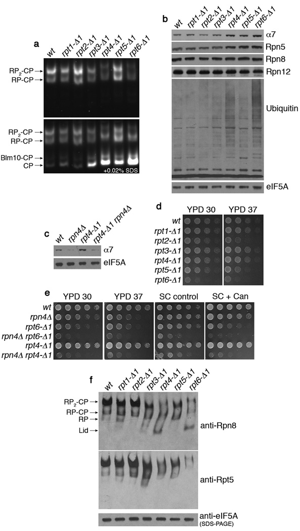
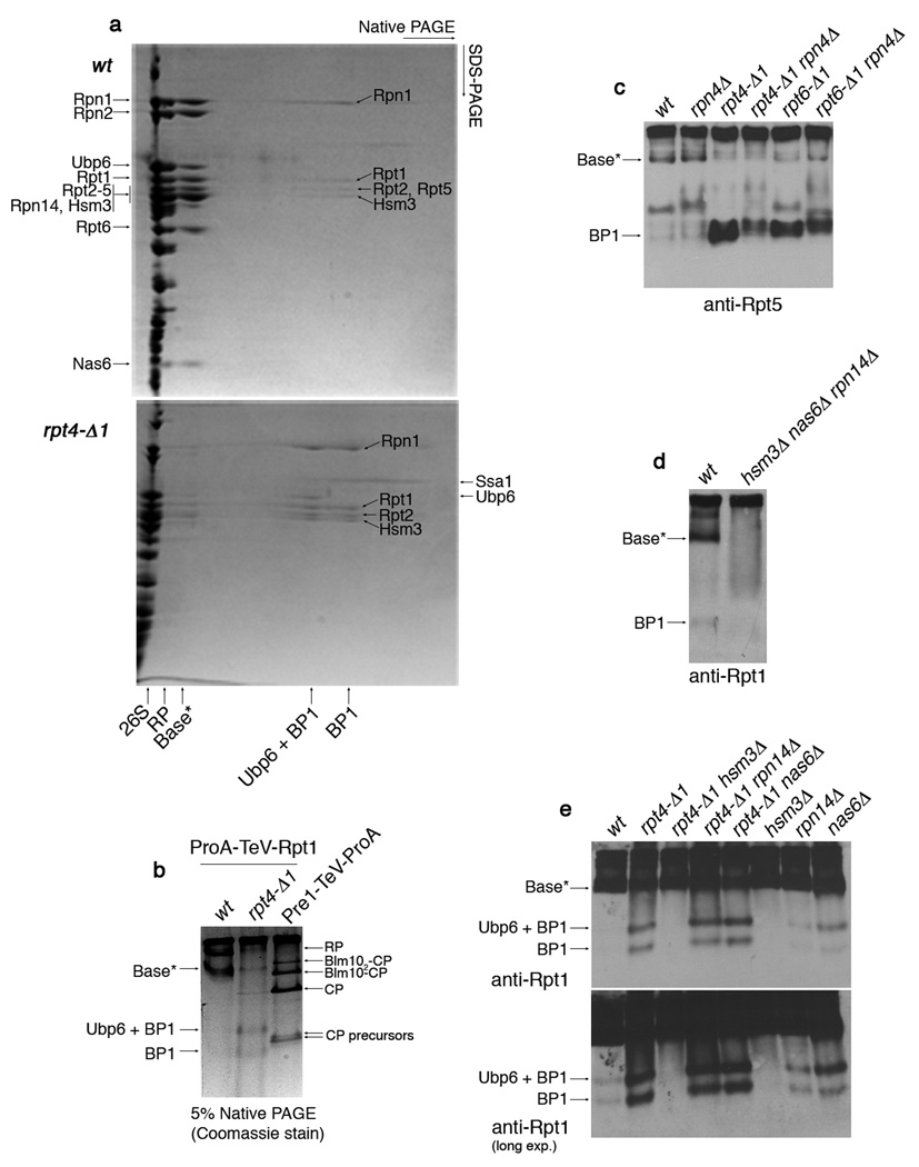
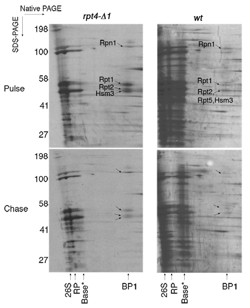
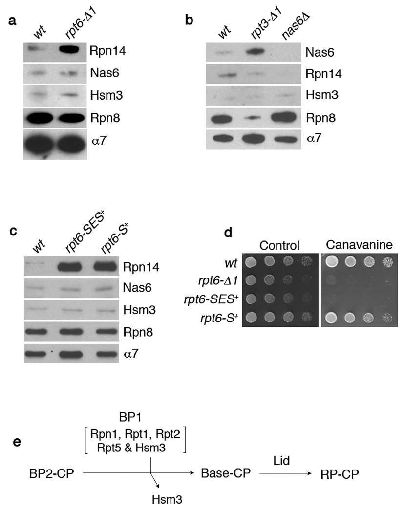
Substrates of the proteasome are recognized and unfolded by the regulatory particle, and then translocated into the core particle (CP) to be degraded. A hetero-hexameric ATPase ring, containing subunits Rpt1-6, is situated within the base subassembly of the regulatory particle. The ATPase ring sits atop the CP, with the Rpt carboxy termini inserted into pockets in the CP. Here we identify a previously unknown function of the Rpt proteins in proteasome biogenesis through deleting the C-terminal residue from each Rpt in the yeast Saccharomyces cerevisiae. Our results indicate that assembly of the hexameric ATPase ring is templated on the CP. We have also identified an apparent intermediate in base assembly, BP1, which contains Rpn1, three Rpts and Hsm3, a chaperone for base assembly. The Rpt proteins with the strongest assembly phenotypes, Rpt4 and Rpt6, were absent from BP1. We propose that Rpt4 and Rpt6 form a nucleating complex to initiate base assembly, and that this complex is subsequently joined by BP1 to complete the Rpt ring. Our studies show that assembly of the proteasome base is a rapid yet highly orchestrated process.


Figures

Figure 1
Figure 1. rpt4-Δ1 and rpt6-Δ1 mutants are proteasome hypomorphs with defective proteasome assembly
a, Native PAGE (3.5%) and two consecutive LLVY-AMC assays with whole cell extracts (85 µg). Immediately after the first LLVY-AMC assay (top panel), the second assay was conducted in the presence of 0.02% SDS (bottom panel). b, c, SDS-PAGE and immunoblotting with whole cell extracts. eIF5A is a loading control. d, e, Growth phenotypes of rpt mutants. Cells were spotted in 4-fold dilutions on YPD, synthetic medium (control), or synthetic medium containing canavanine at 0.5 µg/ml. Plates were incubated for 2–4 days at 30°C or at 37°C (YPD 37). f, 3.5% native PAGE and immunoblotting of whole cell extracts (85 µg) against Rpn8 (lid subunit) and Rpt5 (base subunit). Extracts (20 µg) were resolved by SDS-PAGE and immunoblotting for loading control.
Figure 2
Figure 2. Identification of a base assembly intermediate
a, 2-D native/SDS-PAGE of affinity-purifications with ProA-TeV-Rpt1 (Protein A tag appended to the N-terminus of Rpt1, a base subunit). Following a first-dimension native PAGE (5%), native gel lanes were individually excised and subjected to second-dimension SDS-PAGE (12.5%). Gels were stained with Coomassie blue. Individual spots of the base*, BP1, and Ubp6 + BP1 complexes were excised for mass spectrometry. Labels on the left indicate spots within base*. The presence of Rpn10 and Rpn13 in base* was not determined. Note that Rpt5 is absent from BP1 purified from rpt4-Δ1 mutants, but present in BP1 in untagged rpt4-Δ1 extracts (panel 4c). This appears to reflect a labile association of Rpt5 with BP1 in rpt4-Δ1 mutants. The presence of Ubp6 in BP1 is explained by its interaction with Rpn1. Note: Base* and BP1 species appear comparable between rpt6-Δ1 and rpt4-Δ1 mutants in their level and composition. b, 5% Native PAGE following affinity purification via a ProA-TeV-Rpt1 as in (a) or a Pre1 (CP subunit)-TeV-ProA. Native gels were stained with Coomassie blue. c, d, Immunoblotting following 5% native PAGE of whole cell extracts (100 µg). e, Immunoblotting following 5% native PAGE of affinity-purified samples (2 µg) from indicated strains carrying ProA-TeV-Rpt1.
Figure 3
Figure 3. Pulse-chase analysis of the BP1 base assembly intermediate
Strains were metabolically labeled with S35-methionine in vivo for 4–5 min (pulse) and chased with excess methionine for 30 min (chase). Samples were then subjected to affinity-purification with ProA-TeV-Rpt1 and 2-D native/SDS-PAGE (5% and 4–12%, respectively) followed by autoradiography. BP1, but not Ubp6 + BP1, was labeled with S35-methionine during the pulse in both wild-type and the rpt4-Δ1 mutant.
Figure 4
Figure 4. Interactions between Rpt C-termini and the CP control RP-chaperone release
a-c, SDS-PAGE and immunoblotting followed by affinity purification using a CP tag (Pre1-TeV-ProA). Rpn8 (lid subunit), and α7 (CP subunit) are loading controls. Nas6Δ proteasome (b) is a negative control. The rpt6-SES+ or rpt6-S+ mutants in (c) contain either a three (Ser-Glu-Ser) or one (Ser) residue insertion prior to the fourth residue from the C-terminus. d, Phenotypic analysis of RPT6 insertion mutants. 4-fold serial dilutions were spotted on synthetic medium (control) or canavanine (1 µg/ml) plates. Cells were grown for 2–4 days at 30°C. e, Model for late-stage base assembly. It is proposed that the base is formed by the addition of the BP1 complex, containing Rpn1, Rpt1, Rpt2, Rpt5, and Hsm3, to the putative BP2 complex. Initial contact with the CP is provided by BP2, whereas BP1 exists independently of the CP until it joins the BP2-CP complex. BP2 has not been identified and its exact composition is unknown. However, likely components of BP2 are Rpt4, Rpt6, Rpn2, and perhaps Rpt3. Rpn14 and Nas6 (not shown) may function prior to the formation of BP2-CP.

Comment in

References

    1. Finley D. Recognition and processing of ubiquitin-protein conjugates by the proteasome. Annu. Rev. Biochem. (In press) - PMC - PubMed
    1. Forster A, Masters EI, Whitby FG, Robinson H, Hill CP. The 1.9 A structure of a proteasome-11S activator complex and implications for proteasome-PAN/PA700 interactions. Mol. Cell. 2005;18:589–599. - PubMed
    1. Rabl J, et al. Mechanism of gate opening in the 20S proteasome by the proteasomal ATPases. Mol. Cell. 2008;30:360–368. - PMC - PubMed
    1. Smith DM, et al. Docking of the proteasomal ATPases' carboxyl termini in the 20S proteasome's alpha ring opens the gate for substrate entry. Mol. Cell. 2007;27:731–744. - PMC - PubMed
    1. Whitby FG, et al. Structural basis for the activation of 20S proteasomes by 11S regulators. Nature. 2000;408:115–120. - PubMed

Publication types

MeSH terms

Substances