. 2009 May;5(5):e1000408.
doi: 10.1371/journal.ppat.1000408. Epub 2009 May 1.

The defective prophage pool of Escherichia coli O157: prophage-prophage interactions potentiate horizontal transfer of virulence determinants

Affiliations

The defective prophage pool of Escherichia coli O157: prophage-prophage interactions potentiate horizontal transfer of virulence determinants

Md Asadulghani et al. PLoS Pathog. 2009 May.

Abstract

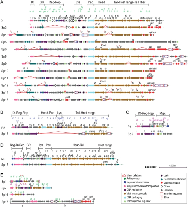
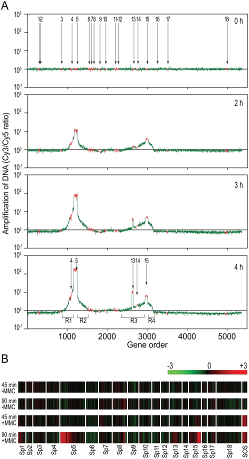
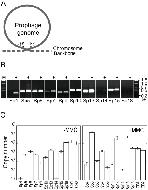
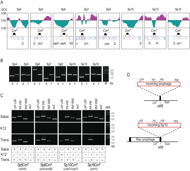
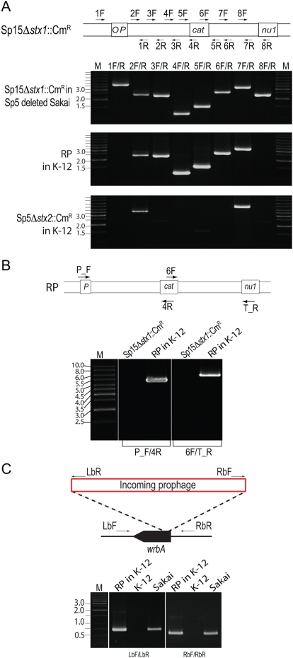
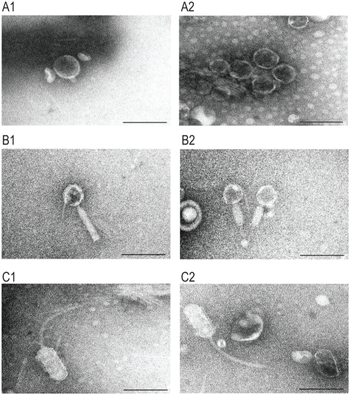
Bacteriophages are major genetic factors promoting horizontal gene transfer (HGT) between bacteria. Their roles in dynamic bacterial genome evolution have been increasingly highlighted by the fact that many sequenced bacterial genomes contain multiple prophages carrying a wide range of genes. Enterohemorrhagic Escherichia coli O157 is the most striking case. A sequenced strain (O157 Sakai) possesses 18 prophages (Sp1-Sp18) that encode numerous genes related to O157 virulence, including those for two potent cytotoxins, Shiga toxins (Stx) 1 and 2. However, most of these prophages appeared to contain multiple genetic defects. To understand whether these defective prophages have the potential to act as mobile genetic elements to spread virulence determinants, we looked closely at the Sp1-Sp18 sequences, defined the genetic defects of each Sp, and then systematically analyzed all Sps for their biological activities. We show that many of the defective prophages, including the Stx1 phage, are inducible and released from O157 cells as particulate DNA. In fact, some prophages can even be transferred to other E. coli strains. We also show that new Stx1 phages are generated by recombination between the Stx1 and Stx2 phage genomes. The results indicate that these defective prophages are not simply genetic remnants generated in the course of O157 evolution, but rather genetic elements with a high potential for disseminating virulence-related genes and other genetic traits to other bacteria. We speculate that recombination and various other types of inter-prophage interactions in the O157 prophage pool potentiate such activities. Our data provide new insights into the potential activities of the defective prophages embedded in bacterial genomes and lead to the formulation of a novel concept of inter-prophage interactions in defective prophage communities.


Conflict of interest statement

The authors have declared that no competing interests exist.

Figures

Figure 1
Figure 1. Alignment of 18 Sakai prophages (Sps) with the corresponding prototype phage genomes.
Genomic organization of lambdoid Sps (A), P2-like Sp (B), P4-like Sp (C), and Mu-like Sp (D) are shown with corresponding prototype phage genomes. Genomic organization of other Sps are shown in (E). Genes on the prototypes are serially numbered from left to right according to their orf numbers ,,,. Only the regions in Sps showing variation in gene organization compared with the prototype genomes are numbered according to the prototype gene. Gene names on the prototypes are shown in green (λ), blue (P2), purple (P4), and black (Mu). Asterisks indicate the genes that are predicted to be non-functional. Phage genomic regions involved in integration/excision (IX), transposition (Tn), regulation (Reg), replication (Rep), general recombination (GR), packaging (Pac), cell lysis (Lys), morphogenesis (Portal, Head, Tail, and Host range) and miscellaneous (Misc) functions are indicated in each prototype genome. An enlarged version with more detailed information is presented in Figure S1.
Figure 2
Figure 2. Microarray analysis of the DNA amplification and transcriptional changes of prophages induced by MMC treatment.
(A) Prophage DNA amplification induced by MMC. Total cellular DNA was prepared from O157 Sakai cells treated with MMC and amplification of prophage regions was analyzed using the oligo DNA microarray. Genes on the O157 Sakai chromosome are shown on the x-axes according to their genomic context. The y-axes indicate the ratios of hybridization signals of test DNA preparations relative to that from the reference DNA for the 0-h sample. The test DNA was prepared from the O157 Sakai cells collected at the indicated time points after MMC was added to the culture. The data from the probes representing prophage and backbone genes on the O157 Sakai chromosome are shown in red and green, respectively. All prophage regions (Sp1–Sp18) on the O157 Sakai chromosome are indicated in the 0-h plot, and selectively enriched prophage regions are indicated in the 4-h plot. Chromosome regions amplified by the regional replication of Sp5 and Sp15 (R1 and R2 by Sp5, R3 and R4 by Sp15) are also indicated in the 4-h plot. (B) Transcriptional changes of the prophage genes induced by MMC. The color bar indicates relative expression levels. Total RNA was prepared from the cells collected at the 45-min and the 90-min time points after the addition of MMC. RNA prepared from the cells collected at the 0-min point was used as the reference. RNA prepared from cells left untreated with MMC was also analyzed with the same protocol (−MMC). Only the data for genes in the 18 prophage regions (Sp1–Sp18) are shown according to their genomic context, along with those for the 10 chromosome genes (sulA, dinI, hlyE, umuD, umuC, recX, recA, dinD, lexA, and dinF) that are known to be induced by MMC treatment (SOS) ,. Average values obtained from two independent experiments are shown. Note that specific probes were unable to be designed for many genes on lambdoid phages because they have nearly identical sequences. These genes were excluded from this analysis.
Figure 3
Figure 3. Excision, circularization, and replication of prophages.
(A) Schematic representation of the strategy to detect excised and circularized prophage genomes by PCR. (B) Detection of PCR products derived from excised and circularized prophage genomes by agarose gel electrophoresis. Total cellular DNA isolated from untreated (−) and MMC-treated cells (+) was analyzed. We examined all Sps, but only the data from the nine prophages that gave positive results are shown. The data of Sp18, a Mu-like phage whose DNA is not circularized, are shown as a negative control. (C) Quantification of circularized phage genomes in the total cellular DNA isolated from untreated (−MMC) and MMC-treated cells (+MMC) using quantitative PCR (qPCR). The data were obtained from three independent analyses, and average copy numbers for each prophage genome are shown. Bars indicate standard deviations. The Sp18 DNA and the chromosomal DNA from two chromosome regions (CB1 and CB2) were quantified as controls.
Figure 4
Figure 4. Phage DNA packaged into phage particles.
(A) FIGE analysis of packaged phage DNA in untreated (−MMC) and MMC-treated (+MMC) cultures. Phage particles were collected from culture supernatants by PEG/NaCl precipitation. DNA preparations applied to the FIGE gel were obtained from 50-ml (untreated) and 1-ml (MMC-treated) cultures. (B) Quantification of packaged phage DNA in the culture supernatants obtained from untreated (−MMC) and MMC-treated (+MMC) O157 Sakai cells. DNase-resistant phage DNA was quantified by qPCR using the primers that were used in Figure 3. DNA preparations equivalent to the 1-ml culture supernatant were used as template DNA. The data were obtained from three independent analyses, and the average copy numbers for each prophage genome are shown. Bars indicate standard deviations. The Sp18 DNA and the chromosomal DNA from two chromosome regions (CB1 and CB2) were monitored as controls.
Figure 5
Figure 5. Construction of CmR-marked prophages and analysis of their transfer and lysogenization into K-12 strains.
(A) Strategy to construct CmR-marked prophages. The gene organizations and GC contents of the regions flanking each target gene that was replaced by the CmR gene cassette are shown. (B) Verification of gene replacement using PCR. PCR products obtained from the O157 Sakai parent strain (p) and mutant strains carrying the CmR cassette in each prophage (c) are shown. (C,D) PCR examination of the transfer of CmR-marked prophages from O157 Sakai to K-12. PCR products obtained from O157 Sakai (Sakai) and K-12 (K12) cells and the transductants (Trans) of CmR-marked Sp5, Sp6, Sp10, and Sp15 are shown in (C). Absence of the stx2 gene in all transductants was also confirmed. Integration sites of each prophage in O157 Sakai are indicated in parentheses. Integration patterns of each prophage in K-12 and positions of PCR primers used are shown in (D). The chromosomal attachment site (attB) for Sp10 was occupied by the Rac prophage, and incoming Sp10 was integrated into the K-12 chromosome using the attR site of the Rac prophage. In the Sp15-transductant, the CmR-marked Sp15 was not found at the yehV locus, where Sp15 is integrated in O157 Sakai. Our preliminary result with the Sp15 transductants indicates that the CmR gene on Sp15 was transferred to K-12 by a chimeric phage generated by recombination between the Sp5 and the CmR-marked Sp15 genomes (see text).
Figure 6
Figure 6. Generation of a chimeric phage by recombination between the CmR-marked Sp15 derivative and Sp5.
(A) PCR scanning analysis of the stx1-flanking region of a chimeric recombinant phage (RP) in a K-12 Sp15Δstx1::CmR transductant. Positions of the PCR primers on the Sp15Δstx1::CmR genome are schematically shown at the top. For comparison, the stx1-flanking region of Sp15Δstx1::CmR in the donor strain O157 Sakai and the corresponding stx2-flanking region of Sp5Δstx2::CmR in its K-12 transductant were analyzed using the same set of primers. In this analysis, a spontaneous Sp5-deletion mutant of the Sp15Δstx1::CmR-containg O157 Sakai was used because Sp15 and Sp5 genomic regions between the P gene and the stx1 (or stx2) gene and between the stx1 (or stx2) gene and the nu1 gene contain highly homologous sequences (see Figure S7 for more details). (B) PCR analysis of the chimeric phage. The stx1-flanking region of the Sp5/Sp15 recombinant phage in K-12 was analyzed using two primer pairs. The positions of the primers on the chimeric phage genome are schematically shown at the top. Primers P_F and T_R are specific to the P and nu1 genes of Sp5, respectively. As a control, an O157 Sakai-derivative containing Sp15Δstx1::CmR was analyzed. PCR products were obtained by the two primer pairs (4.9 kb and 6.4 kb in size, respectively) only from the K-12 derivative carrying the recombinant phage (RP in K-12). (C) PCR analysis of the wrbA locus of a K-12 derivative carrying the chimeric phage (RP in K-12), K-12, and O157 Sakai. Integration of an Sp5-like phage into the wrbA locus (the integration site of Sp5 in O157 Sakai) in the K-12 derivative was confirmed by PCR using two primer pairs. Positions of the primer are schematically shown at the top. Primers LbR and RbF are specific to the left and right ends of the Sp5 genome, respectively.
Figure 7
Figure 7. Electron micrographs of phage particles produced by O157 Sakai.
Electron micrographs of three types of phage particles detected in the culture supernatant of O157 Sakai are shown. (A) Phage particles with a short tail. (B) Phage particles with a contractile and non-flexible tail. (C) Phage particles with an elongated head and a long flexible tail. Phage particles shown in (A) are derived from Sp5 because K-12 strains lysogenized by CmR-marked Sp5 produced phage particles with the identical morphology. The similarity to phage Mu suggests that the phage particles shown in (B) are derived from Sp18. Electron micrographs were taken at magnifications of 60 K or 80 K using a transmission electron microscope and negatively contrasted with 2% uranyl acetate dihydrate. Bar, 100 nm.

References

    1. Brüssow H, Canchaya C, Hardt W D. Phages and the Evolution of Bacterial Pathogens: from Genomic Rearrangements to Lysogenic Conversion. Microbiol Mol Biol Rev. 2004;68:560–602. - PMC - PubMed
    1. Filee J, Forterre P, Laurent J. The role played by viruses in the evolution of their hosts: A view based on informational protein phylogenies. Res Microbiol. 2003;154:237–243. - PubMed
    1. Canchaya C, Proux C, Fournous G, Bruttin A, Brüssow H. Prophage Genomics. Microbiol Mol Biol Rev. 2003;67:238–276. - PMC - PubMed
    1. Freeman VJ. Studies on the virulence of bacteriophage-infected strains of Corynebacterium diphtheriae. J Bacteriol. 1951;61:675–688. - PMC - PubMed
    1. Casjens S. Prophages and bacterial genomics: What have we learned so far? Mol Microbiol. 2003;49:277–300. - PubMed

Publication types

MeSH terms