. 2009 May;5(5):e1000409.
doi: 10.1371/journal.ppat.1000409. Epub 2009 May 1.

Immunogenicity and protective efficacy of a live attenuated H5N1 vaccine in nonhuman primates

Affiliations

Immunogenicity and protective efficacy of a live attenuated H5N1 vaccine in nonhuman primates

Shufang Fan et al. PLoS Pathog. 2009 May.

Abstract

The continued spread of highly pathogenic H5N1 influenza viruses among poultry and wild birds, together with the emergence of drug-resistant variants and the possibility of human-to-human transmission, has spurred attempts to develop an effective vaccine. Inactivated subvirion or whole-virion H5N1 vaccines have shown promising immunogenicity in clinical trials, but their ability to elicit protective immunity in unprimed human populations remains unknown. A cold-adapted, live attenuated vaccine with the hemagglutinin (HA) and neuraminidase (NA) genes of an H5N1 virus A/VN/1203/2004 (clade 1) was protective against the pulmonary replication of homologous and heterologous wild-type H5N1 viruses in mice and ferrets. In this study, we used reverse genetics to produce a cold-adapted, live attenuated H5N1 vaccine (AH/AAca) that contains HA and NA genes from a recent H5N1 isolate, A/Anhui/2/05 virus (AH/05) (clade 2.3), and the backbone of the cold-adapted influenza H2N2 A/AnnArbor/6/60 virus (AAca). AH/AAca was attenuated in chickens, mice, and monkeys, and it induced robust neutralizing antibody responses as well as HA-specific CD4+ T cell immune responses in rhesus macaques immunized twice intranasally. Importantly, the vaccinated macaques were fully protected from challenge with either the homologous AH/05 virus or a heterologous H5N1 virus, A/bar-headed goose/Qinghai/3/05 (BHG/05; clade 2.2). These results demonstrate for the first time that a cold-adapted H5N1 vaccine can elicit protective immunity against highly pathogenic H5N1 virus infection in a nonhuman primate model and provide a compelling argument for further testing of double immunization with live attenuated H5N1 vaccines in human trials.


Conflict of interest statement

The authors have declared that no competing interests exist.

Figures

Figure 1
Figure 1. Vaccine efficacy of the AH/AAca virus in mice.
(A) HI and (B) NT antibody responses to homologous (AH/05) and heterologous (BHG/05) viruses after intranasal vaccination with 106 EID50 of AH/AAca in a 50-µl volume. Serum samples were collected on day 0 prevaccination (blue), 4 weeks after the first (orange) and 4 weeks after the second vaccination (pink). Asterisks indicate a statistically significant difference from antibody titers measured at the preceding time point: **, P<0.01, *, P<0.05. (C–H) Protective efficacy against challenge with the AH/05 (C–E) or BHG/05 virus (F–H). Weight changes (D and G) and survival rates (E and H) are shown only for the groups that were immunized once. The data in panels A–C and F are reported as means±s.d.; the dashed blue lines in these panels indicate the lower limit of detection. p.c., postchallenge.
Figure 2
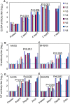
Figure 2. Antibody responses of nonhuman primates.
(A) H5N1-specific antibody levels assessed by ELISA. Hemagglutination-inhibition (HI) antibody to AH/05 (B) and BHG/05 (C) with chicken erythrocytes, and microneutralization (NT) antibody to the AH/05 (D) and BHG/05 (E) viruses. The HI antibody titers with horse erythrocytes were 4- to 8-fold higher than those with chicken erythrocytes (data not shown). Titers are reported for individual vaccinated animals. Blue dashed lines indicate the lower limit of detection. Wpd1/2, week postvaccination dose 1 or 2; wpc, week postchallenge. The P values indicate the antibody titers with a significant increase from the preceding time point. In the control animals, the HI and NT antibodies at all time points are the same as the pretested values, only the ELISA antibody titers, ranged 400–800, were detected at two weeks postchallenge (data not shown).
Figure 3
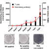
Figure 3. T cell responses against HA antigens in rhesus macaques and its relationship to the neutralizing antibody.
T cell responses in the PBMC samples were measured by overlapping peptides and ex-vivo IFN-γ ELISPOT assay. Data shown are the mean numbers ±s.d. of spot-forming cells (SFC) per 106 PBMCs to HA peptide pools in the vaccinated animals at different time points after vaccination and challenge. Neutralizing antibody in the serum samples was measured by the microneutralization method using AH/05 virus isolate, data shown are mean titers ±s.d. The T cell response and the neutralizing antibody were not detected in the control monkeys at all time points tested (data not shown).
Figure 4
Figure 4. Body temperature of nonhuman primates after challenge with AH/05 virus.
Change in body temperature in nonhuman primates after challenge with AH/05 virus (A) or BHG/05 virus (B). Changes were calculated by subtracting the mean temperature 3 days before challenge from the temperature recorded on the indicated day.
Figure 5
Figure 5. Vaccine efficacy in nonhuman primates assessed on the basis of lung lesions.
Eight macaques were euthanized on day 3 postchallenge with AH/05 virus (A–D) or BHG/05 virus (E–H). Vaccinated animals (A,B,E,F) had less extensive bronchopneumonia (i.e., smaller foci of consolidation) than did unvaccinated animals (C,D,G,H). The vaccinated animals also showed prominent peribronchial lymph follicles (a, e; arrows), and their consolidated lung areas lacked viral antigen-positive cells (B,F). By contrast, the unvaccinated animals had lung lesions of moderate size with a wide consolidated area (C,G; outlined by yellow dashes), smaller and less abundant peribronchial lymph follicles (C,G; arrows), and pneumonic lesions containing many antigen-positive cells (D,H; brown pigment). (I) Schematic diagrams indicating distribution of pathologic lesions in the lungs of animals vaccinated and challenged with AH/05 (V1 and V2); nonvaccinated and challenged with AH/05 (C1 and C2); vaccinated and challenged with BHG/05 (V3 and V4); and nonvaccinated and challenged with BHG/05 (C3 and C4). In vaccinated animals, scant-to-moderate bronchopneumonia was present in each lobe, but viral antigens were not detected in the lesions (V1, V2, V3, and V4; purple). By contrast, more severe bronchopneumonia was observed in nonvaccinated macaque lungs (C1, C2, C3, and C4). Moreover, viral antigens were prominent in the pneumonic lesions in the most affected lung lobes (C1, C2, C3, and C4; red). One lung lobe was entirely affected by pneumonia after infection with the BHG/05 virus (C3). Purple, bronchopneumonia without viral antigen; red, bronchopneumonia with viral antigen.
Figure 6
Figure 6. Viral replication in nonhuman primates.
Virus titers were determined in embryonated eggs injected with tissue homogenates on day 3 (A,B) and day 15 (C) postchallenge with AH/05 virus or BHG/05 virus. Virus was not detected in tissues harvested day 15 postchallenge from animals challenged with the BHG/05 virus (data not shown). Titers are reported for tissues from individual animals, as log10 EID50/g tissue. The dashed blue lines indicate the lower limit of detection. (D) Nasal swabs were collected from all living animals on days 2, 4, and 6 postchallenge for virus isolation in eggs.

References

    1. Chen H, Deng G, Li Z, Tian G, Li Y, et al. The evolution of H5N1 influenza viruses in ducks in southern China. Proc Natl Acad Sci U S A. 2004;101:10452–10457. - PMC - PubMed
    1. Shortridge KF, Zhou NN, Guan Y, Gao P, Ito T, et al. Characterization of avian H5N1 influenza viruses from poultry in Hong Kong. Virology. 1998;252:331–342. - PubMed
    1. Claas EC, Osterhaus AD, van Beek R, De Jong JC, Rimmelzwaan GF, et al. Human influenza A H5N1 virus related to a highly pathogenic avian influenza virus. Lancet. 1998;351:472–477. - PubMed
    1. Subbarao K, Klimov A, Katz J, Regnery H, Lim W, et al. Characterization of an avian influenza A (H5N1) virus isolated from a child with a fatal respiratory illness. Science. 1998;279:393–396. - PubMed
    1. de Jong MD, Tran TT, Truong HK, Vo MH, Smith GJ, et al. Oseltamivir resistance during treatment of influenza A (H5N1) infection. N Engl J Med. 2005;353:2667–2672. - PubMed

Publication types

MeSH terms