The HLA Region and Autoimmune Disease: Associations and Mechanisms of Action
- PMID: 19412418
- PMCID: PMC2647156
- DOI: 10.2174/138920207783591690
The HLA Region and Autoimmune Disease: Associations and Mechanisms of Action
Abstract
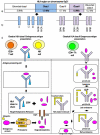
The HLA region encodes several molecules that play key roles in the immune system. Strong association between the HLA region and autoimmune disease (AID) has been established for over fifty years. Association of components of the HLA class II encoded HLA-DRB1-DQA1-DQB1 haplotype has been detected with several AIDs, including rheumatoid arthritis, type 1 diabetes and Graves' disease. Molecules encoded by this region play a key role in exogenous antigen presentation to CD4+ Th cells, indicating the importance of this pathway in AID initiation and progression. Although other components of the HLA class I and III regions have also been investigated for association with AID, apart from the association of HLA-B*27 with ankylosing spondylitis, it has been difficult to determine additional susceptibility loci independent of the strong linkage disequilibrium (LD) with the HLA class II genes. Recent advances in the statistical analysis of LD and the recruitment of large AID datasets have allowed investigation of the HLA class I and III regions to be re-visited. Association of the HLA class I region, independent of known HLA class II effects, has now been detected for several AIDs, including strong association of HLA-B with type 1 diabetes and HLA-C with multiple sclerosis and Graves' disease. These results provide further evidence of a possible role for bacterial or viral infection and CD8+ T cells in AID onset. The advances being made in determining the primary associations within the HLA region and AIDs will not only increase our understanding of the mechanisms behind disease pathogenesis but may also aid in the development of novel therapeutic targets in the future.
Keywords: Genes; autoimmunity & HLA.
Figures

References
-
- Thorsby E., Lie B.A. HLA associated genetic predisposition to autoimmune diseases Genes involved and possible mechanisms. Transpl. Immunol. 2005;14:175–182. - PubMed
-
- Simmonds M.J., Gough S.C. Genetic insights into disease mechanisms of autoimmunity. Br. Med. Bull. 2004;71:93–113. - PubMed
-
- Horton R., Wilming L., Rand V., Lovering R.C., Bruford E.A., Khodiyar V.K., Lush M.J., Povey S., Talbot CC Jr., Wright M.W., Wain H.M., Trowsdale J., Ziegler A., Beck S. Gene map of the extended human MHC. Nat. Rev. Genet. 2004;5:889–899. - PubMed
-
- Mungall A.J., Palmer S.A., Sims S.K., Edwards C.A., Ashurst J.L., Wilming L., Jones M.C., Horton R., Hunt S.E., Scott C.E., Gilbert J.G., Clamp M.E., Bethel G., Milne S., Ainscough R., Almeida J.P., Ambrose K.D., Andrews T.D., Ashwell R.I., Babbage A.K., Bagguley C.L., Bailey J., Banerjee R., Barker D.J., Barlow K.F., Bates K., Beare D.M., Beasley H., Beasley O., Bird C.P., Blakey S., Bray-Allen S., Brook J., Brown A.J., Brown J.Y., Burford D.C., Burrill W., Burton J., Carder C., Carter N.P., Chapman J.C., Clark S.Y., Clark G., Clee C.M., Clegg S., Cobley V., Collier R.E., Collins J.E., Colman L.K., Corby N.R., Coville G.J., Culley K.M., Dhami P., Davies J., Dunn M., Earthrowl M.E., Ellington A.E., Evans K.A., Faulkner L., Francis M.D., Frankish A., Frankland J., French L., Garner P., Garnett J., Ghori M.J., Gilby L.M., Gillson C.J., Glithero R.J., Grafham D.V., Grant M., Gribble S., Griffiths C., Griffiths M., Hall R., Halls K.S., Hammond S., Harley J.L., Hart E.A., Heath P.D., Heathcott R., Holmes S.J., Howden P.J., Howe K.L., Howell G.R., Huckle E., Humphray S.J., Humphries M.D., Hunt A.R., Johnson C.M., Joy A.A., Kay M., Keenan S.J., Kimberley A.M., King A., Laird G.K., Langford C., Lawlor S., Leongamornlert D.A., Leversha M., Lloyd C.R., Lloyd D.M., Loveland J.E., Lovell J., Martin S., Mashreghi-Mohammadi M., Maslen G.L., Matthews L., McCann O.T., McLaren S.J., McLay K., McMurray A., Moore M.J., Mullikin J.C., Niblett D., Nickerson T., Novik K.L., Oliver K., Overton-Larty E.K., Parker A., Patel R., Pearce A.V., Peck A.I., Phillimore B., Phillips S., Plumb R.W., Porter K.M., Ramsey Y., Ranby S.A., Rice C.M., Ross M.T., Searle S.M., Sehra H.K., Sheridan E., Skuce C.D., Smith S., Smith M., Spraggon L., Squares S.L., Steward C.A., Sycamore N., Tamlyn-Hall G., Tester J., Theaker A.J., Thomas D.W., Thorpe A., Tracey A., Tromans A., Tubby B., Wall M., Wallis J.M., West A.P., White S.S., Whitehead S.L., Whittaker H., Wild A., Willey D.J., Wilmer T.E., Wood J.M., Wray P.W., Wyatt J.C., Young L., Younger R.M., Bentley D.R., Coulson A., Durbin R., Hubbard T., Sulston J.E., Dunham I., Rogers J., Beck S. The DNA sequence and analysis of human chromosome 6. Nature. 2003;425:805–811. - PubMed
-
- Shiina T., Inoko H., Kulski J.K. An update of the HLA genomic region, locus information and disease associations 2004. Tissue Antigens. 2004;64:631–649. - PubMed
Grants and funding
LinkOut - more resources
Full Text Sources
Other Literature Sources
Research Materials
