. 2009;4(5):e5421.
doi: 10.1371/journal.pone.0005421. Epub 2009 May 1.

Characterization of granulations of calcium and apatite in serum as pleomorphic mineralo-protein complexes and as precursors of putative nanobacteria

Affiliations

Characterization of granulations of calcium and apatite in serum as pleomorphic mineralo-protein complexes and as precursors of putative nanobacteria

John D Young et al. PLoS One. 2009.

Abstract

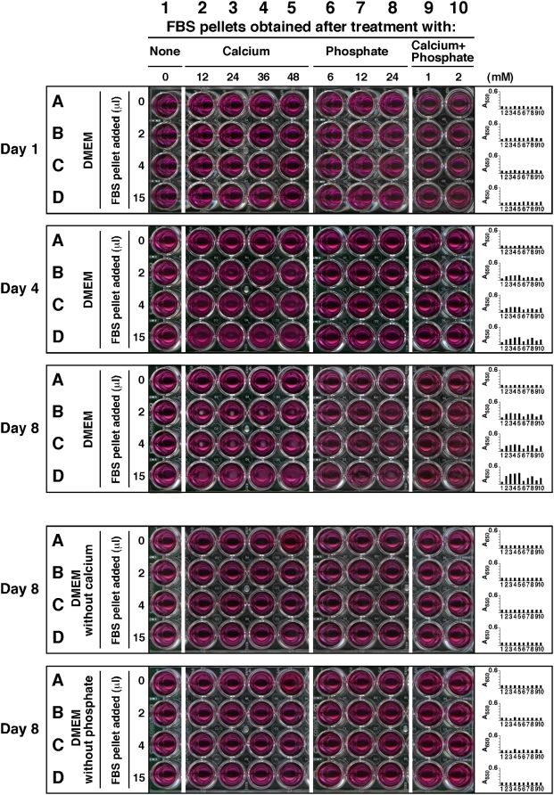
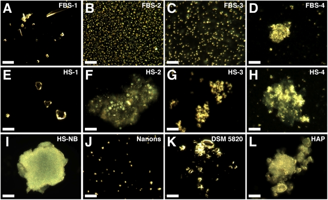
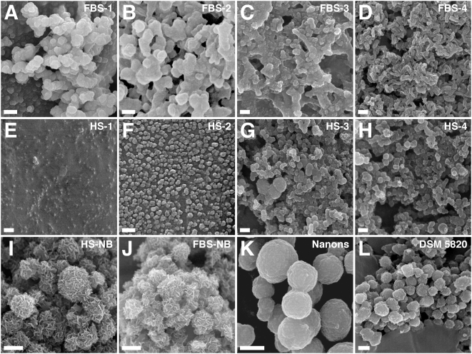
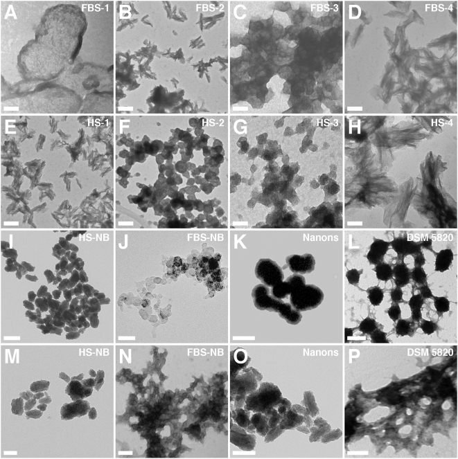
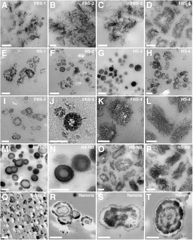
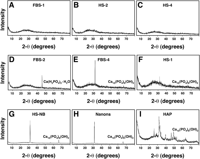
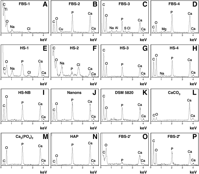
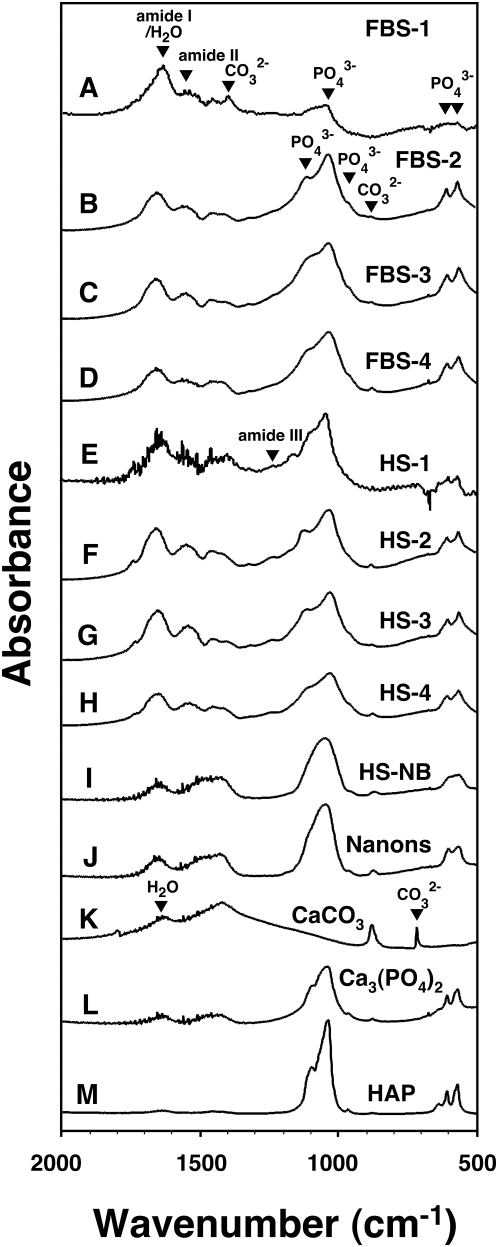
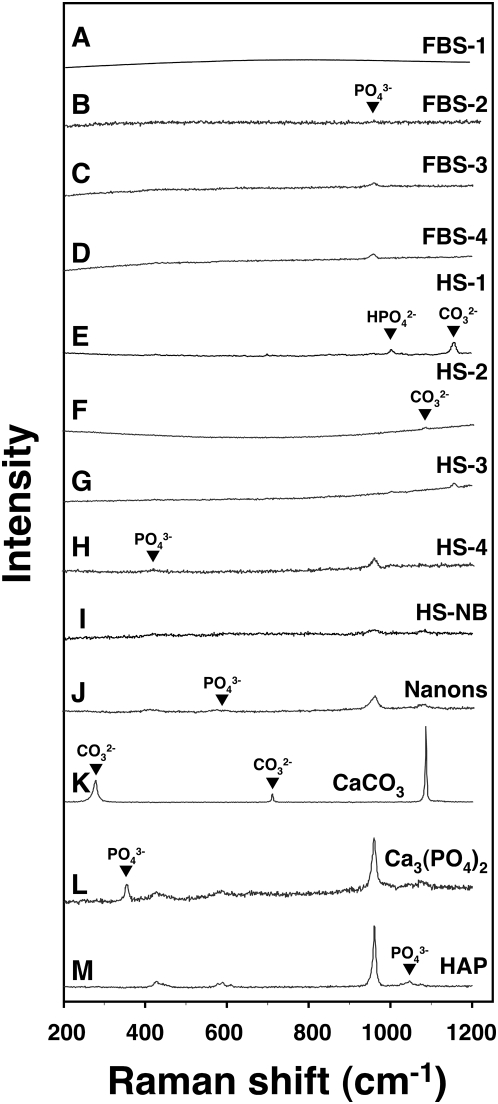
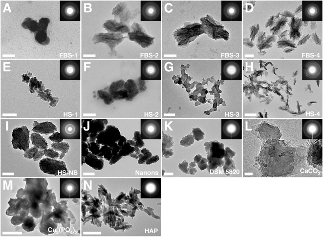
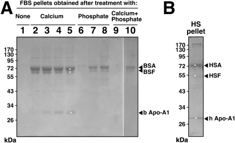
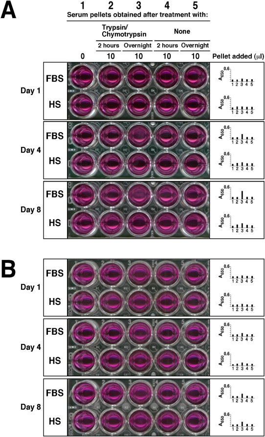
Calcium and apatite granulations are demonstrated here to form in both human and fetal bovine serum in response to the simple addition of either calcium or phosphate, or a combination of both. These granulations are shown to represent precipitating complexes of protein and hydroxyapatite (HAP) that display marked pleomorphism, appearing as round, laminated particles, spindles, and films. These same complexes can be found in normal untreated serum, albeit at much lower amounts, and appear to result from the progressive binding of serum proteins with apatite until reaching saturation, upon which the mineralo-protein complexes precipitate. Chemically and morphologically, these complexes are virtually identical to the so-called nanobacteria (NB) implicated in numerous diseases and considered unusual for their small size, pleomorphism, and the presence of HAP. Like NB, serum granulations can seed particles upon transfer to serum-free medium, and their main protein constituents include albumin, complement components 3 and 4A, fetuin-A, and apolipoproteins A1 and B100, as well as other calcium and apatite binding proteins found in the serum. However, these serum mineralo-protein complexes are formed from the direct chemical binding of inorganic and organic phases, bypassing the need for any biological processes, including the long cultivation in cell culture conditions deemed necessary for the demonstration of NB. Thus, these serum granulations may result from physiologically inherent processes that become amplified with calcium phosphate loading or when subjected to culturing in medium. They may be viewed as simple mineralo-protein complexes formed from the deployment of calcification-inhibitory pathways used by the body to cope with excess calcium phosphate so as to prevent unwarranted calcification. Rather than representing novel pathophysiological mechanisms or exotic lifeforms, these results indicate that the entities described earlier as NB most likely originate from calcium and apatite binding factors in the serum, presumably calcification inhibitors, that upon saturation, form seeds for HAP deposition and growth. These calcium granulations are similar to those found in organisms throughout nature and may represent the products of more general calcium regulation pathways involved in the control of calcium storage, retrieval, tissue deposition, and disposal.


Conflict of interest statement

Competing Interests: The authors have declared that no competing interests exist.

Figures

Figure 1
Figure 1. Calcium and phosphate added to serum produce particle-seeding pellets.
Serum pellets were obtained by adding calcium and/or phosphate to FBS to the amounts indicated at the top and incubated overnight at room temperature. Treatment of serum was divided into 4 groups of ions added: no exogenous ions (column 1, “None”); CaCl2 (columns 2–5, referred as “Calcium,” 12–48 mM); Na2HPO4 (columns 6–8, “Phosphate,” 6–24 mM); and a combination of both CaCl2 and Na2HPO4 (columns 9 and 10, “Calcium+Phosphate,” at either 1 mM or 2 mM each). After overnight incubation, the pellets were obtained and processed as described in the Materials and Methods . The resuspended pellets were inoculated into either serum-free DMEM (rows A–D, top 3 panels), calcium-free DMEM (rows A–D, 4th panel from top), or phosphate-free DMEM (rows A–D, bottom panel) in the same order, as three exact replicas. The amount of resuspended pellet volume inoculated into each well is depicted on the left. Observations were done on days 1, 4, and 8 for the top three panels and on day 8 for the bottom two. All incubations were done at 37°C in cell culture conditions. The wells in column 1 corresponded to inoculation with serum pellets obtained from FBS without any ion treatment, while the wells in row A represented control DMEM, without any pellet inoculation. These controls showed no turbidity by visual inspection. On the other hand, turbidity and the presence of precipitates could be seen in all the other wells of the “Day 4” and “Day 8” panels. The wells of the 4th panel, corresponding to incubation in DMEM without calcium, did not show any precipitate whereas the bottom panel wells (DMEM without phosphate) produced small amounts of precipitates.
Figure 2
Figure 2. Serum pellets and NB have similar pleomorphic morphologies under dark field microscopy.
Serum pellets were prepared from untreated serum (A and E) or following addition of either 48 mM CaCl2 (B and F), 24 mM Na2HPO4 (C and G), or a combination of both 2 mM CaCl2 and 2 mM Na2HPO4 (D and H) to the indicated serum, followed by incubation at room temperature overnight, centrifugation, and washing steps as described in the Materials and Methods . Low amounts of large heterogeneous particles were noted in the untreated serum pellets (A and E) while the other serum pellets (B–D, F–H) produced smaller and more homogeneous round particles. Such round particles tended to aggregate forming clumps and granular patches (D, F–H). Note that individual granules can be discerned against a background of aggregated material. Similar morphologies were noted in the controls, with HS-NB (I) showing an aggregated diffuse mass in which granules are embedded; “nanons” (J) as dispersed round particles; and DSM 5820 (K) as clumps of highly refringent particles. (L) HAP is shown for comparison. Scale bars: 10 µm.
Figure 3
Figure 3. Morphology of serum pellet granulations seen under SEM demonstrates resemblance to NB.
Serum pellets were prepared as in Fig. 2 from untreated serum (A and E) or after addition of either CaCl2 (B and F), Na2HPO4 (C and G), or a combination of both CaCl2 and Na2HPO4 (D and H) to the indicated serum (amounts of ions added as described in Materials and Methods ). Serum pellets primarily harbored round particles in FBS pellets (A–C) and these particles tended to be smaller in HS (E–G). Treatment with both CaCl2 and Na2HPO4 produced particles undergoing different stages of crystallization and film coalescence as well as needle-like projections or spindles (D and H). The morphologies of the serum pellets were similar to the NB controls obtained from either 10% HS (I) or 10% FBS (J) as well as to the NB strains “nanons” (K) and DSM 5820 (L), even though the sizes of the serum pellets particles tended to be smaller than NB. Note that while both “nanons” and DSM 5820 showed smooth surfaces, HS-NB and FBS-NB appeared with rough surfaces containing needle-like crystalline projections. Scale bars: 100 nm (B); 250 nm (C–E, G, H); 300 nm (F, L); 500 nm (I, J); 600 nm (K); 1 µm (A).
Figure 4
Figure 4. Morphology of unstained serum pellet particles observed by TEM shows similarity to NB.
Serum pellets were prepared from untreated serum (A and E) or after addition of either CaCl2 (B and F), Na2HPO4 (C and G), or both (D and H) to the indicated serum, followed by preparation for TEM. Samples were viewed without fixation or staining. The serum pellets displayed various morphologies including round particles (A, C, F, and G) while other samples harbored spindles with more crystalline appearance (B, D, E, and H). In general, the combination of calcium and phosphate tended to produce more readily spindles with needle-like crystalline projections (D and H). The various NB controls shown in the bottom two rows were displayed to show comparable morphologies with predominant round particle shapes (3rd row) or more crystalline aggregates (4th row). However, morphological variations can still be seen within each row. Thus, NB cultured directly from 10% HS (I) or 10% FBS (J) displayed predominantly chain-linked ovoid or spherical shapes resembling dividing bacteria. In contrast, the NB strains “nanons”(K) and DSM 5820 (L) shown here appear further along in their crystallization and while they have retained ovoid shapes, they also show more pronounced fusion and aggregation. In the case of “nanons” (K), there are noticeable thick walls that appear less electron-dense than the core, presumably formed of minerals. For DSM 5820 (L), crystalline bridges can be seen connecting the fused ovoid particles. (M–P) show further progression in aggregation and crystallization, with the appearance of coalesced films (more noticeable in N and P). Film-like aggregation is generally seen with longer periods of incubation or by reducing the amount of serum in the culture medium (to less than 3%). Scale bars: 50 nm (C, G, H, J); 100 nm (A, D, F, N, P); 200 nm (B, E, I, K–M, O).
Figure 5
Figure 5. Morphological resemblance between serum pellets and NB as seen by thin section TEM.
Serum pellets obtained from untreated serum (A and E) or after addition of either CaCl2 (B and F), Na2HPO4 (C and G), or a combination of both (D, H–L) to the indicated serum, were processed for thin section TEM as described in the Materials and Methods . The FBS pellets (A–D) show spindles in various stages of crystallization and aggregation, with (A and B) consisting of smaller filamentous stacks while (C and D) show spindles in the process of fusion and coalescence, presumably to form crystallized films. On the other hand, the HS granulations (E–H) show predominantly round particles with a diameter between 20 to 100 nm. (I–L) represent magnified images of serum granulation specimens to illustrate marked pleomorphism and heterogeneity within the samples prepared in FBS (I–K) or HS (L). (I and J) show FBS-4 round particles appearing with thick, single walls, which appear to represent precursors to the larger fused ovoid particles containing crystallized needle-like projections (K). These appear to coalesce further until they form the spindles appearing as stacks of crystalline filaments arranged co-linearly, shown here for HS-4 (L); note that the various HS samples appear predominantly as round shapes, thus this image was shown to emphasize the phenomenon of pleomorphism. Large structures with ring-like formations were characteristic of NB cultured directly from 10% HS (M–P) and “nanons” cultured in 10% FBS (Q–T). In (S), “nanons” can be seen to be fusing to form stacks of filaments. Note the presence of spherical shapes with two (Q), three (R and S), and four (T) concentric rings of electron-dense material. In general, “nanons” (Q–T) tended to be larger than the similar formations seen in the serum pellets (K and L). Scale bars: 50 nm (E, I–L); 100 nm (B–D, F–H, O, P), 200 nm (A, M, N, S); 300 nm (T); 500 nm (Q, R).
Figure 6
Figure 6. Powder X-ray diffraction spectra of serum granulations demonstrating both amorphous and crystalline patterns.
Serum granulations (pellets) were obtained from either untreated serum (A and F) or following treatments with CaCl2 (B and D) or a combination of CaCl2 and Na2HPO4 (C and E) to the indicated serum, and were dried prior to XRD analysis. Note that the XRD spectra include amorphous patterns (A–C) and peaks corresponding to crystalline compounds of Ca(H2PO4)2·H2O (D) and Ca10(PO4)6(OH)2 (E and F). Crystalline patterns were seen associated with both calcium (D) and calcium phosphate-treated sera (E) as well as untreated serum (F), whereas amorphous patterns were seen not only with untreated serum (A), but also with calcium-treated (B) and calcium phosphate-treated sera (C). XRD spectra corresponding to Ca10(PO4)6(OH)2 were obtained for NB cultured from 10% HS (G) or 10% FBS (H) as well as commercially available HAP used for comparison.
Figure 7
Figure 7. Energy-dispersive X-ray spectroscopy of the serum granulations reveals a broad spectrum of compositions similar to NB.
Serum granulations (pellets) were obtained as before from untreated serum (A and E) or after addition of either CaCl2 (B and F), Na2HPO4 (C and G), or a combination of both (D and H) to the indicated serum. EDX spectra were also obtained for NB cultured from HS (I) or for two NB strains cultured in FBS (J and K). Commercial reagents of CaCO3 (L), calcium phosphate labeled as Ca3(PO4)2 (M), and HAP (N) were used for comparisons. Serum granulations were also prepared by adding CaCl2 to FBS followed by incubation for 1 hour (O) or overnight (P) at room temperature. The following Ca/P ratios were obtained: (B) 1.31; (C–E) 1.33; (F) 1.86; (G) 1.3; (H) 1.24; (I) 1.28; (J) 1.36; (K) 1.31; (M) 1.31; (N) 1.63; (O) 5.42; and (P) 1.24. In (A) and (L), Ca and P were not detected. The presence of C in some samples might be attributed in part to the formvar carbon-coated grids used as support for the analysis.
Figure 8
Figure 8. Fourier-transformed infrared spectroscopy of serum granulations reveals the presence of carbonate and phosphate.
Serum granulations (pellets) were prepared from untreated serum (A and E) or following addition of either CaCl2 (B and F), Na2HPO4 (C and G), or a combination of both (D and H) to the indicated serum. The FTIR spectra of the serum granulations revealed peaks characteristic of phosphate at 575 cm−1, 605 cm−1, 960 cm−1, and 1,000–1,150 cm−1 as well as carbonate at 875 cm−1 and 1,400–1,430 cm−1. Peaks corresponding to amide I, II, and III near 1,660 cm−1, 1,550 cm−1, and 1,250 cm−1, respectively, were also noted in the serum granulations (A–H) probably corresponding to the presence of proteins from the serum used. NB cultured from 10% healthy HS incubated in DMEM for one month (I) and the NB strain “nanons” cultured in 10% FBS (J) showed FTIR spectra identical to those of serum granulations except for the amide peaks which were of lower intensity for amide I and were absent for amide II. Note that the presence of residual water seen in the controls (K–M) at 1,650 cm−1 could also contribute to the intensity of the peak corresponding to amide I. Commercially available CaCO3 (K), Ca3(PO4)2 (L), and HAP (M) were included for comparison.
Figure 9
Figure 9. Micro-Raman spectroscopy shows similar chemical compositions for the serum granulations and NB.
Serum granulations (pellets) obtained from untreated serum (A and E) or following addition of either CaCl2 (B and F), Na2HPO4 (C and G), or a combination of both (D and H) to the indicated serum, were submitted to micron-Raman spectroscopy. NB cultured from 10% healthy HS in DMEM (I) and the NB strain “nanons” cultured in 10% FBS (J) were also included for comparison. The various serum granulation/pellet and NB specimens showed marked variability of peaks with phosphate ions being obtained more consistently. Phosphate peaks were seen at 361 cm−1, 440 cm−1, 581 cm−1, 962 cm−1, 1,002 cm−1 (HPO4 2−), and 1,048 cm−1 whereas carbonate peaks were noticed at 280 cm−1, 712 cm−1, 1,080 cm−1, and 1,150 cm−1. Commercially available CaCO3 (K), Ca3(PO4)2 (L), and HAP (M) were included for comparison.
Figure 10
Figure 10. Serum granulations and NB specimens show similar electron diffraction patterns.
Serum granulations (pellets) obtained from untreated serum (A and E) or following addition of either CaCl2 (B and F), Na2HPO4 (C and G), or a combination of both (D and H) to the indicated serum, were processed for TEM without fixation or staining. NB cultured from 10% HS (I) as well as the NB strains “nanons” (J) and DSM 5820 (K) cultured in 10% FBS were also included for comparison. The electron diffraction patterns shown in the insets indicated that the serum granulations and the NB samples consisted of polycrystalline minerals either with a low degree of crystallinity as shown by the fuzzy rings (A, B, E–G) or with a more crystalline mineral phase as shown by the presence of arrays of dots on the corresponding diffraction patterns (C, D, H). In comparison, the commercial reagents CaCO3 (L), Ca3(PO4)2 (M), and HAP (N), used as controls, more consistently displayed a high degree of crystallinity as shown by the presence of dots on their electron diffraction patterns. Scale bars: 50 nm (A, L); 100 nm (B–E, G, I, J); 200 nm (F, H, K, N); 500 nm (M).
Figure 11
Figure 11. SDS-PAGE profiles of proteins bound to serum granulations and protein identification.
(A) Serum granulations (pellets) were prepared exactly as in Fig. 1, from either untreated FBS (referred as “None”) or through the addition of the indicated amounts of CaCl2 (“Calcium”) and/or Na2HPO4 (“Phosphate”) to FBS. The indicated sequence of the lanes depicted in the heading matches exactly with the sequence of wells seen in Fig. 1. No protein band was found in the pellet of untreated FBS (lane 1), whereas the other lanes showed dose-dependence with respect to the precipitating reagents used. The white marks represent the positions of the bands excised for identification by MALDI-TOF mass fingerprint analysis whereas the identified proteins are shown on the right. While bovine serum albumin (BSA) and apolipoprotein A1 (b Apo-A1) were the only proteins identified in their respective positions, other proteins besides bovine fetuin-A (BSF) were also identified in the 52–65 kDa range (see the text for details). (B) This HS pellet was obtained with 30 mM CaCl2 and processed exactly as done with the FBS pellets. Abbreviations used: HSA, human serum albumin; HSF, human serum fetuin-A; and h Apo-A1, human apolipoprotein A1.
Figure 12
Figure 12. Trypsin and chymotrypsin treatment of FBS and HS generates pellets that have particle-seeding activity when inoculated into serum-free medium.
Seeding-pellets were prepared by adding trypsin (A) or chymotrypsin (B) at 0.5% to either FBS or HS, followed by incubation at 37°C for 2 hours or overnight, then centrifugation and washing as described in the Materials and Methods . 10 µl aliquots of these resuspended pellets (wells 2 and 3, with well 2 corresponding to 2 hours of serum incubation after protease treatment and well 3 corresponding to overnight incubation of the same protease treatment) were then inoculated into 1 ml of DMEM and incubated in cell culture conditions for the time indicated on the left. “Day 1” refers to the day of pellet inoculation. As control, sera that received no addition of proteases were also incubated, centrifuged, and washed the same way, after which the pellets (barely visible in most control specimens) were inoculated into DMEM and incubated exactly as before (wells 4 and 5, described as “None”). As an additional control, DMEM alone was incubated without any inoculation (well 1). Note the time-dependent increase in turbidity associated with either protease treatment, more noticeable with the pellets that had been obtained from sera treated with proteases overnight as contrasted with 2 hour treatment (compare between visuals obtained for wells 3 and 2). Turbidity was usually higher with the FBS pellets compared to HS pellets. Note also the different amounts of turbidity seen with trypsin (A) vs. chymotrypsin (B) treatments; turbidities were generally higher with trypsin treatment. Control wells inoculated with pellets obtained from serum that received no protease treatment (wells 4 and 5), or containing DMEM alone (well 1), produced either little or no visible turbidity.

References

    1. Kajander EO, Kuronen I, Åkerman K, Pelttari A, Çiftçioglu N. Nanobacteria from blood, the smallest culturable autonomously replicating agent on Earth. Proc Soc Photo Opt Instrum Eng. 1997;3111:420–428.
    1. Kajander EO, Çiftçioglu N. Nanobacteria: an alternative mechanism for pathogenic intra- and extracellular calcification and stone formation. Proc Natl Acad Sci U S A. 1998;95:8274–8279. - PMC - PubMed
    1. Kajander EO. Nanobacteria: propagating calcifying nanoparticles. Lett Appl Microbiol. 2006;42:549–552. - PubMed
    1. Ciftcioglu N, McKay DS, Mathew G, Kajander EO. Nanobacteria: fact or fiction? Characteristics, detection, and medical importance of novel self-replicating, calcifying nanoparticles. J Investig Med. 2006;54:385–394. - PubMed
    1. Çiftçioglu N, Björklund M, Kuorikoski K, Bergström K, Kajander EO. Nanobacteria: an infectious cause for kidney stone formation. Kidney Int. 1999;56:1893–1898. - PubMed

Publication types

MeSH terms