. 2009 Jul 10;515(2):181-96.
doi: 10.1002/cne.22059.

Hippocampal injury, atrophy, synaptic reorganization, and epileptogenesis after perforant pathway stimulation-induced status epilepticus in the mouse

Affiliations

Hippocampal injury, atrophy, synaptic reorganization, and epileptogenesis after perforant pathway stimulation-induced status epilepticus in the mouse

Friederike Kienzler et al. J Comp Neurol. .

Abstract

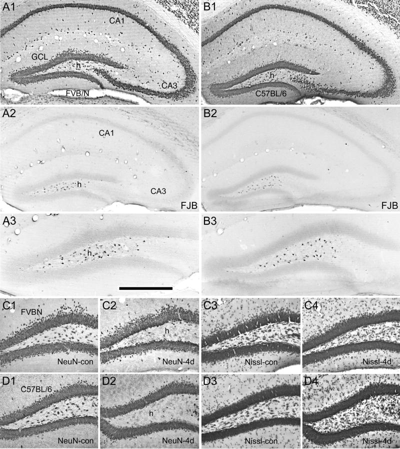
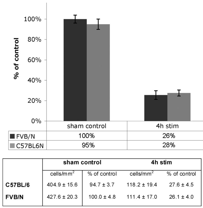
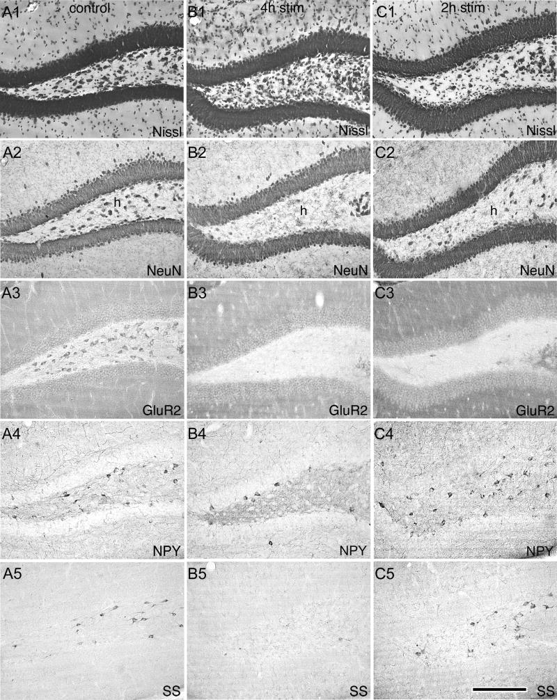
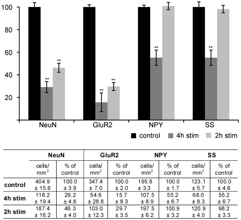
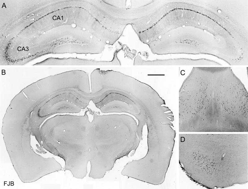
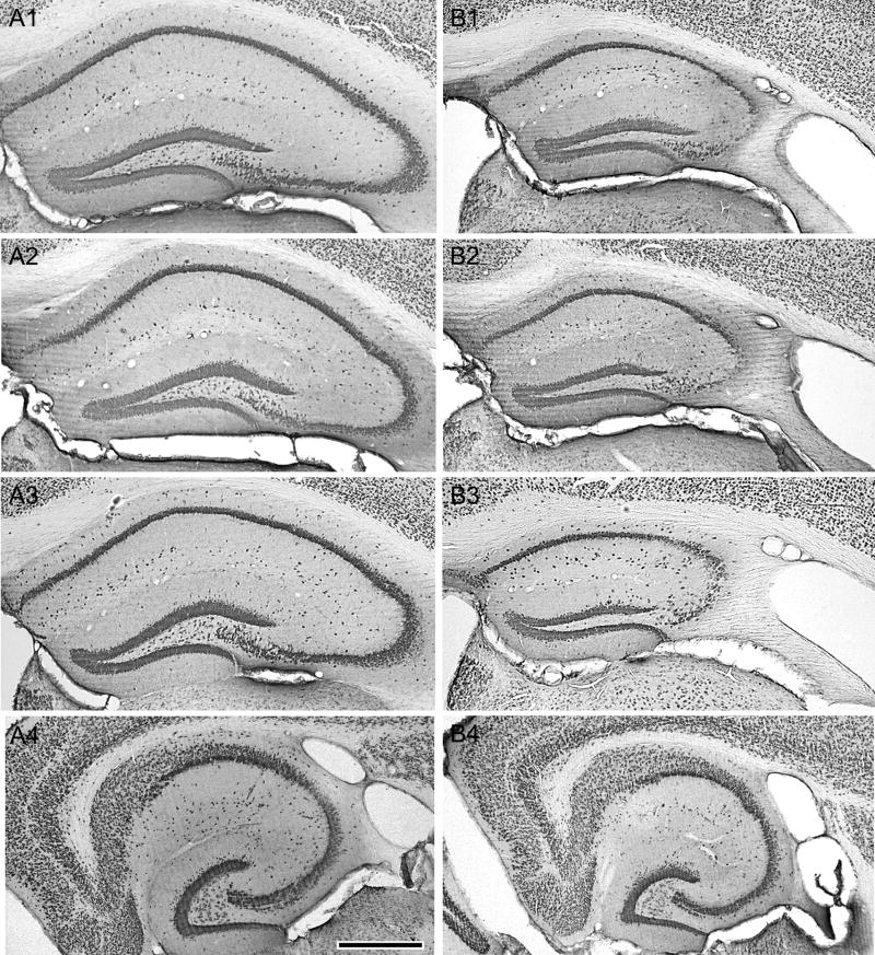
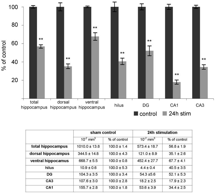
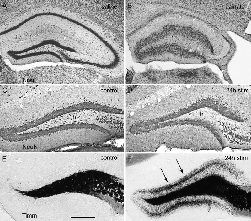
Prolonged dentate granule cell discharges produce hippocampal injury and chronic epilepsy in rats. In preparing to study this epileptogenic process in genetically altered mice, we determined whether the background strain used to generate most genetically altered mice, the C57BL/6 mouse, is vulnerable to stimulation-induced seizure-induced injury. This was necessary because C57BL/6 mice are reportedly resistant to the neurotoxic effects of kainate-induced seizures, which we hypothesized to be related to strain differences in kainate's effects, rather than genetic differences in intrinsic neuronal vulnerability. Bilateral perforant pathway stimulation-induced granule cell discharge for 4 hours under urethane anesthesia produced degeneration of glutamate receptor subunit 2 (GluR2)-positive hilar mossy cells and peptide-containing interneurons in both FVB/N (kainate-vulnerable) and C57BL/6 (kainate-resistant) mice, indicating no strain differences in neuronal vulnerability to seizure activity. Granule cell discharge for 2 hours in C57BL/6 mice destroyed most GluR2-positive dentate hilar mossy cells, but not peptide-containing hilar interneurons, indicating that mossy cells are the neurons most vulnerable to this insult. Stimulation for 24 hours caused extensive hippocampal neuron loss and injury to the septum and entorhinal cortex, but no other detectable damage. Mice stimulated for 24 hours developed hippocampal sclerosis, granule cell mossy fiber sprouting, and chronic epilepsy, but not the granule cell layer hypertrophy (granule cell dispersion) produced by intrahippocampal kainate. These results demonstrate that perforant pathway stimulation in mice reliably reproduces the defining features of human mesial temporal lobe epilepsy with hippocampal sclerosis. Experimental studies in transgenic or knockout mice are feasible if electrical stimulation is used to produce controlled epileptogenic insults.


Figures

Figure 1
Figure 1
Acute neurodegeneration in the FVB/N and C57BL/6 mouse hippocampus one and four days after four hours of bilateral perforant pathway stimulation. (A1 and B1) NeuN-immunoreactivity in the normal FVB/N (A1) and C57BL/6 (B1) hippocampus showing normal dorsal hippocampal anatomy. (A2 and B2) FluoroJade-B (FJB) staining one day after bilateral perforant pathway stimulation for four hours. Note that both mouse strains exhibited numerous FJB-positive neurons, but only in the hilus of the dentate gyrus (h), with no detectable FJB staining detected in any other hippocampal subregion. (A3 and A4) FJB-positive cells in the hilus at higher magnification. (C and D) Hilar neuron loss four days after four hours of perforant pathway stimulation in FVB/N and C57BL/6 mice. (C1) NeuN-immunostaining in the sham control FVB/N dentate gyrus. (C2) NeuN-immunoreactivity showing absence of NeuN-positive hilar neurons (h) after four hours of perforant pathway stimulation. (C3) Nissl-stained section from the sham control FVB/N dentate gyrus. (C4) Nissl-stained section after four hours of perforant pathway stimulation showing that the loss of NeuN-positive hilar neurons reflects neuronal death rather than loss of NeuN immunoreactivity alone. (D1-4) the identical series of photographs in C57BL/6 mice showing similar hilar neuron loss regardless of genetic strain. Note that green FJB fluorescence was photographed, converted to grayscale, and then inverted to produce the images of black FJB-positive, acutely degenerating neurons on a white background. Calibration bar: 600 um in A1-B2; 330 um in A3-B3; 350 um in C and D.
Figure 2
Figure 2
Hilar neuron loss four days after four hours of perforant pathway stimulation in FVB/N (kainate-vulnerable) and C57BL/6 (kainate-resistant) mice. The area of the hilus was measured in square millimeters using the Adobe Photoshop CS3 Extended Measurement program that calculates the area bounded by an irregular border. The number of NeuN-positive hilar neuron somata with a visible nucleus was counted within this area and the numbers of neurons were expressed per square millimeter of area. Note that both strains exhibited a similar seizure-induced hilar neuron loss following electrical stimulation-induced seizure activity. Both stimulated groups were significantly different from their respective controls (P<0.01), but not from each other (P>0.05) by Student’s t-test.
Figure 3
Figure 3
Hilar neuron loss after two or four hours of bilateral perforant pathway stimulation in C57BL/6 mice. (A1-A5) Sections from a sham control mouse showing the normal patterns of Nissl staining (A1), and immunoreactivities in the C57BL/6 mouse for NeuN (A2), GluR2 subunit (A3), neuropeptide Y (A4), and somatostatin (A5). (B1-B5) Four days after four hours of perforant pathway stimulation, extensive loss of all hilar subtypes is apparent. (C1-C5) Four days after two hours of perforant pathway stimulation, GluR2-positive hilar neurons are extensively affected (C-3), but neuropeptide Y- and somatostatin-positive hilar neurons in adjacent sections survive. Calibration bar: 210 um in all panels.
Figure 4
Figure 4
Selective hilar neuron loss after two or four hours of perforant pathway stimulation. Note that four hours of stimulation-induced granule cell discharges decreased the numbers of all three hilar neuron subtypes (glutamate receptor subtype 2 (GluR2)-, Neuropeptide Y (NPY)-, and somatostatin (SS)-positive hilar interneurons, whereas two hours of stimulation selectively decreased the number of GluR2 subunit-positive hilar neurons. All four-hour means were significantly different from their respective controls (P<0.01), as were the NeuN and GluR2 means in the two-hour groups. The NPY and SS groups in the two-hour group were not significantly different from control (P>0.05) by Student’s t-test. The area of the hilus was measured in square millimeters using the Adobe Photoshop CS3 Extended Measurement program that calculates the area bounded by an irregular border. The number of immunoreactive hilar neuron somata was counted within this area and these numbers were expressed per square millimeter of area. Note that only cells with a visible nucleus were counted. Therefore, the means indicate the approximate extent of cell loss in experimental animals, but not the total number of neurons present.
Figure 5
Figure 5
FluoroJade-B staining one day after 24 hours of bilateral perforant pathway stimulation in the urethane-anesthetized C57BL/6 mouse. (A) Bilateral afferent stimulation resulted in extensive acute degeneration of hilar neurons, and CA1 and CA3 pyramidal cells. (B) A coronal section of the whole mouse brain illustrates the selectivity of the damage evoked by afferent stimulation. Despite confirmed hippocampal granule cell discharging throughout the 24 hours stimulation period, acutely FJB-positive neurons were restricted to the hippocampus (A,B), septum (C), and the entorhinal cortex, in which the FJB-positive cells were only detectable in horizontal sections (D). Note that green FJB fluorescence was photographed, converted to grayscale, and then inverted. Calibration bar: 450 um in A; 1mm in B; 400 um in C and D.
Figure 6
Figure 6
FluoroJade-B staining throughout the longitudinal axis of the hippocampus one day after 24 hours of bilateral perforant pathway stimulation in the urethane-anesthetized C57BL/6 mouse. Note that acute degeneration of dentate hilar neurons and hippocampal pyramidal cells was consistently present in coronal sections at all levels throughout the dorsal hippocampus (A-C), and decreased in extent in the ventral hippocampus (D) with most FJB-positive cells in the hilus. The presence of acutely degenerating neurons in the entorhinal cortex of horizontal sections (D) was a highly localized (arrows) and consistent feature following 24 hours of afferent stimulation. Asterisks in (D) mark the stimulating electrode tracks. Note that green FJB fluorescence was converted to grayscale and inverted. Calibration bar: 400 um in A-C; 300 um in D.
Figure 7
Figure 7
Hippocampal atrophy in dorsal and ventral hippocampus eight months after 24 hours of bilateral perforant pathway stimulation in the urethane-anesthetized C57BL/6 mouse. NeuN immunostaining in all sections. (A1-A3) Normal NeuN immunostaining in three different sham-stimulated mice that survived for eight months following sham treatment. (B1-B3) Identical NeuN immunostaining in co-processed sections from three stimulated mice. Note the loss of dentate hilar neurons and hippocampal pyramidal cells, and the resulting hippocampal atrophy. This pattern of hippocampal neuron loss was present throughout the dorsal hippocampus in all stimulated animals. (A4 and A5) Ventral hippocampal sections showing less extensive hippocampal neuron loss and hippocampal atrophy in the ventral hippocampus (these sections are from the mouse shown in panels A2 and B2. Calibration bar: 500 um.
Figure 8
Figure 8
Hippocampal volume changes in dorsal and ventral hippocampus six months after 24 hours of bilateral perforant pathway stimulation in the urethane-anesthetized C57BL/6 mouse. The areas of every other section throughout the entire rostro-caudal extent of the hippocampus were measured using the Adobe Photoshop CS3 Extended Measurement program that calculates the area bounded by an irregular border. Five areas in coronal sections 4 stimulated- and 3 sham-stimulated mice were measured, including the entire hippocampus, the dentate granule cell and molecular layers (DG), the hilus of the dentate gyrus, and areas CA1 and CA3. Note that hippocampal shrinkage was greater in the dorsal than the ventral hippocampus. Asterisks denote the experimental group means that were significantly different from their respective controls (** P<0.01; *P<0.05, by Student’s t-test).
Figure 9
Figure 9
Absence of granule cell layer hypertrophy and presence of mossy fiber sprouting after 24 hours of bilateral perforant pathway stimulation in the C57BL/6 mouse. (A) Nissl-stained section from a control animal two months after intrahippocampal saline injection. (B) Nissl-stained section from an experimental animal two months after intrahippocampal kainic acid (0.2 ug). Note granule cell layer hypertrophy. (C and D) NeuN-immunostaining in the dentate gyrus from a sham-stimulated control mouse (C) and a stimulated mouse (D), showing that despite confirmed granule cell discharging and extensive hilar neuron loss (h), the granule cell hypertrophy caused by kainic acid (B) was not reproduced by perforant pathway stimulation. (E and F) Timm staining in the dentate gyrus from the same sham control (E) and stimulated (F) mice, showing the presence of granule cell mossy fiber sprouting above the granule cell layer (arrows) eight months post-stimulation. Calibration bar: 600 um in A and B; 180 um in C-F.

Similar articles

Cited by

References

    1. Abercrombie M. Estimation of nuclear population from microtome sections. Anat Rec. 1946;94:239–247. - PubMed
    1. Andersen P, Holmqvist B, Voorhoeve PE. Entorhinal activation of dentate granule cells. Acta Physiol Scand. 1966;66:448–460. - PubMed
    1. Bakst I, Avendano C, Morrison JH, Amaral DG. An experimental analysis of the origins of somatostatin-like immunoreactivity in the dentate gyrus of the rat. J Neurosci. 1986;6:1452–1462. - PMC - PubMed
    1. Blümcke I, Suter B, Behle K, Kuhn R, Schramm J, Elger CE, Wiestler OD. Loss of hilar mossy cells in Ammon’s horn sclerosis. Epilepsia. 2000;41(Suppl 6):S174–S180. - PubMed
    1. Blümcke I, Zuschratter W, Schewe JC, Suter B, Lie AA, Riederer BM, Meyer B, Schramm J, Elger CE, Wiestler OD. Cellular pathology of hilar neurons in Ammon’s horn sclerosis. J Comp Neurol. 1999;414:437–453. - PubMed

Publication types

MeSH terms