Review
. 2009:11:235-57.
doi: 10.1146/annurev.bioeng.10.061807.160502.

Living-cell microarrays

Affiliations
Review

Living-cell microarrays

Martin L Yarmush et al. Annu Rev Biomed Eng. 2009.

Abstract

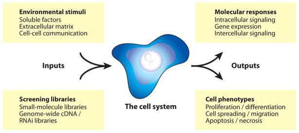
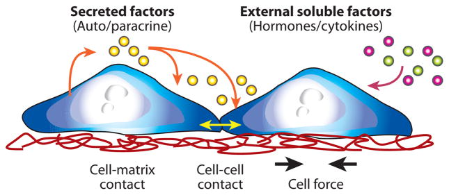
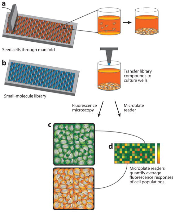
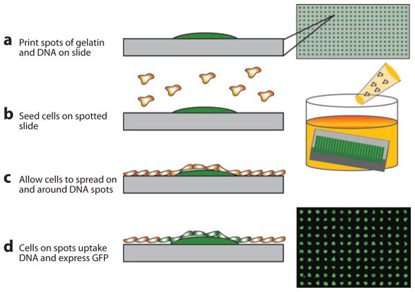
Living cells are remarkably complex. To unravel this complexity, living-cell assays have been developed that allow delivery of experimental stimuli and measurement of the resulting cellular responses. High-throughput adaptations of these assays, known as living-cell microarrays, which are based on microtiter plates, high-density spotting, microfabrication, and microfluidics technologies, are being developed for two general applications: (a) to screen large-scale chemical and genomic libraries and (b) to systematically investigate the local cellular microenvironment. These emerging experimental platforms offer exciting opportunities to rapidly identify genetic determinants of disease, to discover modulators of cellular function, and to probe the complex and dynamic relationships between cells and their local environment.


Figures

Figure 1
Figure 1
Attributes of a cell assay.
Figure 2
Figure 2
The cell microenvironment.
Figure 3
Figure 3
High-throughput screening. (a) Cells are seeded through a manifold into wells of a high-density microtiter plate and are allowed to sediment and attach. (b) Concentrated compounds from the small-molecule library are transferred from their storage in library microtiter plates to the wells of the cell-seeded microtiter plates. Cells in each well are then exposed to a well-calibrated stimulus, (c) responses are captured by fluorescence microscopy, quantified by automated image analysis, and (d) displayed as a heatmap to identify potential hits from the molecular library.
Figure 4
Figure 4
Spotted cell microarray technology. (a) DNA is mixed with gelatin and spotted as a high-density array on a glass slide, and it is dried and stored until needed for an experiment. (b) To perform an experiment, the array is rehydrated, and cells are seeded uniformly across the array. (c) Cells attach and spread across the array, and (d) those cells residing above DNA spots take up the underlying nucleotides and become transfected in a spatially localized fashion (42).
Figure 5
Figure 5
Microfabrication and soft lithography. (a) Photomasks are drawn using a computer-aided design tool and are printed by a high-resolution printer on mylar transparency films. (b) Photoresist-coated silicon wafers are exposed through the photomask and (c) developed to translate the photomask design to the silicon surface. (d) The silicon then serves as a master mold for casting transparent polydimethylsiloxane (PDMS) polymer replicas, which can be (e) drilled to create inlets and outlets and bonded to a glass substrate to create closed microfluidic devices or (f) used to create stencils with micropatterned through-holes.
Figure 6
Figure 6
Examples of microfabricated cell arrays. (a) An array of mechanical cell traps captures single cells and allows dynamic exposure to compounds in solution (72). (b) An array of mechanical traps captures individual cells from two different populations (one green, one red) and places them in contact for investigation of cell fusion. Inset illustrates cell pairing method (73). (c) Chemical surface patterning is used to control cell location and spread area. Cell area (inset top) was found to correlate with cell phenotype (apotosis and growth) (inset bottom) (116). (d) Surface patterning is used to create pairs of cells in contact or with controlled gaps to dissect contact-dependent and -independent behavior (75). (e) The hepatocyte-fibroblast contact area is systematically varied while holding the cell ratio constant by patterning hepatocyte islands of different diameters and densities surrounded by fibroblasts. Albumin synthesis (red) was preferentially induced in hepatocytes at island edges where they were in contact with adjacent fibroblasts. Inset shows a single island (80). (f) Fibroblasts are patterned by dielectrophoresis and embedded in a flexible hydrogel film. Inset shows close-up of the embedded cell array elements (89).
Figure 7
Figure 7
Examples of microfluidic cell arrays. (a) A microfluidic array delivers a concentration gradient of fluorescent dextran in columns to independently seeded rows of fluorescently labeled cells. The figure illustrates the full device (top left), the 64-element cell culture array (right), and a single element (lower left) (110). (b) A microfluidic device uses flow-encoded switching to control parallel delivery of different stimulus timing regimens to preseeded GFP reporter cells. The figure illustrates device design (top left), dye filled channels at different time points (top middle), channels seeded with fluorescent GFP reporter cells (top right), example excitation sequences (black) and the resulting dynamic exposures of 4 channels (green) varying pulse duration, width, and frequency (109). (c) A microfluidic valve-controlled array enables dynamic gene expression profiling. The figure illustrates device design, valve design, reporter cell seeding, stimulation strategy (left), phase and fluorescence images of GFP reporter cells in an array element (top right), and a heat map quantifying dynamic GFP responses across the array (bottom right) (111). (d) A microfluidic array for toxin screening. The figure illustrates device design (top left), close-up of valve-controlled array elements (top right), and fluorescence images of cell viability (green, live: red, dead) across the array of cell lines and putative toxins (bottom) (113).

References

    1. Carrel A, Burrows MT. Cultivation of tissues in vitro and its technique. J Exp Med. 1911;14:244–47. - PMC - PubMed
    1. Wynn TA. Cellular and molecular mechanisms of fibrosis. J Pathol. 2008;214:199–210. - PMC - PubMed
    1. Ingber DE. Mechanical control of tissue morphogenesis during embryological development. Int J Dev Biol. 2006;50:255–66. - PubMed
    1. Werner S, Krieg T, Smola H. Keratinocyte-fibroblast interactions in wound healing. J Investig Dermatol. 2007;127:998–1008. - PubMed
    1. Saez JC, Berthoud VM, Branes MC, Martinez AD, Beyer EC. Plasma membrane channels formed by connexins: their regulation and functions. Physiol Rev. 2003;83:1359–400. - PubMed

Publication types

Substances

LinkOut - more resources