Review
. 2009 Aug;21(8):796-806.
doi: 10.1111/j.1365-2982.2009.01311.x. Epub 2009 Apr 22.

High-resolution manometry in clinical practice: utilizing pressure topography to classify oesophageal motility abnormalities

Affiliations
Review

High-resolution manometry in clinical practice: utilizing pressure topography to classify oesophageal motility abnormalities

J E Pandolfino et al. Neurogastroenterol Motil. 2009 Aug.

Abstract

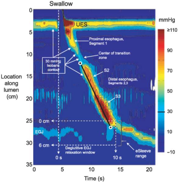
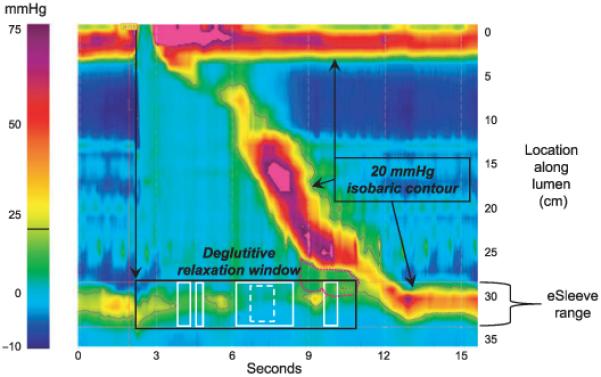
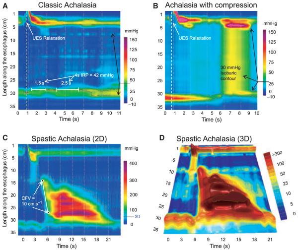
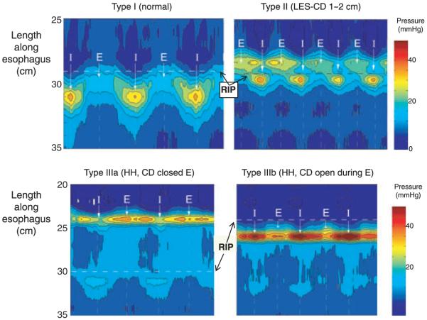
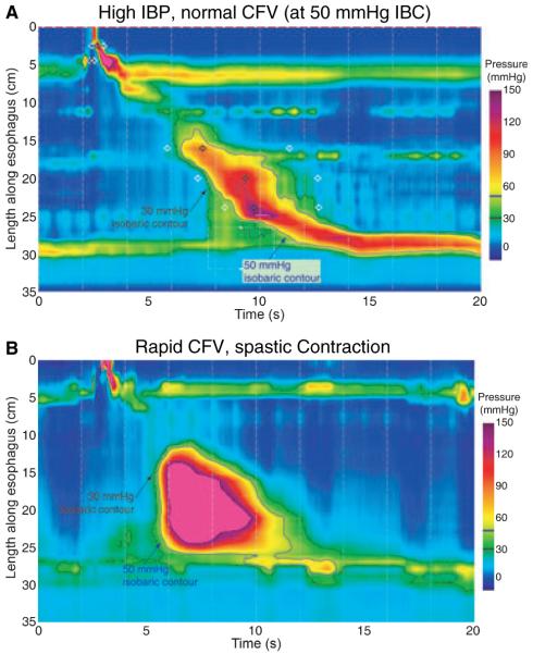
High-resolution manometry capable of pressure monitoring from the pharynx to the stomach together with pressure topography plotting represents an unquestionable evolution in oesophageal manometry. However, with this advanced technology come challenges and one of those is devising the optimal scheme to apply high-resolution oesophageal pressure topography (HROPT) to the clinical evaluation of patients. The first iteration of the Chicago classification was based on a systematic analysis of motility patterns in 75 control subjects and 400 consecutive patients. This review summarizes the analysis process as it has evolved. Individual swallows are analysed in a stepwise fashion for the morphology of the oesophagogastric junction (OGJ), the extent of OGJ relaxation, the propagation velocity of peristalsis, the vigour of the peristaltic contraction, and abnormalities of intrabolus pressure utilizing metrics that have now been customized to HROPT. These results are then synthesized into a comprehensive diagnosis that, although based on conventional manometry criteria, is also customized to HROPT measures. The resultant classification objectifies the identification of three unique subtypes of achalasia. Additionally, it provides enhanced detail in the description of distal oesophageal spasm, nutcracker oesophagus subtypes, and OGJ obstruction. It is our expectation that modification of this classification scheme will continue to occur and this should further clarify the utility of pressure topography plotting in assessing oesophageal motility disorders.


Figures

Figure 1
Figure 1
Typical swallow pressure topography spanning from the pharynx (locations 0–2 cm) to stomach (locations 29–35 cm) of a normal subject with normal peristalsis and normal oesophagogastric junction (OGJ) relaxation. The transition zone, demarcating the end of the proximal oesophageal segment (striated muscle) and the beginning of the distal oesophageal segment (smooth muscle), is readily identified as a pressure minimum. Note that the distal segment, in fact, has three sub-segments within it, each with an identifiable pressure peak. The most distal sub-segment, the lower oesophageal sphincter, contracts at the termination of peristalsis and then descends back to the level of the crural diaphragm as the period of swallow-related oesophageal shortening ends. The onset of the deglutitive relaxation window is at the onset of upper sphincter relaxation while the offset is 10 s later. The spatial domain within which OGJ relaxation is assessed (the eSleeve™ range) is user defined, spanning at least 6 cm, depending on the extent of oesophageal shortening after the swallow. The characteristics of the distal oesophageal contraction are defined by the isobaric contour tool set at 30 mmHg (highlighted with arrows). The isobaric contour can then be utilized to measure the contractile front velocity (CFV) and identify breaks in the contractile wavefront. The CFV is the slope of the line connecting points (black circles) on the 30 mmHg isobaric contour at the proximal margin and the distal margin of the smooth muscle oesophagus (CFV = 3 cm s−1).
Figure 2
Figure 2
The integrated relaxation pressure (IRP) is a more complex metric of oesophagogastric junction (OGJ) relaxation than a simple end-expiratory measurement of OGJ pressure after a swallow. The IRP requires persistence of OGJ relaxation for 4 s within the relaxation window (black box) but the actual time periods that go into its calculation (solid white boxes) can be contiguous or, as in this example, non-contiguous. By finding the periods of lowest pressure within the relaxation window, the IRP reduces, but does not eliminate, influences of intrabolus pressure (IBP) (pink dotted contour) or crural diaphragm (CD) contractions. The black isobaric contour is set at 20 mmHg. Note that a purer measure of intrinsic LOS relaxation would isolate a briefer time period within the relaxation window and isolate times least influenced by IBP and CD contractility. For example, a 1-s IRP (dashed box) in this example is 7.2 mmHg compared to the standard (4-s) IRP of 9.6 mmHg. The 4-s IRP was selected as the standard metric because it best differentiated the impaired OGJ relaxation in achalasia from non-achalasic individuals.
Figure 3
Figure 3
Achalasia subtypes are distinguished by three distinct manometric patterns of oesophageal body contractility. In classic achalasia (Panel A), there is no significant pressurization within the body of the oesophagus and impaired oesophagogastric junction relaxation (integrated relaxation pressure of 42 mmHg in this example). Panel B represents a swallow from a patient with the ‘achalasia with compression’ subtype exhibiting rapid pan-oesophageal pressurization. Panel C illustrates a pressure topography plot typical of spastic achalasia. Although this swallow is also associated with rapidly propagated pressurization, the pressurization is attributable to an abnormal lumen obliterating contraction. The three dimensional rendering of these pressure data (Panel D) illustrates the peaks and valleys of that spastic contraction and this swallow would likely appear as a rosary-bead pattern on fluoroscopy. Modified from: Pandolfino JE, Kwiatek MA, Nealis T, Bulsiewicz W, Post J, Kahrilas PJ. Achalasia: a new clinically relevant classification by high-resolution manometry. Gastroenterology 2008; 135: 1526.
Figure 4
Figure 4
Pressure topography plots of oesophagogastric junction (OGJ) pressure morphology subtypes primarily distinguished by the extent of lower oesophageal sphincter-crural diaphragm (LOS-CD). separation during several respiratory cycles. The pressure scale is shown at the right. Instantsof peak inspiration are marked I with mid expiration (E) indicated midway between inspirations. The locus of the respiratory inversion point (RIP) is indicated by a horizontal dashed line. Type I is characterized by complete overlap of the CD and the LOS. The RIP lies at the proximal margin of the OGJ. Type II is characterized by minimal, but discernible, LOS-CD separation, but the nadir pressure between the LOS and CD was still greater than gastric pressure. The RIP is within the OGJ at the proximal margin of the CD. OGJ type III is the high-resolution oesophageal pressure topography signature of hiatus hernia. Two subtypes are discernible, IIIa and IIIb, with the distinction being that the respiratory inversion point was proximal to the CD with IIIa and proximal to the LOS in IIIb. The shift in respiratory inversion point is likely indicative of a grossly patulous hiatus, open throughout the respiratory cycle. Modified from Ref. (20).
Figure 5
Figure 5
Differentiating increased intrabolus pressure (top) from a rapidly propagated contraction (bottom). The upper panel illustrates a swallow with functional obstruction at the oesophagogastric junction (OGJ). Note that the 30 mmHg isobaric contour line (black) deviates quickly from the 50 mmHg isobaric contour line (blue). In this case the contractile front velocity (CFV) is normal, reflecting the propagation velocity of 50 mmHg isobaric contour rather than the 30 mmHg isobaric contour. In contrast, the lower panel represents a swallow with rapid CFV attributable to spasm. OGJ relaxation is normal and the 30 mmHg and 50 mmHg isobaric contours parallel each other indicating that no compartmentalized oesophageal pressurization has occurred. The entire distal oesophagus is contracting simultaneously. Modified from Ref. (6).

References

    1. Clouse RE, Staiano A. Topography of the esophageal peristaltic pressure wave. Am J Physiol. 1991;261:G677–84. - PubMed
    1. Grubel C, Hiscock R, Hebbard G. Value of spatiotemporal representation of manometric data. Clin Gastroenterol Hepatol. 2008;6:525–30. - PubMed
    1. Pandolfino JE, Ghosh SK, Zhang Q, Jarosz A, Shah N, Kahrilas PJ. Quantifying OGJ morphology and relaxation with high-resolution manometry: a study of 75 asymptomatic volunteers. Am J Physiol Gastrointest Liver Physiol. 2006;290:G988–97. - PubMed
    1. Ghosh SK, Pandolfino JE, Zhang Q, Jarosz A, Shah N, Kahrilas PJ. Quantifying esophageal peristalsis with high-resolution manometry: a study of 75 asymptomatic volunteers. Am J Physiol Gastrointest Liver Physiol. 2006;290:G988–97. - PubMed
    1. Ghosh SK, Pandolfino JE, Rice J, Clarke JO, Kwiatek M, Kahrilas PJ. Impaired deglutitive OGJ relaxation in clinical esophageal manometry: a quantitative analysis of 400 patients and 75 controls. Am J Physiol Gastrointest Liver Physiol. 2007;293:G878–85. - PubMed

Publication types