

Localized SDF-1alpha gene release mediated by collagen substrate induces CD117 stem cells homing
- PMID: 19413887
- PMCID: PMC3837586
- DOI: 10.1111/j.1582-4934.2008.00624.x
Localized SDF-1alpha gene release mediated by collagen substrate induces CD117 stem cells homing
Abstract
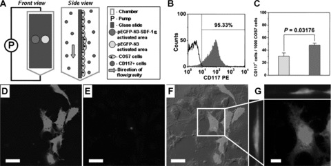
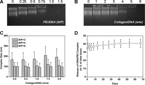
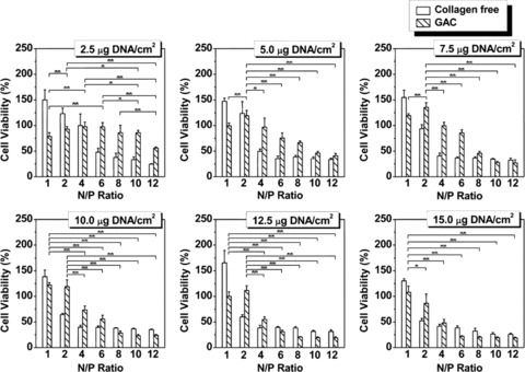
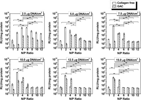
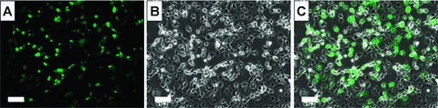
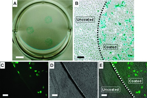
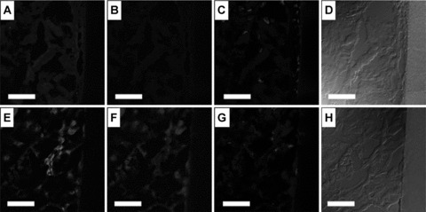
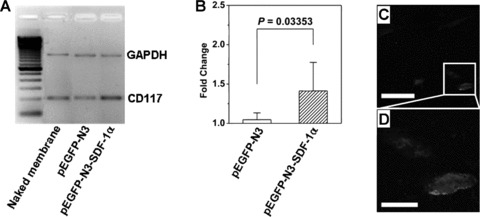
Stromal cell-derived factor-1alpha (SDF-1alpha) mediated mobilization and homing of stem cells showed promising potential in stem cell based tissue engineering and regenerative medicine. However local and sustained release of SDF-1alpha is indispensable for stem cell mediated regenerative process due to its short half-life under inflammatory conditions. In this study, a gene activated collagen substrate (GAC) was formed via assembly of plasmid encoding SDF-1alpha into a collagen substrate to create a microenvironment favoring stem cell homing. Local release of SDF-1alpha from the transfected cells on GAC and its effect on CD117(+) stem cell homing were investigated. Non-viral poly-ethyleneimine (25kDa PEI)/DNA complexes were mixed with rat tail collagen solution to form the GAC. Optimization of GAC was carried out based on collagen effects on the PEI/DNA complexes, viability and luciferase expression of COS7 cells on GAC. CD117(+) stem cells homing in response to SDF-1alpha local expression from transfected cells on GAC were investigated in a flow chamber in vitro and in a mouse hind limb model in vivo. The gene expression, migration of CD117(+) stem cells and the induced inflammation were investigated with immunostaining, reverse transcription polymerase chain reaction (RT-PCR) and H&E staining. The optimized parameters for GAC were DNA dosage 10 microg/cm(2), molar ratio of PEI nitrogen in primary amine to DNA phosphate (N/P ratio) 4 and mass ratio of collagen to DNA (C/D ratio) 1.0. It kept cell viability above 75% and transfection efficiency around 5.8 x 10(5) RLU/mg protein. GAC allowed the sustained gene release up to 60 days. GAC mediated SDF-1alpha gene release induced migration and homing of CD117(+) stem cells in vitro and in vivo significantly, and the inflammation of GAC reduced significantly two weeks after transplantation. GAC is a promising stem cell based therapeutic strategy for regenerative medicine.
Figures
Similar articles
-
Localized and sustained SDF-1 gene release mediated by fibronectin films: A potential method for recruiting stem cells.Int J Artif Organs. 2009 Mar;32(3):141-9. doi: 10.1177/039139880903200304. Int J Artif Organs. 2009. PMID: 19440989
-
Controlled release of stromal cell-derived factor-1 alpha in situ increases c-kit+ cell homing to the infarcted heart.Tissue Eng. 2007 Aug;13(8):2063-71. doi: 10.1089/ten.2006.0013. Tissue Eng. 2007. PMID: 17518719
-
Mobilizing of haematopoietic stem cells to ischemic myocardium by plasmid mediated stromal-cell-derived factor-1alpha (SDF-1alpha) treatment.Regul Pept. 2005 Feb 15;125(1-3):1-8. doi: 10.1016/j.regpep.2004.10.014. Regul Pept. 2005. PMID: 15582707
-
SDF-1α as a therapeutic stem cell homing factor in myocardial infarction.Pharmacol Ther. 2011 Jan;129(1):97-108. doi: 10.1016/j.pharmthera.2010.09.011. Epub 2010 Oct 20. Pharmacol Ther. 2011. PMID: 20965212 Review.
-
Unraveling the interplay between inflammation and stem cell mobilization or homing: Implications for tissue repair and therapeutics.Tzu Chi Med J. 2024 Sep 5;36(4):349-359. doi: 10.4103/tcmj.tcmj_100_24. eCollection 2024 Oct-Dec. Tzu Chi Med J. 2024. PMID: 39421490 Free PMC article. Review.
Cited by
-
Fabrication of Stromal Cell-Derived Factor-1 Contained in Gelatin/Hyaluronate Copolymer Mixed with Hydroxyapatite for Use in Traumatic Bone Defects.Micromachines (Basel). 2021 Jul 14;12(7):822. doi: 10.3390/mi12070822. Micromachines (Basel). 2021. PMID: 34357232 Free PMC article.
-
Intracardiac injection of matrigel induces stem cell recruitment and improves cardiac functions in a rat myocardial infarction model.J Cell Mol Med. 2011 Jun;15(6):1310-8. doi: 10.1111/j.1582-4934.2010.01086.x. Epub 2010 May 14. J Cell Mol Med. 2011. PMID: 20477905 Free PMC article.
-
Identification and functional characterization of CD133+GFAP+CD117+Sca1+ neural stem cells.Mol Cell Biochem. 2022 Mar;477(3):897-914. doi: 10.1007/s11010-021-04339-3. Epub 2022 Jan 26. Mol Cell Biochem. 2022. PMID: 35079926
-
Local activation of cardiac stem cells for post-myocardial infarction cardiac repair.J Cell Mol Med. 2012 Nov;16(11):2549-63. doi: 10.1111/j.1582-4934.2012.01589.x. J Cell Mol Med. 2012. PMID: 22613044 Free PMC article. Review.
-
Animal models of cardiac disease and stem cell therapy.Open Cardiovasc Med J. 2010 Nov 26;4:231-9. doi: 10.2174/1874192401004010231. Open Cardiovasc Med J. 2010. PMID: 21258568 Free PMC article.
References
Publication types
MeSH terms
Substances
LinkOut - more resources
Full Text Sources
Miscellaneous
