. 2009 Jul;13(7):1302-13.
doi: 10.1111/j.1582-4934.2008.00633.x. Epub 2008 Dec 24.

Cellular repressor of E1A-stimulated genes attenuates cardiac hypertrophy and fibrosis

Affiliations

Cellular repressor of E1A-stimulated genes attenuates cardiac hypertrophy and fibrosis

Zhouyan Bian et al. J Cell Mol Med. 2009 Jul.

Abstract

Cellular repressor of E1A-stimulated genes (CREG) is a secreted glycoprotein of 220 amino acids. It has been proposed that CREG acts as a ligand that enhances differentiation and/or reduces cell proliferation. CREG has been shown previously to attenuate cardiac hypertrophy in vitro. However, such a role has not been determined in vivo. In the present study, we tested the hypothesis that overexpression of CREG in the murine heart would protect against cardiac hypertrophy and fibrosis in vivo. The effects of constitutive human CREG expression on cardiac hypertrophy were investigated using both in vitro and in vivo models. Cardiac hypertrophy was produced by aortic banding and infusion of angiotensin II in CREG transgenic mice and control animals. The extent of cardiac hypertrophy was quantitated by two-dimensional and M-mode echocardiography as well as by molecular and pathological analyses of heart samples. Constitutive over-expression of human CREG in the murine heart attenuated the hypertrophic response, markedly reduced inflammation. Cardiac function was also preserved in hearts with increased CREG levels in response to hypertrophic stimuli. These beneficial effects were associated with attenuation of the mitogen-activated protein kinase (MAPK)-extracellular signal-regulated kinase 1 (MEK-ERK1)/2-dependent signalling cascade. In addition, CREG expression blocked fibrosis and collagen synthesis through blocking MEK-ERK1/2-dependent Smad 2/3 activation in vitro and in vivo. Therefore, the expression of CREG improves cardiac functions and inhibits cardiac hypertrophy, inflammation and fibrosis through blocking MEK-ERK1/2-dependent signalling.


Figures

Figure 1
Figure 1
Characterization of human CREG transgenic (TG) mice. (A) Representative Western blot of human CREG protein from different tissue of TG mice as indicated. (B) Representative Western blots of human CREG protein in the heart tissue from 4 lines of both TG and WT mice. (C) Representative Western blots of TG CREG and endogenous CREG protein levels in the heart from TG mice. (D) Representative Western blots of mouse and human CREG protein in the heart tissue from TG mice after aortic banding at time-points indicated.
Figure 2
Figure 2
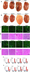
The effects of CREG on cardiac hypertrophy in vivo. (A and B) Gross hearts of sham and AB mice at 8 weeks after surgery or saline- or Ang II-infused mice at 4weeks of infusion. (C and D) Haematoxylin and eosin and wheat germ agglutinin (WGA) staining of sham and AB mice at 8 weeks after surgery. (E and F) Haematoxylin and eosin and WGA staining of 4 weeks of saline- and Ang II-infused mice. (G and H) Analysis of hypertrophic markers. Total RNA was isolated from hearts of mice of the indicated groups, and expression of transcripts for ANP, BNP, Myh-7 and Acta1 induced by AB or Ang II infusion were determined by real-time PCR analysis. Data represent typical results of three to four different experiments as mean ± S.E.M. (n= 4 to 6 mice/per group). *P < 0.01 was obtained for the WT/sham or WT/saline values.
Figure 3
Figure 3
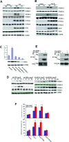
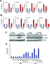
The effect of CREG on MEK-ERK1/2 signalling pathway. (A and B) Representative blots of MEK1/2, ERK1/2, p38 and JNK phosphorylation and their total protein expression at 8 weeks after AB surgery or at 4 weeks after Ang II infusion in WT and TG mice. (C) The protein expression level of CREG after infection with Ad-CREG or Ad-shCREG. Upper, quantitative results. Bottom, Representative blots. Values are mean ± S.E.M. *P < 0.01 for difference from Ad-GFP group values. (D) Representative blots of MEK1/2 and ERK1/2 activation after treated with Ang II for indicated time in different adenovirus infected primary cardiac myocytes. The results were reproducible in three separate experiments. (E) Immunoprecipitation analysis showed that CREG directly interacted with MEK1. (F) The effect of CREG on the enlargement of myocytes area and [3H]-leucine incorporation induced by Ang II after infection with ade-novirus. Cardiac myocytes were pre-treated with 1 μm U0126 or PBS and then treated with Ang II for 48 hrs after infection with Ad-shRNA or Ad-shCREG for 24 hrs. The results were reproducible in three separate experiments as mean ± S.E.M. *P < 0.01 was obtained for the control groups; §P < 0.01 was obtained for Ad-shRNA infection group.
Figure 4
Figure 4
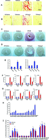
The effects of CREG on fibrosis in vivo and in vitro. (A and B) Picrosirius red (PSR) staining on histological sections of the left ventricle was performed on indicated groups 8 weeks after AB or 4 weeks after Ang II infusion. (C and D) Representative PSR staining of microscopical images of whole heart cross sections. (E) Fibrotic areas/whole left ventricle area from histological sections was quantified using an image-analysing system. (F and G) Real-time PCR analyses of Tgfβ1, Col1α1, Col3α1, Ctgf were performed to determine mRNA expression levels in indicated groups. GAPDH was used as the sample loading control. Data represent typical results of 3 different experiments as mean ± S.E.M. (n= 4 to 6 mice/per group). *P < 0.01 was obtained for the WT/sham or WT/saline values. (H and I) CREG was shown to inhibit TGF-β1-induced [3H] proline incorporation and the promoter activities of COL1A2 and CTGF. [3H] proline incorporation and luciferase assay were performed as described in ‘Materials and methods’. Values are mean ± S.E.M. The results were reproducible in three separate experiments. *P < 0.01 was obtained for Ad-GFP infection group.
Figure 5
Figure 5
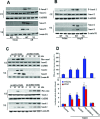
The effect of CREG on TGF-β/Smad signalling. (A and B) Representative blots of Smad-2 phos-phorylation and Smad-2/3 transloca-tion from indicated groups 8 weeks after AB (n= 3) or 4 weeks of Ang II infusion (n= 4). The results were reproducible in three separate experiments (CE, cytoplasmic extract; NE, nuclear extract). (C) Representative blots of Smad-2 phosphorylation and Smad-2/3 translocation induced by TGF-β1 in cardiac fibroblasts after infection with different adenovirus. (D) The effect of ERK1/2 activation on collagen synthesis along with promoter activities of COL1A2 and CTGF. Cells were infected with or without indicated adenovirus for 24 hrs, and then incubated with 15 ng/ml TGF-β1 for up to 48 hrs. [3H] proline incorporation and luciferase assay were performed as described in ‘Materials and methods’. *P < 0.01 was obtained for control group. The results were reproducible in three separate experiments.
Figure 6
Figure 6
The effect of CREG on inflammation in vivo and in vitro. (A and B) Realtime PCR analysis of TNF-α, IL-1β, IL-6 and MCP-1 mRNA expression in the myocardium obtained from indicated groups at 8 weeks AB (A, n= 4) or 4 weeks Ang II infusion (B, n= 6). Each assay was performed in triplicate. *P < 0.05 for difference from WT/sham values or WT/saline values. (C) Western blot analysis of IκBα and IKK-β phosphorylation of the myocardium was obtained from indicated animals at 8 weeks AB (A, n= 5) or 4 weeks Ang II infusion (B, n= 4). (D) The effects of CREG expression and ERK1/2 activation on Ang II-induced NF-κB transcriptional activity. Luciferase assay were performed as described in ‘Materials and methods’. Values are mean ± S.E.M. *P < 0.01 was obtained for PBS/control group. The results were reproducible in three separate experiments.

References

    1. Liehn EA, Merx MW, Postea O, et al. Ccr1 deficiency reduces inflammatory remodeling and preserves left ventricular function after myocardial infarction. J Cell Mol Med. 2008;12:496–506. - PMC - PubMed
    1. Das DK, Maulik N, Engelman RM. Redox regulation of angiotensin II signaling in the heart. J Cell Mol Med. 2004;8:144–52. - PMC - PubMed
    1. Das M, Das S, Das DK. Caveolin and MAP kinase interaction in angiotensin II preconditioning of the myocardium. J Cell Mol Med. 2007;11:788–97. - PMC - PubMed
    1. Turnell AS, Mymryk JS. Roles for the coactivators CBP and p300 and the APC/C E3 ubiquitin ligase in E1A-dependent cell transformation. Br J Cancer. 2006;95:555–60. - PMC - PubMed
    1. Shindoh M, Higashino F, Kohgo T. E1AF, an etsoncogene family transcription factor. Cancer Lett. 2004;216:1–8. - PubMed

Publication types

MeSH terms

Substances