. 2009 May 6;96(9):3832-9.
doi: 10.1016/j.bpj.2008.11.075.

Spatio-temporal plasticity in chromatin organization in mouse cell differentiation and during Drosophila embryogenesis

Affiliations

Spatio-temporal plasticity in chromatin organization in mouse cell differentiation and during Drosophila embryogenesis

Dipanjan Bhattacharya et al. Biophys J. .

Abstract

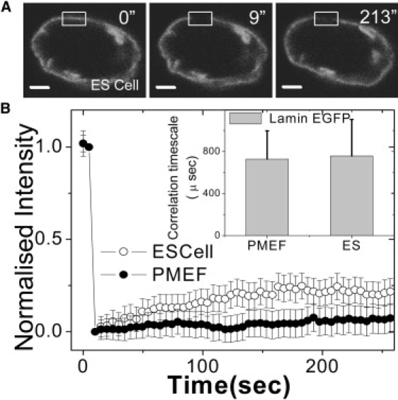
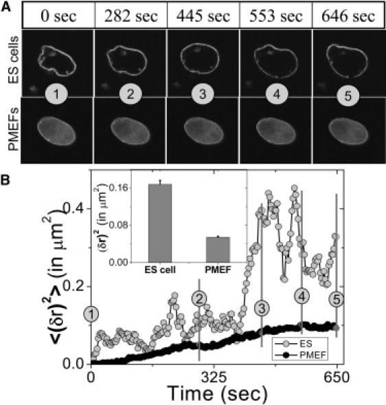
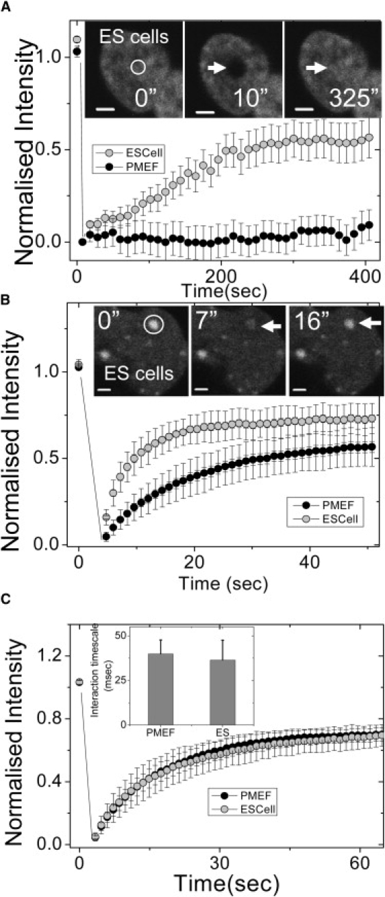
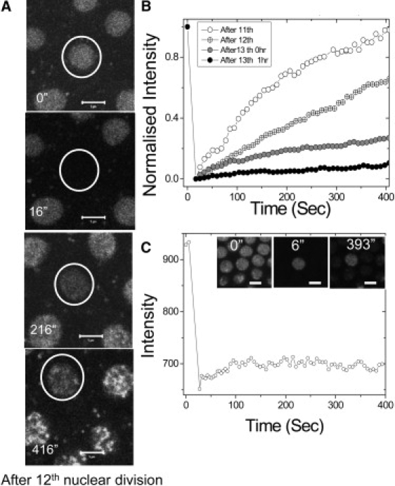
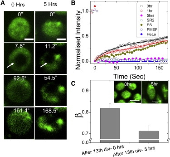
Cellular differentiation and developmental programs require changing patterns of gene expression. Recent experiments have revealed that chromatin organization is highly dynamic within living cells, suggesting possible mechanisms to alter gene expression programs, yet the physical basis of this organization is unclear. In this article, we contrast the differences in the dynamic organization of nuclear architecture between undifferentiated mouse embryonic stem cells and terminally differentiated primary mouse embryonic fibroblasts. Live-cell confocal tracking of nuclear lamina evidences highly flexible nuclear architecture within embryonic stem cells as compared to primary mouse embryonic fibroblasts. These cells also exhibit significant changes in histone and heterochromatin binding proteins correlated with their distinct epigenetic signatures as quantified by immunofluorescence analysis. Further, we follow histone dynamics during the development of the Drosophila melanogaster embryo, which gives an insight into spatio-temporal evolution of chromatin plasticity in an organismal context. Core histone dynamics visualized by fluorescence recovery after photobleaching, fluorescence correlation spectroscopy, and fluorescence anisotropy within the developing embryo, revealed an intriguing transition from plastic to frozen chromatin assembly synchronous with cellular differentiation. In the embryo, core histone proteins are highly mobile before cellularization, actively exchanging with the pool in the yolk. This hyperdynamic mobility decreases as cellularization and differentiation programs set in. These findings reveal a direct correlation between the dynamic transitions in chromatin assembly with the onset of cellular differentiation and developmental programs.


Figures

Figure 1
Figure 1
Plasticity in nuclear organization I: FRAP/FCS of lamin proteins in mouse ES and PMEF cells. (A) Fluorescent images of an ES cell nucleus transiently expressing EGFP-LaminB1. Images indicate prebleach (0 s), immediately after bleach (9 s), and 213 s after bleach. The scale bar is 2 μm. The bleach region is shown by the rectangle. (B) Normalized fluorescence intensities of EGFP-LaminB1 transiently expressed in both ES cells and in PMEF cells. PMEF cells show lower recovery in the shown time interval compared to ES cells (n = 20). (Inset) Diffusion timescales of EGFP-Lamin-B1 in PMEF and ES cell nuclei obtained using FCS show no significant difference in the two systems. The error bars are standard deviations.
Figure 2
Figure 2
Plasticity in nuclear organization II: nuclear envelope fluctuations in mouse ES and PMEF cells. (A) Nuclear envelope fluctuations of ES cell and PMEF expressing EGFP-LaminB1 are shown for the time points indicated above the panel. The numbers in circles indicate corresponding time points. (B) Time series of the mean-square fluctuation of the nuclear membrane (EGFP-LaminB1) in ES cells and in PMEFs. (Inset) The mean and the standard error of mean-square fluctuations of the nuclear membrane, showing a more fluidic structure in ES cells compared to PMEFs.
Figure 3
Figure 3
Dynamics of core, linker histones, and HP1 proteins in mouse ES and PMEF cells. (A) Normalized fluorescence intensities of core histone (H2B-EGFP) in ES and PMEF cells. A high fluorescence recovery was observed in the nuclei of ES cells, with negligible recovery in PMEFs in similar timescales (n = 20). (Inset) Images of H2B-EGFP expressing ES cells prebleach (0 s), after bleach (10 s), and after recovery of fluorescence (325 s) are shown. The scale bar is 2 μm. (B) FRAP of heterochromatin protein-1 (HP1α-EGFP) transiently expressed in ES cells and PMEFs showed a higher and faster fluorescence recovery in the nuclei of ES cells compared to PMEFs (n = 18). (Inset) Images of HP1α-EGFP expressing ES cells prebleach (0 s), after bleach (7 s), and after recovery of fluorescence (16 s) are shown. The scale bar is 2 μm. (C) Normalized fluorescence intensities of linker histone H1.5-EGFP in ES cells and PMEFs show no difference in dynamics (n = 15). (Inset) The graph indicates similar interaction timescales of H1.5-EGFP in ES cells and PMEFs revealed by FCS experiments.
Figure 4
Figure 4
Large exchange of core histones between the nuclei and the yolk in the Drosophila melanogaster syncytial blastoderm. (A) A nucleus before photobleaching and subsequent frames after photobleaching (0 s, 200 s, and 400 s) before cellularization (after the 12th nuclear division) is shown. The scale bar is 5 μm. (B) Normalized fluorescence intensities of typical nuclei at different time points (after the 11th, 12th, and 13th nuclear divisions and 1 h from the 13th nuclear division), showing progressive fall in recovery. (C) Quantified fluorescence intensity indicates no fluorescence loss in a nucleus in the time after photobleaching of its adjacent nuclei, indicating exchange of the core histones between the yolk and the nucleus. (Inset) FLIP images after the 13th nuclear division (before cellularization) at indicated time points. The scale bar is 5 μm.
Figure 5
Figure 5
Plasticity in chromatin assembly in the early Drosophila embryo, postcellularization. (A) Time-lapse images of a small bleached region inside a cell nucleus at two stages of development (0 and 5 h after the 13th nuclear division). (B) Quantitation of images as in panel A, showing a high fluorescence recovery in the nuclei at 0 h from the 13th nuclear division and after 1 h from the 13th nuclear division (after completion of cellularization) and is similar to the recovery observed in ES cells. This fractional recovery comes down 5 h from the 13th nuclear division, and is comparable to FRAP recovery of H2B-EGFP in human HeLa cells, Drosophila SR2+ cells, and PMEFs. (C) The mean and the standard deviation of the β-factor inside cell nuclei at 0 h from the 13th nuclear division and 5 h from the 13th nuclear division. The decrease in β-factor indicates the condensation of the chromosomal mesh at 5 h from the 13th nuclear division (n = 30, each). (Inset) Confocal fluorescence images of the nuclei at 0 h from the 13th nuclear division and 5 h after the 13th nuclear division. Heterogeneous patches of chromatin condensation after 5 h from the 13th nuclear division can be seen.
Figure 6
Figure 6
Evolution of rigidity maps of the chromatin in the Drosophila embryo using fluorescence anisotropy. (A) Color-coded anisotropy maps are shown for typical nuclei in the anterior and posterior of the embryo at 0 min (defined at 5 min after the completion of the 13th mitotic cycle), and at 70 min when nuclei of higher and more variegated chromatin rigidity states have emerged. The bar to the right shows anisotropy values corresponding to specific colors. (B) The anisotropy rise at the indicated time points are computed at each pixel and the plot shows the rise in the mean anisotropy (p < 0.001). (Inset) Corresponding small changes in standard deviation of anisotropy over pixels.

References

    1. Misteli T. Beyond the sequence: cellular organization of genome function. Cell. 2007;128:787–800. - PubMed
    1. Lanctot C., Cheutin T., Cremer M., Cavalli G., Cremer T. Dynamic genome architecture in the nuclear space: regulation of gene expression in three dimensions. Nat. Rev. Genet. 2007;8:104–115. - PubMed
    1. Khorasanizadeh S. The nucleosome: from genomic organization to genomic regulation. Cell. 2004;116:259–272. - PubMed
    1. Luger K., Hansen J.C. Nucleosome and chromatin fiber dynamics. Curr. Opin. Struct. Biol. 2005;15:188–196. - PubMed
    1. Gruenbaum Y., Margalit A., Goldman R.D., Shumaker D.K., Wilson K.L. The nuclear lamina comes of age. Nat. Rev. Mol. Cell Biol. 2005;6:21–31. - PubMed

Publication types

LinkOut - more resources