. 2009 Jun 13;367(1896):2293-310.
doi: 10.1098/rsta.2008.0314.

Subject-specific, multiscale simulation of electrophysiology: a software pipeline for image-based models and application examples

Affiliations

Subject-specific, multiscale simulation of electrophysiology: a software pipeline for image-based models and application examples

R S MacLeod et al. Philos Trans A Math Phys Eng Sci. .

Abstract

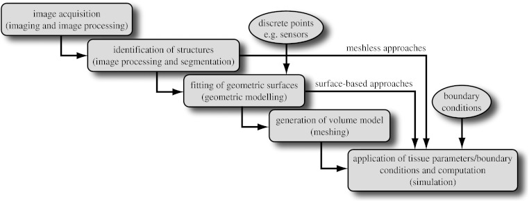
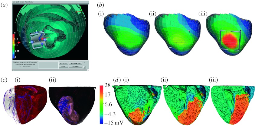
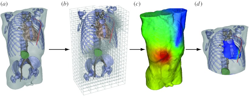
Many simulation studies in biomedicine are based on a similar sequence of processing steps, starting from images and running through geometric model generation, assignment of tissue properties, numerical simulation and visualization of the results--a process known as image-based geometric modelling and simulation. We present an overview of software systems for implementing such a sequence both within highly integrated problem-solving environments and in the form of loosely integrated pipelines. Loose integration in this case indicates that individual programs function largely independently but communicate through files of a common format and support simple scripting, so as to automate multiple executions wherever possible. We then describe three specific applications of such pipelines to translational biomedical research in electrophysiology.


Figures

Figure 1
Figure 1
Schematic of a simulation pipeline. Each element has a functional title and then, in parentheses, the technical description of the associated task.
Figure 2
Figure 2
Example of volume rendering with ImageVis3D of a torso model based on a high-resolution CT scan (512×512×3172 with a voxel size of 0.51×0.51×0.50 mm, courtesy of Siemens Corporate Research, Princeton). By controlling transfer functions, it is possible to identify different systems (e.g. skeleton, vasculature) and organs (e.g. heart, kidneys and bladder).
Figure 3
Figure 3
Example of meshing of the head in a paediatric epilepsy patient. (a) The particle distribution over the head surface and highlight of the variation in particle size, the adaptivity of the particles over the skin. (b) The associated tetrahedral mesh and (c) another higher resolution view of the mesh highlighting the cortex and cerebrospinal fluid.
Figure 4
Figure 4
(a–d) Illustration of simulation of electromagnetic field propagation in a patient-specific brain model. The figure shows a finite-element method discretization of Poisson's equation with a patient-specific, five-compartment, geometrical model derived from a segmentation of brain magnetic resonance imaging. The solid lines in the simulation images indicate isopotentials and the small white lines are electrical current streamlines.
Figure 5
Figure 5
Whole-heart electrical model of ischaemia with a realistic ischaemic zone. (a) A single image from an interactive session using SCIRun with the three-dimensional heart geometry cut away to reveal the location of the interactive ischaemic region tool. (b) The associated computed epicardial potentials of a simulation of subendocardial ischaemia of progressing transmural extent ((i) 40, (ii) 70 and (iii) 90%). (c(i)(ii)) A volume rendering of gadolinium-enhanced images of an animal heart illustrating the coronary vessels and the perfusion bed for this heart, which we used to create subject-specific models. (d(i–iii)) Slices of the heart model with colour indicating the electric potential from a simulation of ischaemia in the subject-specific geometric model.
Figure 6
Figure 6
Pipeline for computing defibrillation potentials in children. The figures shows the steps ((a) setting electrode configuration, (b) refinement of hexahedral mesh for electrode locations, (c) finite-element solution of potentials and (d) analysis of potentials at the heart to predict defibrillation effectiveness) required to place electrodes and then compute and visualize the resulting cardiac potentials.

Similar articles

Cited by

References

    1. Besier T., Gold G., Delp S., Fredericson M., Beaupre G. The influence of femoral internal and external rotation on cartilage stresses within the patellofemoral joint. J. Orthop. Res. 2008;26:1627–1635. doi: 10.1002/jor.20663. - DOI - PubMed
    1. Blackett S.A., Bullivant D., Stevens C., Hunter P.J. Open source software infrastructure for computational biology and visualization. Conf. Proc. IEEE Eng. Med. Biol. Soc. 2005;6:6079–6080. doi: 10.1109/IEMBS.2005.1615879. - DOI - PubMed
    1. Blemker S., Asakawa D., Gold G., Delp S. Image-based musculoskeletal modeling: applications, advances, and future opportunities. J. Magn. Reson. Imaging. 2007;25:441–451. doi: 10.1002/jmri.20805. - DOI - PubMed
    1. Bowman G., Huang X., Yao Y., Sun J., Carlsson G., Guibas L., Pande V. Structural insight into RNA hairpin folding intermediates. J. Am. Chem. Soc. 2008;130:9676–9678. doi: 10.1021/ja8032857. - DOI - PMC - PubMed
    1. Callahan, M., Cole, M., Shepherd, J., Stinstra, J. & Johnson, C. 2009 A meshing pipeline for biomedical computing. Eng. Comput 25, 115–130. (10.1007/s00366-008-0106-1) - DOI

Publication types

LinkOut - more resources