. 2009 May 11;206(5):1135-47.
doi: 10.1084/jem.20082242. Epub 2009 May 4.

Kallikrein 5 induces atopic dermatitis-like lesions through PAR2-mediated thymic stromal lymphopoietin expression in Netherton syndrome

Affiliations

Kallikrein 5 induces atopic dermatitis-like lesions through PAR2-mediated thymic stromal lymphopoietin expression in Netherton syndrome

Anaïs Briot et al. J Exp Med. .

Abstract

Netherton syndrome (NS) is a severe genetic skin disease with constant atopic manifestations that is caused by mutations in the serine protease inhibitor Kazal-type 5 (SPINK5) gene, which encodes the protease inhibitor lymphoepithelial Kazal-type-related inhibitor (LEKTI). Lack of LEKTI causes stratum corneum detachment secondary to epidermal proteases hyperactivity. This skin barrier defect favors allergen absorption and is generally regarded as the underlying cause for atopy in NS. We show for the first time that the pro-Th2 cytokine thymic stromal lymphopoietin (TSLP), the thymus and activation-regulated chemokine, and the macrophage-derived chemokine are overexpressed in LEKTI-deficient epidermis. This is part of an original biological cascade in which unregulated kallikrein (KLK) 5 directly activates proteinase-activated receptor 2 and induces nuclear factor kappaB-mediated overexpression of TSLP, intercellular adhesion molecule 1, tumor necrosis factor alpha, and IL8. This proinflammatory and proallergic pathway is independent of the primary epithelial failure and is activated under basal conditions in NS keratinocytes. This cell-autonomous process is already established in the epidermis of Spink5(-/-) embryos, and the resulting proinflammatory microenvironment leads to eosinophilic and mast cell infiltration in a skin graft model in nude mice. Collectively, these data establish that uncontrolled KLK5 activity in NS epidermis can trigger atopic dermatitis (AD)-like lesions, independently of the environment and the adaptive immune system. They illustrate the crucial role of protease signaling in skin inflammation and point to new therapeutic targets for NS as well as candidate genes for AD and atopy.


Figures

Figure 1.
Figure 1.
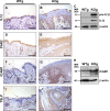
Ichthyosiform phenotype, abnormal differentiation, and asymmetrical desmosomal split in Spink5−/− grafted epidermis. (A and B) Macroscopic observation of WT (WTg) or Spink5 KO (KOg) mice skin 8 wk after grafting on nude recipient mice. (C and D) Hematoxylin/eosin staining of skin graft cross sections. The KOg epidermis shows hyperplasia with papillomatosis (bracket) and spongiosis, nuclei are seen in corneocytes (parakeratosis; arrow), and an inflammatory infiltrate is present in the dermis (asterisks). Pictures are representative of 14 WT and 17 KO independent grafts on nude mice, performed in five independent graft series. (E–J) Immunohistochemistry of epidermal differentiation markers performed on WTg and KOg grafted skin. (K and L) Desmoglein-1, detected by immunohistochemistry in the whole epidermis of WTg, is markedly decreased in the GR of KOg epidermis. (M and N) Desmosomes analyzed by transmission electron microscopy in the GR-SC intercellular space (red arrows). Asymmetrical split desmosomes were observed in KOg epidermis. Pictures are representative of two independent experiments, each including three WTg and three KOg. HF, hair follicle. Bars: (C and D) 29.4 µm; (E–L) 23.5 µm; (M and N) 0.1 µm.
Figure 2.
Figure 2.
Inflammatory cells infiltrate in Spink5−/− grafted skin. (A–C) Hematoxylin/eosin (HE) staining shows a high number of polynuclear eosinophilic cells (yellow arrows) in the upper dermis and the basal layer of KO grafted epidermis (KOg). (D–F) Numerous mast cells are colored by Toluidine blue (TB) in KOg dermis (black arrows) contrary to WTg dermis. (G–I) Immunohistochemistry staining and Western blotting confirms increased production of the 34-kD β1-tryptase monomer in KOg compared with WTg. Three independent stainings were performed on four WTg and four KOg. Western blotting is representative of two independent experiments, each including two WTg and two KOg. C and F are enlargements of B and E, respectively. Bars: (A and B) 9.4 µm; (C) 2 µm; (D and E) 11.75 µm; (F) 2.5 µm; (G and H) 23.5 µm.
Figure 3.
Figure 3.
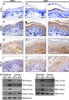
Proinflammatory mediators in Spink5−/− graft. (A and B) IL-1β immunohistochemistry staining is increased in Spink5−/− grafted epidermis (KOg) compared with WTg. (C) Western blotting reveals the presence of high amounts of the proform (31 kD) and active form (17 kD) of IL-1β in KOg epidermis. (D–J) Immunohistochemistry experiments on skin graft cross sections. (D and E) In WTg epidermis, PAR2 is localized in the GR. In KOg, PAR2 staining is increased in the GR and is extended to the basal layer. (F and G) ICAM1 is restricted to the basal cells in the WTg epidermis, whereas in the KOg it is extended to the membrane of suprabasal and GR cells. (H) Western blotting confirms the increase of ICAM1 expression in KOg epidermis. (I and J) TSLP is expressed in all epidermal layers of KOg but is not detected in WTg epidermis. Immunostaining panels and Western blotting are representative of two independent experiments, each including three WTg and three KOg, and two WTg and two KOg, respectively. Bars: (A, B, and D–G) 11.5 µm; (I and J) 23 µm.
Figure 4.
Figure 4.
Early inflammatory events in Spink5−/− embryos. (A–L) Immunohistochemistry on E19.5 embryos skin cross sections. (A–C) IL-1β immunostaining is more intense and sharp in the GR of Spink5 KO embryo epidermis (KOe) compared with WT embryo (WTe). (D–F) Compared with WTe, PAR2 staining in KOe is increased and juxtanuclear in suprabasal keratinocytes. (G–I) In WTe, ICAM1 is restricted to the basal keratinocytes and to the dermis, whereas it is extended to suprabasal layers in KOe epidermis. (J–L) Contrary to WTe, TSLP staining is detected in the suprabasal and GR of KOe epidermis. Immunostaining panels are representative of two independent experiments, each including three WTe and three KOe. (M) mRNA expression level of Il-1β, Tnf-α, Par2, Icam1, Tslp, Tarc, Mdc, and Hprt were measured by quantitative RT-PCR from WTe and KOe epidermis and dermis. Data are representative of two independent experiments in which two WTe and two KOe were analyzed. C, F, I, and L are enlargements of B, E, H, and K, respectively. Bars: (A, B, D, E, G, H, J, and K) 11.5 µm; (C, F, I, and L) 4.9 µm.
Figure 5.
Figure 5.
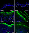
Overexpression of TSLP in skin of NS patients. Immunofluorescence experiments performed on skin cryosections using anti-TSLP antibody (green). TSLP expression is not detectable in two healthy control skins but, in contrast, this cytokine is strongly expressed in the skin of four NS patients (NS1–NS4). The signal is cytoplasmic in the granular keratinocytes and decreases through the spinous layers. The pictures are representative of two independent experiments in which two healthy and four NS patients were included. Dashed white lines represent epidermis/dermis junction. Blue staining (DAPI) indicates nuclei. Bars, 11.5 µm.
Figure 6.
Figure 6.
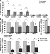
KLK5 up-regulates TSLP and proinflammatory molecules in keratinocytes. (A) NHKs were stimulated with 400 nM of recombinant KLK5 for 3 h. TSLP, ICAM1, IL8, and TNF-α transcripts are increased by seven-, five-, four-, and fivefold, respectively. No difference was observed for IL-1β, TARC, and MDC (n = 5). (B) NHK transfected with 60 pmol PAR2 siRNA and stimulated with KLK5 showed PAR2 mRNA down-regulation (by 56%) 4 d after transfection. In PAR2 siRNA-transfected cells, KLK5 is less efficient in inducing TSLP, ICAM1, IL8, and TNF-α mRNA (48, 55, 60, and 62% of control, respectively; n = 4). (C) In cells treated with 10 µM NF-κB pathway inhibitor (BAY11-7082), KLK5 efficiency to up-regulate TSLP, ICAM1, IL8, and TNF-α mRNA is decreased by 81, 70, 59, and 40%, respectively (n = 3). For each experiment, the mRNAs of interest were measured by quantitative RT-PCR. Each point is the mean of triplicate amplification for at least three independent experiments (n). *, P < 0.05; **, P < 0.01; ***, P < 0.001.
Figure 7.
Figure 7.
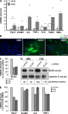
TSLP and proinflammatory molecules are induced by KLK5 in keratinocytes of NS patients. (A) Quantitative RT-PCR on cultured primary keratinocytes reveals that TSLP is significantly overexpressed by fivefold in three NS patients (NSK) compared with three healthy controls (NHK). ICAM1, IL8, TNF-α, TARC, and MDC mRNA are overexpressed in two out of three NSKs but not IL-1β mRNA. Each point represents the mean of two independent experiments for each individual. *, P < 0.05. (B) NHK and NSK were treated with 10 µg/ml brefeldin A, and TSLP was detected by immunofluorescence (green). Blue staining is DAPI. Pictures are representative of two independent brefeldin A treatments, each realized on two NHKs and two NSKs. (C and D) NSKs were transfected with KLK5 siRNA and its efficient down-regulation was confirmed by quantitative RT-PCR, Western blotting (laminin 5 used as loading control), and ELISA, 48 and 72 h after transfection. Results are representative of three independent experiments. (E) KLK5 knockdown induces a decrease of TSLP measured by quantitative RT-PCR. ICAM1, IL8, and TNF-α mRNAs were less efficiently reduced. Data are the mean ± SD of one experiment, which is representative of three independent experiments. Bars, 49 µm
Figure 8.
Figure 8.
KLK5 triggers proinflammatory and proallergic microenvironment independently of external stimuli. Lack of KLK5 inhibition by LEKTI initiates proinflammatory and proallergic cascades independently of environmental factors. In LEKTI-deficient keratinocytes, hyperactive KLK5 directly induces TSLP, IL8, and TNF-α overexpression through PAR2 and NF-κB pathway activation. Unregulated KLK5 also degrades desmosomes at the interface between the GR and the SC leading to defective SC adhesion and IL-1β, IL8, and TNF-α secretion by mechanically stressed keratinocytes. In addition, all these cytokines could induce TARC and MDC chemokines secretion by keratinocytes and dermal fibroblasts. These proinflammatory mediators trigger eosinophilic and mast cell recruitment and activation. TSLP has been reported to activate resident LCs, which migrate to draining lymph nodes and promote the differentiation of naive T cells (Th0) into Th2 cells. Collectively, activated keratinocytes together with eosinophilic and mast cells induce pro-Th2 microenvironment favoring the development of an AD-like phenotype.

References

    1. Candi E., Schmidt R., Melino G. 2005. The cornified envelope: a model of cell death in the skin.Nat. Rev. Mol. Cell Biol. 6:328–340 - PubMed
    1. Swartzendruber D.C., Wertz P.W., Kitko D.J., Madison K.C., Downing D.T. 1989. Molecular models of the intercellular lipid lamellae in mammalian stratum corneum.J. Invest. Dermatol. 92:251–257 - PubMed
    1. Green K.J., Simpson C.L. 2007. Desmosomes: new perspectives on a classic.J. Invest. Dermatol. 127:2499–2515 - PubMed
    1. Egelrud T. 2000. Desquamation in the stratum corneum.Acta Derm. Venereol. Suppl. (Stockh.). 208:44–45 - PubMed
    1. Elias P.M., Williams M.L., Holleran W.M., Jiang Y.J., Schmuth M. 2008. Pathogenesis of permeability barrier abnormalities in the ichthyoses: Inherited disorders of lipid metabolism.J. Lipid Res. 49:697–714 - PMC - PubMed

Publication types