. 2009 May 11;206(5):1117-34.
doi: 10.1084/jem.20082831. Epub 2009 May 4.

Low-dose rectal inoculation of rhesus macaques by SIVsmE660 or SIVmac251 recapitulates human mucosal infection by HIV-1

Affiliations

Low-dose rectal inoculation of rhesus macaques by SIVsmE660 or SIVmac251 recapitulates human mucosal infection by HIV-1

Brandon F Keele et al. J Exp Med. .

Abstract

We recently developed a novel strategy to identify transmitted HIV-1 genomes in acutely infected humans using single-genome amplification and a model of random virus evolution. Here, we used this approach to determine the molecular features of simian immunodeficiency virus (SIV) transmission in 18 experimentally infected Indian rhesus macaques. Animals were inoculated intrarectally (i.r.) or intravenously (i.v.) with stocks of SIVmac251 or SIVsmE660 that exhibited sequence diversity typical of early-chronic HIV-1 infection. 987 full-length SIV env sequences (median of 48 per animal) were determined from plasma virion RNA 1-5 wk after infection. i.r. inoculation was followed by productive infection by one or a few viruses (median 1; range 1-5) that diversified randomly with near starlike phylogeny and a Poisson distribution of mutations. Consensus viral sequences from ramp-up and peak viremia were identical to viruses found in the inocula or differed from them by only one or a few nucleotides, providing direct evidence that early plasma viral sequences coalesce to transmitted/founder viruses. i.v. infection was >2,000-fold more efficient than i.r. infection, and viruses transmitted by either route represented the full genetic spectra of the inocula. These findings identify key similarities in mucosal transmission and early diversification between SIV and HIV-1, and thus validate the SIV-macaque mucosal infection model for HIV-1 vaccine and microbicide research.


Figures

Figure 1.
Figure 1.
Viral kinetics after i.r. or i.v. infection. Groups of three animals received i.r. inoculations of SIVsmE660 or SIVmac251 in dosages indicated at weeks 0, 1, 2, 3, 4, and 5 (underlined) or by the i.v. route at week 0 (only) of the fourth cycle. Plasma was sampled at ramp-up (green symbols) or at peak (blue symbols) viremia for env sequence analysis. Numbers of transmitted/founder viruses found to be responsible for productive infection are indicated in parenthesis. Plasma viral load determinations were previously reported (41).
Figure 2.
Figure 2.
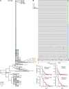
Viral sequence analysis after intrarectal SIVmac251 infection of animal CP1W. (A) NJ tree of sequences from ramp-up (green symbols) or peak (blue symbols) viremia or from the inoculum (black symbols). (B) Highlighter alignment of all CP1W sequences compared with four identical sequences from the inoculum and to the transmitted/founder virus. Nucleotide polymorphisms are indicated by a colored tic mark (thymine in red, guanine in orange, adenine in green, and cytosine in blue). APOBEC-mediated G-to-A mutations are indicated by purple tics, and deletions are indicated by gray tics. (C) Poisson model of early viral diversity. Brown labels indicate sequences with three or more APOBEC-mediated G-to-A mutations compared with consensus.
Figure 3.
Figure 3.
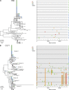
Viral sequence diversity after i.r. or i.v. SIVmac251 infection. (A) NJ and Highlighter analyses of sequences from i.r.-infected animal PBE at ramp-up (green symbols) or peak (blue symbols) viremia compared with inoculum sequences (black symbols). (B) i.v.-infected animal CG71. Brown labels indicate sequences with three or more APOBEC-mediated G-to-A mutations compared with consensus.
Figure 4.
Figure 4.
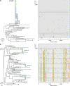
Viral sequence diversity after i.r. or i.v. SIVsmE660 infection. (A) NJ and Highlighter analyses of sequences from i.r.-infected animal CG87 at ramp-up (green symbols) or peak (blue symbols) viremia compared with inoculum sequences (black symbols). (B) i.v.-infected animal CP37. Nine transmitted/founder variant lineages are indicated, although additional minor variants and recombinants are also evident (see text). Bracket indicates a single-nucleotide insertion.
Figure 5.
Figure 5.
i.r. transmission of five viruses, followed by extensive recombination. NJ and Highlighter plots from animal CG7V at ramp-up (green symbols) or peak (blue symbols) viremia with recombinant sequences labeled in purple (#) and minor transmitted/founder viral variants 3, 4, and 5 in red (*). Brown labels indicate sequences with three or more APOBEC-mediated G-to-A mutations compared with consensus.
Figure 6.
Figure 6.
Unequal representation of transmitted viruses. Transmitted variants 1 and 2 in animal CP3C are well represented at ramp-up (green symbols) and peak (blue symbols) viremia compared with a third transmitted variant (red asterisk) that is represented by only a single sequence at ramp-up. Inoculum sequences are indicated by black symbols.
Figure 7.
Figure 7.
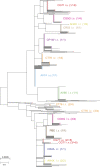
Composite SIVmac251 tree from i.r.- and i.v.-infected animals. The NJ tree depicts numerous transmitted/founder virus lineages distributed throughout a diverse set of inoculum sequences (black symbols). Most i.r.- or i.v.-transmitted viruses are represented by discrete, low-diversity lineages. Animal designation, route of infection, and numbers of transmitted viruses are indicated.

References

    1. Chun T.W., Engel D., Berrey M.M., Shea T., Corey L., Fauci A.S. 1998. Early establishment of a pool of latently infected, resting CD4(+) T cells during primary HIV-1 infection.Proc. Natl. Acad. Sci. USA. 95:8869–8873 - PMC - PubMed
    1. Haase A.T. 2005. Perils at mucosal front lines for HIV and SIV and their hosts.Nat. Rev. Immunol. 5:783–792 - PubMed
    1. Pope M., Haase A.T. 2003. Transmission, acute HIV-1 infection and the quest for strategies to prevent infection.Nat. Med. 9:847–852 - PubMed
    1. Shattock R.J., Moore J.P. 2003. Inhibiting sexual transmission of HIV-1 infection.Nat. Rev. Microbiol. 1:25–34 - PubMed
    1. Keele B.F., Giorgi E.E., Salazar-Gonzalez J.F., Decker J.M., Pham K.T., Salazar M.G., Sun C., Grayson T., Wang S., Li H., et al. 2008. Identification and characterization of transmitted and early founder virus envelopes in primary HIV-1 infection.Proc. Natl. Acad. Sci. USA. 105:7552–7557 - PMC - PubMed

Publication types

MeSH terms