. 2009 Jul;29(14):3975-90.
doi: 10.1128/MCB.01867-08. Epub 2009 May 4.

The Nek6 and Nek7 protein kinases are required for robust mitotic spindle formation and cytokinesis

Affiliations

The Nek6 and Nek7 protein kinases are required for robust mitotic spindle formation and cytokinesis

Laura O'Regan et al. Mol Cell Biol. 2009 Jul.

Abstract

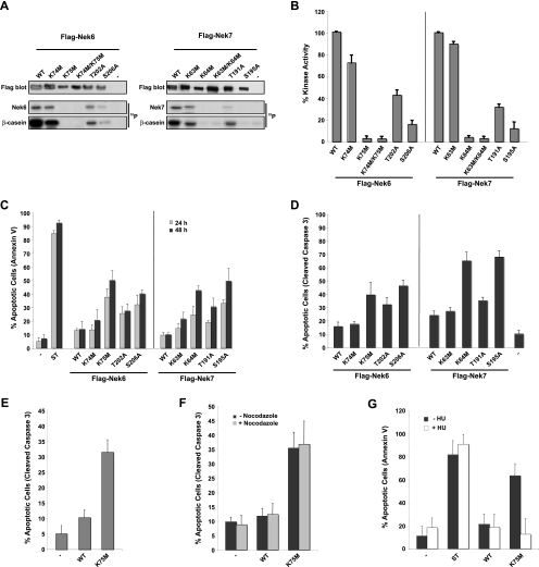
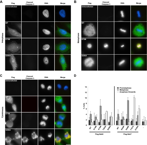
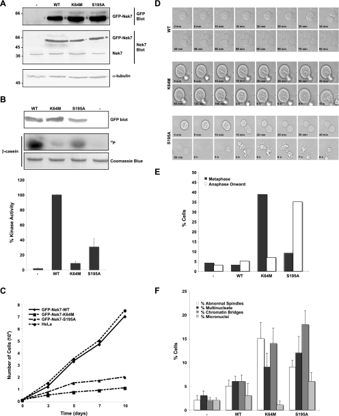
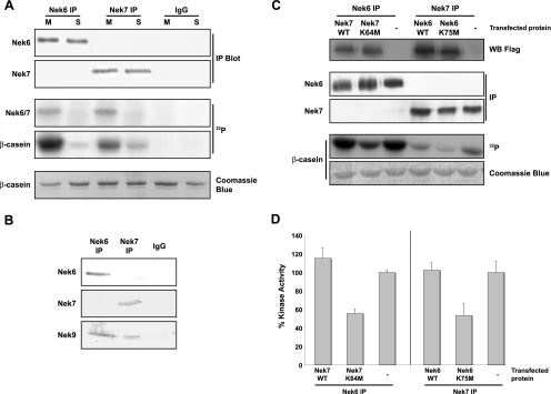
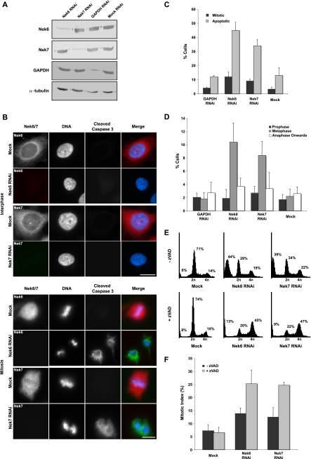
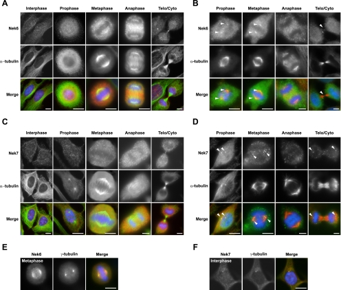
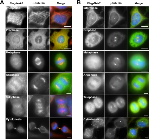
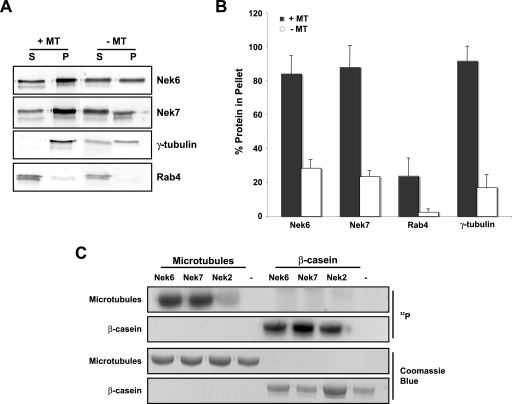
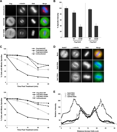
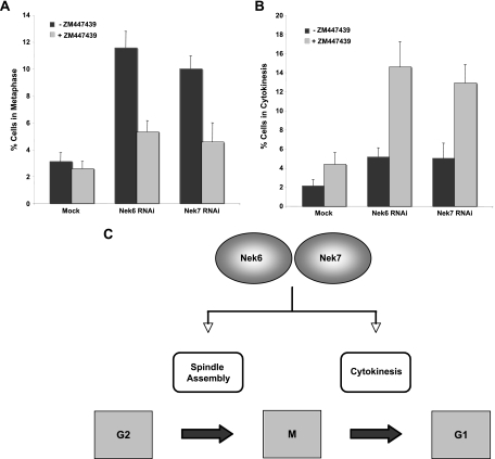
Nek6 and Nek7 are members of the NIMA-related serine/threonine kinase family. Previous work showed that they contribute to mitotic progression downstream of another NIMA-related kinase, Nek9, although the roles of these different kinases remain to be defined. Here, we carried out a comprehensive analysis of the regulation and function of Nek6 and Nek7 in human cells. By generating specific antibodies, we show that both Nek6 and Nek7 are activated in mitosis and that interfering with their activity by either depletion or expression of reduced-activity mutants leads to mitotic arrest and apoptosis. Interestingly, while completely inactive mutants and small interfering RNA-mediated depletion delay cells at metaphase with fragile mitotic spindles, hypomorphic mutants or RNA interference treatment combined with a spindle assembly checkpoint inhibitor delays cells at cytokinesis. Importantly, depletion of either Nek6 or Nek7 leads to defective mitotic progression, indicating that although highly similar, they are not redundant. Indeed, while both kinases localize to spindle poles, only Nek6 obviously localizes to spindle microtubules in metaphase and anaphase and to the midbody during cytokinesis. Together, these data lead us to propose that Nek6 and Nek7 play independent roles not only in robust mitotic spindle formation but also potentially in cytokinesis.


Figures

FIG. 1.
FIG. 1.
Expression of kinase-inactive Nek6 and Nek7 mutants induces apoptosis. (A) HEK 293 cells were transiently transfected with FLAG-tagged Nek6 and Nek7 constructs, as indicated, for 24 h before cells were lysed and subjected to immunoprecipitation with anti-Flag antibodies. The amount of kinase precipitated was determined by Western blotting with anti-Flag antibodies and the immunoprecipitates used for kinase assays, with β-casein as a substrate. Autoradiographs (32P) are shown of the Nek6/Nek7 and β-casein proteins. WT, wild type. (B) Activity of the different Nek6 and Nek7 constructs is expressed as a percentage of wild-type activity normalized to the amount of precipitated protein. The data are shown as means ± standard deviations from three separate experiments. (C) HeLa cells were transiently transfected with Flag-tagged Nek6 and Nek7 constructs for 24 and 48 h before being assayed for their ability to take up annexin V/FITC stain via flow cytometry analysis. As a positive control, untransfected cells were treated with 1 μM staurosporine (ST) for 4 h. (D) HeLa cells were transiently transfected with Flag-tagged Nek6 and Nek7 constructs for 48 h before being fixed and stained with Flag antibodies to detect transfected cells and cleaved caspase 3 antibodies to detect apoptotic cells. Data in panels C and D represent means (±standard deviations) from counts of at least 50 cells in three separate experiments. (E) RPE1-hTERT cells were transiently transfected with FLAG-tagged Nek6-WT and Nek6-K75M constructs for 48 h before being fixed and stained with anti-FLAG and anti-cleaved caspase 3 antibodies. Data represent means (±standard deviations) from counts of at least 100 cells in three separate experiments. (F) HeLa cells were transiently transfected with FLAG-tagged Nek6-WT and Nek6-K75M constructs for 48 h with and without treatment with 500 ng/ml nocodazole before being fixed and stained with anti-FLAG antibodies to detect transfected cells and anti-cleaved caspase 3 antibodies to detect apoptotic cells. Data represent means (±standard deviations) from counts of at least 100 cells in three separate experiments. (G) HeLa cells were transiently transfected with Flag-Nek6 constructs for 48 h in the presence or absence of 1 mM hydroxyurea before being assayed for their ability to take up annexin V/FITC via flow cytometry analysis. Data represent means (±standard deviations) from three separate experiments. In all experiments, untransfected cells (−) were used as a negative control.
FIG. 2.
FIG. 2.
Kinase-inactive Nek6 and Nek7 trigger mitotic delay at two stages. (A to C) HeLa cells were either untransfected or transfected, as indicated, with Flag-tagged Nek6-wild type (WT), Nek6-K75M, or Nek6-S206A for 48 h before being fixed and stained with antibodies against Flag (green in merge) to detect transfected cells and cleaved caspase 3 (red in merge) to detect apoptotic cells. DNA was stained with Hoechst 33258 (blue in merge). Cells in interphase (A), metaphase (B), and cytokinesis (C) are shown. Scale bar, 10 μm. (D) HeLa cells were either untransfected (−) or transiently transfected with different Flag-Nek6 and Flag-Nek7 constructs, as indicated, for 24 h before being processed for immunofluorescence microscopy with Flag antibodies to detect transfected cells and α-tubulin antibodies to detect the mitotic spindle. The histogram represents the observed frequency of transfected cells in the different stages of mitosis indicated. Data are means (±standard deviations) from three separate experiments, in which at least 50 cells were counted for each construct.
FIG. 3.
FIG. 3.
Cells stably expressing kinase-inactive Nek7 exhibit defective cell cycle progression. (A) Cell lysates were prepared from parental HeLa cells (−) or HeLa cells stably expressing GFP-Nek7 constructs, as indicated, and were subjected to SDS-PAGE and Western blot analysis with antibodies against GFP, Nek7, and α-tubulin. Molecular masses (kDa) are indicated on the left. WT, wild type. (B) Cell lysates were subjected to immunoprecipitation with anti-GFP antibodies, as described in the legend for Fig. 1A. The amount of kinase precipitated was determined by Western blotting with anti-GFP antibodies and the immunoprecipitates used for kinase assays, with β-casein as a substrate. Products were analyzed by SDS-PAGE, Coomassie blue staining, and autoradiography (32P). Activity levels of the different Nek7 constructs are expressed as a percentage of the wild-type activity normalized to the amount of precipitated protein. The data are shown as means (±standard deviations) from three separate experiments. (C) Growth curves of HeLa cells stably expressing GFP-Nek7 constructs are shown; data represent the means from three separate experiments. (D) Mitotic HeLa cells stably expressing GFP-Nek7 constructs, as indicated, were analyzed by time-lapse bright-field microscopy. Images are presented from metaphase onwards, with time indicated on each panel. (E) The outcome of each live cell imaged as in panel D was scored according to whether they were delayed in metaphase or late mitosis prior to undergoing apoptosis or whether they progressed normally. A total of 40 to 50 cells were scored for each cell line. (F) Parental HeLa cells (−) or HeLa cells stably expressing GFP-Nek7 proteins, as indicated, were fixed and stained with α-tubulin antibodies to detect the microtubule network and Hoechst 33258 to detect DNA. The percentage of cells with abnormal mitotic spindles or cells that were multinucleated, micronucleated, or still attached by chromatin bridges was scored. Data represents means (±standard deviations) from counts of at least 150 cells in three independent experiments.
FIG. 4.
FIG. 4.
Endogenous Nek6 and Nek7 are activated in mitosis. (A) Lysates were prepared from untransfected HEK 293 cells synchronized in S or M phase of the cell cycle with hydroxyurea or nocodazole, respectively, and proteins immunoprecipitated with Nek6 or Nek7 antibodies or control rabbit IgGs. The immunoprecipitates were subjected to Western blotting (IP blot) with Nek6 or Nek7 antibodies and used in kinase assays, with β-casein as a substrate. Samples were analyzed by SDS-PAGE, Coomassie blue staining, and autoradiography (32P). (B) Lysates were prepared from untransfected, asynchronous HEK 293 cells and proteins immunoprecipitated with Nek6 or Nek7 antibodies or control rabbit IgGs. Immunoprecipitates were subjected to Western blotting with Nek6, Nek7, or Nek9 antibodies. (C) HEK 293 cells were either untransfected (−) or transiently transfected with Flag-Nek7-wild type (WT) or Flag-Nek7-K64M and Flag-Nek6-WT or Flag-Nek6-K75M constructs, as indicated. After 24 h, cells lysates were subjected to Western blotting with Flag antibodies to determine transfection efficiency. Lysates were subjected to immunoprecipitation (IP) with Nek6 or Nek7 antibodies and immunoprecipitates used for Western blot analysis and kinase assays, with β-casein as a substrate. Samples were analyzed by SDS-PAGE, Coomassie blue staining, and autoradiography (32P). (D) Activity levels of the immunoprecipitated proteins are expressed as a percentage of wild-type activity in untransfected (−) cells normalized to the amount of precipitated protein. The data are shown as means (±standard deviations) from three separate experiments.
FIG. 5.
FIG. 5.
RNAi depletion of Nek6 or Nek7 induces apoptosis following mitotic arrest. (A) HeLa cells were either mock transfected or transfected with siRNA oligonucleotides targeted against Nek6, Nek7, or GAPDH. After 72 h, extracts were prepared and analyzed by SDS-PAGE and Western blotting with Nek6, Nek7, GAPDH, or α-tubulin antibodies. (B) HeLa cells were transfected with siRNA oligonucleotides, as indicated, for 72 h before being fixed and analyzed by immunofluorescence microscopy with antibodies against Nek6 or Nek7 (red on merge) and cleaved caspase 3 (green on merge). DNA was stained with Hoechst 33258 (blue on merge). Cells in interphase and mitosis are shown. Scale bar, 10 μm. (C) HeLa cells were treated with siRNA oligonucleotides for 72 h, as indicated, and analyzed as described in the legend for panel B. A total of 100 to 300 cells were scored for mitosis or apoptosis in three independent experiments, and means (±standard deviations) are indicated. (D) Following treatment of cells with siRNA oligonucleotides, as indicated, the frequencies of cells in different stages of mitosis were scored. Data represent means (±standard deviations) from three separate experiments, counting at least 100 cells per experiment. (E) HeLa cells were transfected with Nek6 or Nek7 siRNA oligonucleotides or mock transfected for 72 h in the presence or absence of 100 μM zVAD, as indicated. Cells were then harvested and fixed before being stained with propidium iodide and before the cell cycle distribution being analyzed by flow cytometry. The percentages of cells in less than 2n, 2n, and 4n peaks are indicated. (F) HeLa cells were transfected with Nek6 or Nek7 siRNA oligonucleotides or mock transfected for 72 h in the presence or absence of zVAD before being fixed and analyzed by immunofluorescence microscopy, and the frequencies of cells in mitosis were scored. Data represent means (±standard deviations) from three separate experiments, counting at least 100 cells in each.
FIG. 6.
FIG. 6.
In mitosis, Nek6 localizes to spindle microtubules and spindle poles, while Nek7 localizes only with spindle poles. (A) HeLa cells were processed for immunofluorescence microscopy with Nek6 (red in merge) and α-tubulin (green in merge) antibodies. Telo/Cyto, telophase/cytokinesis. (B) HeLa cells were treated with extraction buffer for 30 s before being fixed and permeabilized in methanol and processed for immunofluorescence microscopy, as described in the legend for panel A. Arrowheads denote spindle pole or midbody staining. (C and D) Same process as described in legends for panels A and B, respectively, except using Nek7 (red) antibodies. (E and F) HeLa cells were fixed and processed for immunofluorescence microscopy with Nek6 or Nek7 (red) antibodies, as indicated, and γ-tubulin (green) antibodies. In all panels, DNA was stained with Hoechst 33258 (blue), and merged images are shown. Scale bars, 10 μm.
FIG. 7.
FIG. 7.
HeLa cells were transiently transfected with Flag-tagged Nek6-wild type (A) or Nek7-wild type (B) constructs for 24 h before being processed for immunofluorescence microscopy with Flag (green in merge) and α-tubulin (red in merge) antibodies. DNA was stained with Hoechst 33258 (blue). Scale bars, 10 μm.
FIG. 8.
FIG. 8.
Nek6 and Nek7 interact with and phosphorylate microtubule preparations in vitro. (A) Nek6 and Nek7 and the control proteins Rab4 and γ-tubulin were generated by in vitro translation in the presence of [35S]methionine. Input proteins were incubated with taxol-stabilized microtubules (+MT) or without taxol-stabilized microtubules (−MT). Proteins were spun at 35,000 rpm on a sucrose cushion for 30 min, and supernatant (S) and pellet (P) fractions were collected and analyzed by SDS-PAGE and autoradiography. (B) The histogram represents the percentages of input protein found within the pellet fractions with and without taxol-stabilized microtubules. Data represent means (±standard deviations) from three separate experiments. (C) Kinase assays were performed for 30 min at 30°C with no kinase (−) or purified recombinant His-tagged Nek6, Nek7, or Nek2 kinases and purified microtubules or β-casein as substrates, before analysis by SDS-PAGE, Coomassie blue staining, and autoradiography (32P).
FIG. 9.
FIG. 9.
Nek6 and Nek7 are required for formation of robust mitotic spindles. (A) HeLa cells were either untransfected or transiently transfected with Flag-Nek6-K75M or Flag-Nek7-K64M constructs. After 24 h, cells were processed for immunofluorescence microscopy with Flag antibodies to detect transfected cells (green in merge) and α-tubulin antibodies to detect the microtubule network (red in merge). DNA was stained with Hoechst 33258 (blue). Scale bar, 10 μm. (B) The relative intensity of the metaphase spindle of transfected cells was measured for each construct, using quantitative fluorescence imaging. Data represent means (±standard deviations) from measurements from 50 cells for each construct. The relative spindle intensity is measured as a percentage of the untransfected spindle intensity (−). WT, wild type. (C) HeLa cells were transiently transfected with Flag-Nek6 (top) or Flag-Nek7 (bottom) constructs for 24 h. Cells were then treated with nocodazole before being fixed at the time points indicated. Fixed cells were processed for immunofluorescence microscopy with Flag antibodies to detect transfected cells and α-tubulin antibodies to detect the microtubule network. Transfected mitotic cells were scored for the integrity of their mitotic spindles. Spindles were considered to have collapsed when no clear spindle could be seen within the cell. A total of 30 cells were counted for each construct in three separate experiments. (D) HeLa cells treated with Nek6 or Nek7 siRNA oligonucleotides for 72 h were fixed and analyzed by immunofluorescence microscopy with Nek6 or Nek7 (red in merge) and α-tubulin antibodies (green in merge). DNA was stained with Hoechst 33258 (blue). (E) The relative intensity and length of metaphase spindles in Nek6-, Nek7-, and GAPDH-depleted cells were measured using ImageJ analysis software.
FIG. 10.
FIG. 10.
Overriding the SAC leads to a delay in cytokinesis, in response to Nek6 or Nek7 depletion. (A, B) HeLa cells were mock transfected or transfected with Nek6 or Nek7 siRNA oligonucleotides for 72 h in the presence or absence of the Aurora B inhibitor ZM447439 before being fixed and analyzed by immunofluorescence microscopy. The frequency of cells in metaphase (A) or cytokinesis (B) was scored. Data represent means (±standard deviations) from three separate experiments, with at least 100 cells in each case. (C) Model showing two points of action for the Nek6 and Nek7 kinases in mitotic progression, one in spindle formation and one in cytokinesis. This model is based upon data presented here, showing that depletion of either of the kinases or expression of inactive mutants leads to metaphase arrest, while depletion of either of the kinases in the presence of a SAC inhibitor or expression of hypomorphic mutants leads to a cytokinesis arrest.

References

    1. Bahe, S., Y. D. Stierhof, C. J. Wilkinson, F. Leiss, and E. A. Nigg. 2005. Rootletin forms centriole-associated filaments and functions in centrosome cohesion. J. Cell Biol. 17127-33. - PMC - PubMed
    1. Belham, C., J. Roig, J. A. Caldwell, Y. Aoyama, B. E. Kemp, M. J. Comb, and J. Avruch. 2003. A mitotic cascade of NIMA family kinases. J. Biol. Chem. 27834897-34909. - PubMed
    1. Capra, M., P. G. Nuciforo, S. Confalonieri, M. Quarto, M. Bianchi, M. Nebuloni, R. Boldorini, F. Pallotti, G. Viale, M. L. Gishizky, G. F. Draetta, and P. P. Di Fiore. 2006. Frequent alterations in the expression of serine/threonine kinases in human cancers. Cancer Res. 668147-8154. - PubMed
    1. Chen, J., L. Li, Y. Zhang, H. Yang, Y. Wei, L. Zhang, X. Liu, and L. Yu. 2006. Interaction of Pin1 with Nek6 and characterization of their expression correlation in Chinese hepatocellular carcinoma patients. Biochem. Biophys. Res. Commun. 3411059-1065. - PubMed
    1. Davies, H., C. Hunter, R. Smith, P. Stephens, C. Greenman, G. Bignell, J. Teague, A. Butler, S. Edkins, C. Stevens, A. Parker, S. O'Meara, T. Avis, S. Barthorpe, L. Brackenbury, G. Buck, J. Clements, J. Cole, E. Dicks, K. Edwards, S. Forbes, M. Gorton, K. Gray, K. Halliday, R. Harrison, K. Hills, J. Hinton, D. Jones, V. Kosmidou, R. Laman, R. Lugg, A. Menzies, J. Perry, R. Petty, K. Raine, R. Shepherd, A. Small, H. Solomon, Y. Stephens, C. Tofts, J. Varian, A. Webb, S. West, S. Widaa, A. Yates, F. Brasseur, C. S. Cooper, A. M. Flanagan, A. Green, M. Knowles, S. Y. Leung, L. H. Looijenga, B. Malkowicz, M. A. Pierotti, B. T. Teh, S. T. Yuen, S. R. Lakhani, D. F. Easton, B. L. Weber, P. Goldstraw, A. G. Nicholson, R. Wooster, M. R. Stratton, and P. A. Futreal. 2005. Somatic mutations of the protein kinase gene family in human lung cancer. Cancer Res. 657591-7595. - PubMed

Publication types

MeSH terms

LinkOut - more resources