. 2009 May 15;182(10):6217-25.
doi: 10.4049/jimmunol.0803842.

TLR signals promote IL-6/IL-17-dependent transplant rejection

Affiliations

TLR signals promote IL-6/IL-17-dependent transplant rejection

Luqiu Chen et al. J Immunol. .

Abstract

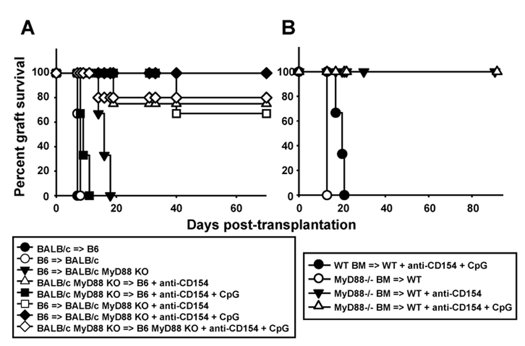
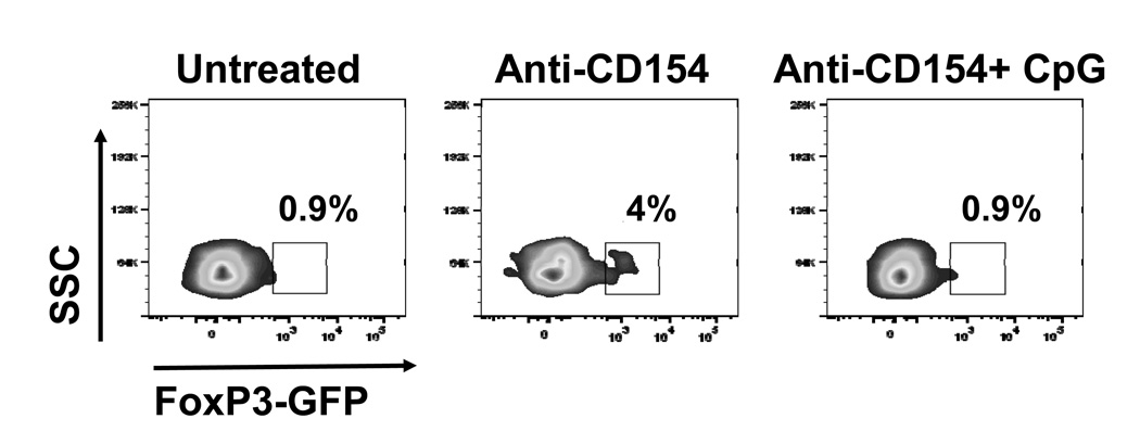
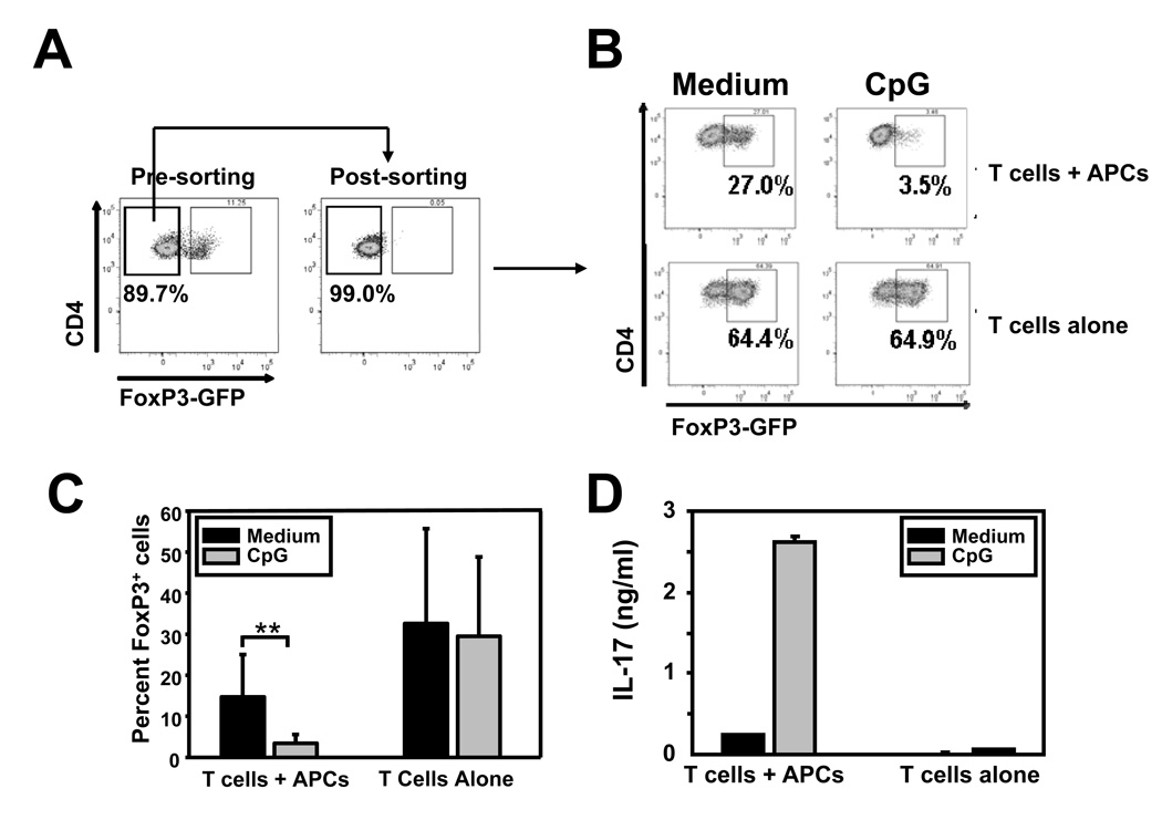
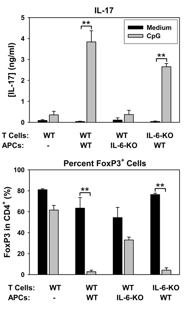
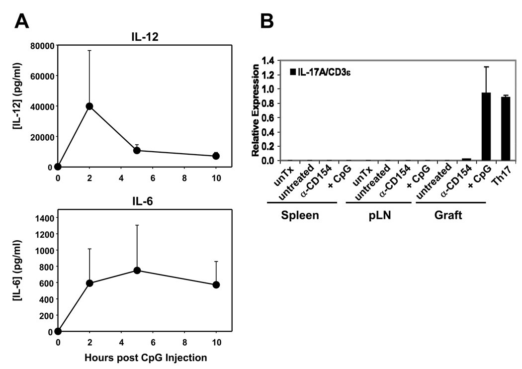
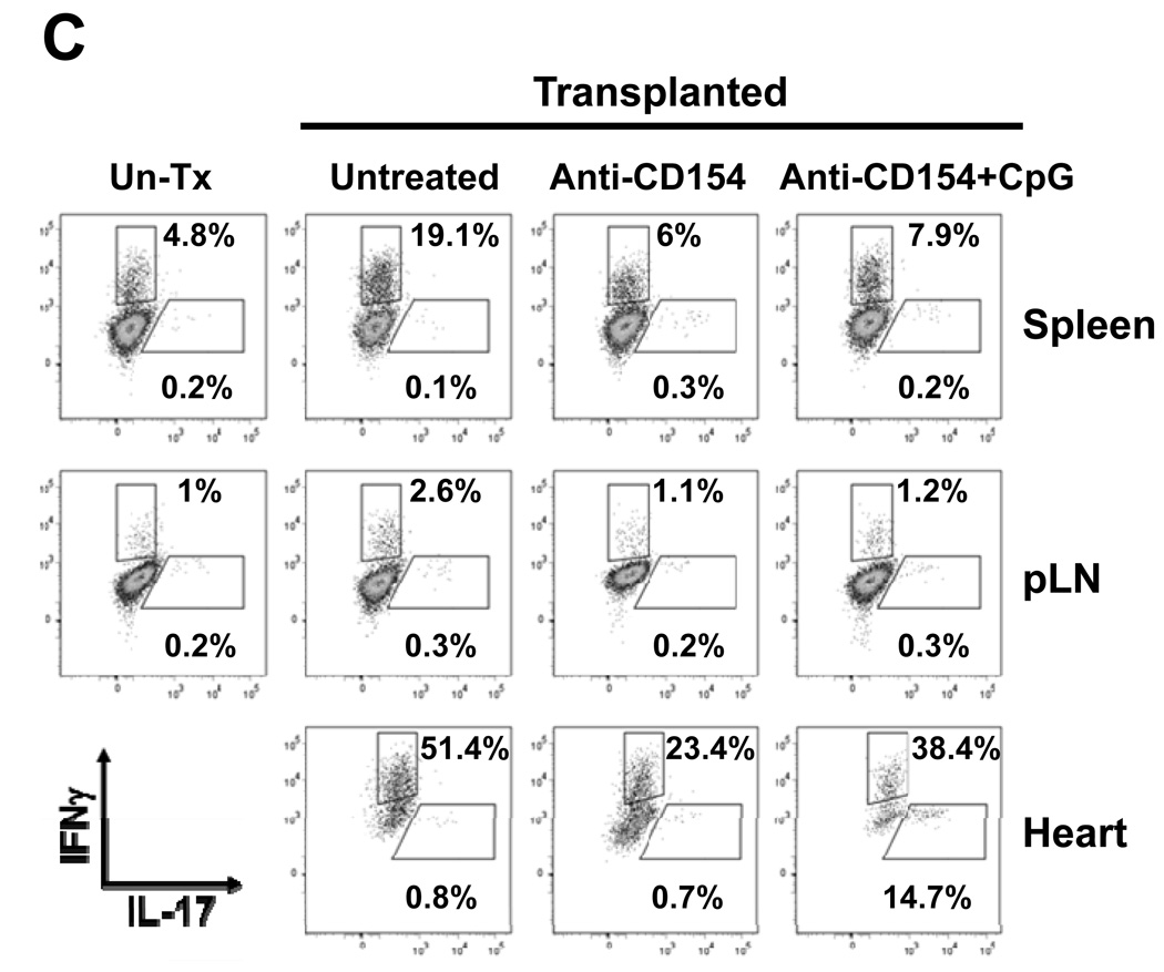
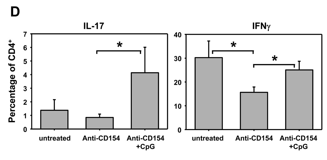
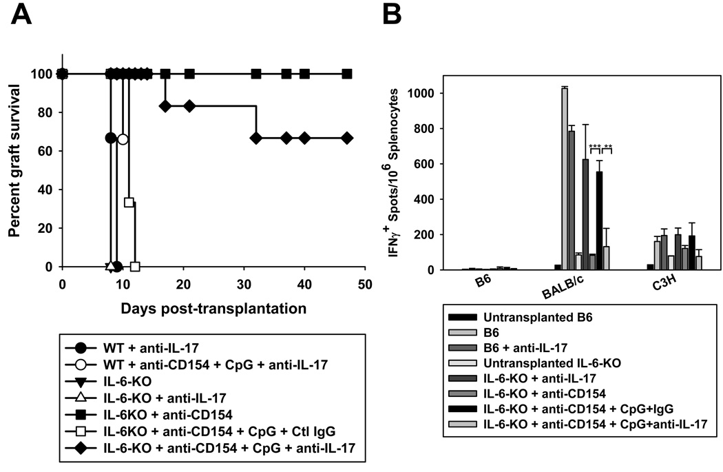
Acute allograft rejection has often been correlated with Th1 differentiation, whereas transplantation tolerance is frequently associated with induction of regulation. The discovery of the Th17 phenotype has prompted its scrutiny in transplant rejection. Although IL-17 has recently been observed in settings of acute allograft rejection and drives rejection in T-bet-deficient mice that have impaired type 1 T cell responses, there is little evidence of its requirement during acute rejection in wild-type animals. We and others have previously shown that TLR9 signaling by exogenous CpG at the time of transplantation is sufficient to abrogate anti-CD154-mediated acceptance of fully mismatched cardiac allografts. In this study, we investigated the mechanism by which acute rejection occurs in this inflammatory context. Our results indicate that CpG targets recipient hemopoietic cells and that its pro-rejection effects correlate both with prevention of anti-CD154-mediated conversion of conventional CD4(+) T cells into induced regulatory T cells and with the expression of IFN-gamma and IL-17 by intragraft CD4(+) T cells. Moreover, the combined elimination of IL-6 and IL-17 signaling abrogated the ability of CpG to promote acute cardiac allograft rejection. Thus, proinflammatory signals at the time of transplantation can change the quality of the effector immune response and reveal a pathogenic function for IL-6 and IL-17 in wild-type recipients.


Figures

Figure 1
Figure 1. The pro-rejection effect of CpG requires MyD88 on recipient hematopoietic cells
A. Wild-type and MyD88-deficient hearts of BALB/c origin were transplanted into wild-type and MyD88−/− mice on the B6 background. Recipients were either left untreated or treated with anti-CD154±CpG and cardiac allograft survival was examined over time (n=3–5 mice/group). B. T-depleted bone marrow cells from wild-type or MyD88−/− mice on a B6 background (CD45.2+) were injected into congenic (CD45.1+) lethally irradiated B6 recipients and allowed to reconstitute the immune system for 8 weeks prior to transplantation with BALB/c hearts. Recipients were either left untreated or treated with anti-CD154±CpG and cardiac allograft survival was examined over time (n=3 mice/group).
Figure 2
Figure 2. CpG prevents conversion of naïve CD4+ T cells into iTregs in vivo
CD4+GFP cells were sorted from TEa-Tg-FoxP3GFP knock-in mice, labeled with PKH26 and injected i.v. (5×10⁶) into syngeneic B6 recipients one day prior to transplantation with BALB/c hearts and indicated treatments. Allografts were harvested on day 10 post transplantation and infiltrating leukocytes analyzed by flow cytometry for expression of GFP among CD4-gated events. Numbers in the plots represent percentages of GFP+ cells among CD4+ cells.
Figure 3
Figure 3. CpG prevents conversion of naïve T cells into iTregs and promotes IL-17 production in an IL-6-dependent manner in vitro
Sorted naïve CD4+ T cells (CD25 CD44lowGFP) from FoxP3GFP-knock-in mice were stimulated with immobilized anti-CD3 and anti-CD28, in the presence of TGF-β and IL-2 and ± CpG for 5 days, with or without irradiated syngeneic splenocytes (APCs). A. Cells were analyzed by flow cytometry for expression of CD4 and GFP before and after cell-sorting. B and C. Cells were analyzed by flow cytometry for expression of CD4 and FoxP3-GFP on day 5 of the culture. Panel B shows a representative flow cytometry profile. Panel C shows the means and standard deviations of the percentage of FoxP3+ cells among CD4+ cells from 5 independent experiments. D. Supernatants were collected on day 5 and analyzed by ELISA for IL-17 content. Results represent means and standard deviations from triplicate determinations and are representative of 4 independent experiments.
Figure 4
Figure 4. CpG-mediated IL-17 production is dependent on APC-but not T cell-derived IL-6
Naïve T cells from wild-type and IL-6−/− mice were sorted as for Figure 3 and stimulated as above except that APCs were syngeneic bone marrow-derived dendritic cells (BMDCs) of either wiltype or IL-6−/− origin. Cells and supernatants were harvested on day 5 for analysis of Foxp3 expression on CD4+ cells by flow cytometry and of IL-17 by ELISA, respectively. Results represent means and standard deviations from triplicate determinations and are representative of 3 independent experiments. **p<0.01 between the groups spanned by the horizontal bars.
Figure 5
Figure 5. CpG promotes expression of IL-17 in cardiac allografts
A. B6 mice were injected with CpG (100µg) i.p. and serum was collected serially at the indicated time points. Concentration of IL-12 and IL-6 were determined by multiplex bead analysis and ELISA, respectively. Results represent mean+SD from 3 animals per group. B. BALB/c hearts were transplanted into B6 or FoxP3GFP-knock-in recipients that were either left untreated or received anti-CD154 (MR1) or anti-CD154+CpG (CpG). Spleen, pLN and cardiac allografts were harvested 7–14 days post-transplantation. An untransplantated B6 mouse and Th17 cells differentiated in vitro from naïve CD4+ T cells were used as controls. Leukocytes were isolated from the different tissues and mRNA expression for IL-17 and CD3ε was determined by real time RT-PCR. The figure shows the ratio of IL-17A:CD3ε expression levels and the mean+SD of triplicate determinations. The plot is representative of 4 independent experiments. C. Spleen, pLN and heart allografts were harvested 8–12 days post transplantation. Leukocytes were isolated, stimulated for 6h with PMA and ionomycin in the presence of GolgiPlug and stained for analysis by flow cytometry. The plot represents intracellular expression of IL-17 and IFN-γ on CD4-gated events and is representative of 3 independent experiments. D. The percentage of IL-17 and IFN-g-producing cells among CD4-gated events is represented as mean+SD of 3 independent experiments and 5–7 mice per goup. *p<0.05 between the groups indicated by the horizontal bars.
Figure 6
Figure 6. IL-6 and IL-17 are required for CpG to prevent anti-CD154-mediated cardiac allograft acceptance
Wild-type B6 (n=3 per control group) or IL-6−/− mice on a B6 background were transplanted with BALB/c hearts and left untreated or treated with anti-CD154 +/− CpG (n=3–5 for all control groups) in the presence of anti-IL-17 (n=6) or control IgG (n=3 but similar results were obtained in the absence of control isotype, n=5, not shown). A. Graft survival was examined over time. B. Animals were sacrificed on day 50–80 post-transplantation and splenocytes were restimulated with irradiated syngeneic (B6), donor (BALB/c) or third party (C3H) splenocytes for detection of IFN-γ-producing cells by ELISpot. ***p<0.001, **p<0.01, between the groups spanned by the horizontal bars.

References

    1. Weaver CT, Hatton RD, Mangan PR, Harrington LE. IL-17 family cytokines and the expanding diversity of effector T cell lineages. Annu Rev Immunol. 2007;25:821–852. - PubMed
    1. Chen Y, Wood KJ. Interleukin-23 and TH17 cells in transplantation immunity: does 23+17 equal rejection? Transplantation. 2007;84:1071–1074. - PubMed
    1. Zhou P, Szot GL, Guo Z, Kim O, He G, Wang J, Grusby MJ, Newell KA, Thistlethwaite JR, Bluestone JA, Alegre ML. Role of STAT4 and STAT6 signaling in allograft rejection and CTLA4-Ig-mediated tolerance. J Immunol. 2000;165:5580–5587. - PubMed
    1. Kishimoto K, Dong VM, Issazadeh S, Fedoseyeva EV, Waaga AM, Yamada A, Sho M, Benichou G, Auchincloss H, Jr, Grusby MJ, Khoury SJ, Sayegh MH. The role of CD154-CD40 versus CD28-B7 costimulatory pathways in regulating allogeneic Th1 and Th2 responses in vivo. J Clin Invest. 2000;106:63–72. - PMC - PubMed
    1. Loong CC, Hsieh HG, Lui WY, Chen A, Lin CY. Evidence for the early involvement of interleukin 17 in human and experimental renal allograft rejection. J Pathol. 2002;197:322–332. - PubMed

Publication types

MeSH terms