. 2009 May 15;182(10):6369-78.
doi: 10.4049/jimmunol.0804287.

Proximal glycans outside of the epitopes regulate the presentation of HIV-1 envelope gp120 helper epitopes

Affiliations

Proximal glycans outside of the epitopes regulate the presentation of HIV-1 envelope gp120 helper epitopes

Hualin Li et al. J Immunol. .

Abstract

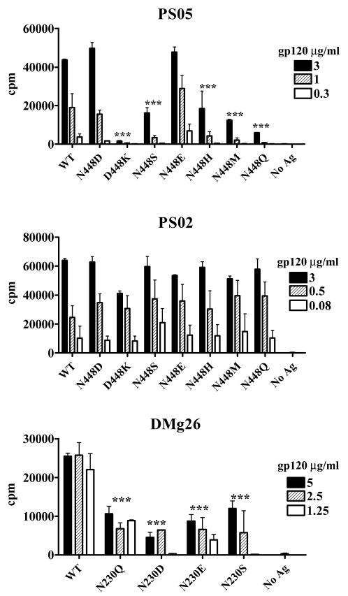
Glycosylation of HIV-1 envelope gp120 determines not only the proper structure, but also the immune responses against this Ag. Although glycans may be part of specific epitopes or shield other epitopes from T cells and Abs, this study provides evidence for a different immunomodulatory function of glycans associated with gp120 residues N230 and N448. These glycans are required for efficient MHC class II-restricted presentation of nearby CD4 T cell epitopes, even though they are not part of the epitopes. The glycans do not affect CD4 T cell recognition of more distant epitopes and are not essential for the proper folding and function of gp120. Data on CD4 T cell recognition of N448 mutants combined with proteolysis analyses and surface electrostatic potential calculation around residue N448 support the notion that N448 glycan near the epitope's C terminus renders the site to be surface accessible and allows its efficient processing. In contrast, the N230 glycan contributes to the nearby epitope presentation at a step other than the proteolytic processing of the epitope. Hence, N-glycans can determine CD4 T cell recognition of nearby gp120 epitopes by regulating the different steps in the MHC class II processing and presentation pathway after APCs acquire the intact gp120 Ag exogenously. Modifications of amino acids bearing glycans at the C termini of gp120 helper epitopes may prove to be a useful strategy for enhancing the immunogenicity of HIV-1 envelope gp120.


Figures

FIGURE 1
FIGURE 1. Site-specific N-linked glycan deletions introduced to gp120 and their positions relative to CD4 T cell epitopes examined in the study
Amino acid substitutions were introduced into gp120BH10 to remove N-glycans linked with residues 197, 230, 234, 406, 448, or 463 (marked by circles). The mutated proteins were tested for recognition by gp120-specific CD4 T cell lines PS02, DMg26, and PS05. Locations of the epitopes recognized by these T cell lines were highlighted by circles of different colors: pink for PS02 and blue for DMg26 in the C2 region (overlap was highlighted with both pink and blue), and red for PS05 in the C4 region. This diagram (modified from Leonard et al. (46)) shows the relatively conserved (C1-C5) and variable (V1-V5) regions of the gp120 protein, while the 30-amino acid signal peptide at the N terminus was omitted.
FIGURE 2
FIGURE 2. Recognition of gp120 mutants lacking specific N-glycans by CD4 T cells and mAbs
A. N448-glycan is required for CD4 T cell recognition of the neighboring PS05 epitope. CD4 T cell recognition of gp120 mutants lacking glycans at N448 (N448Q or T450A), N406 (N406Q), or N463 (N463Q) was evaluated using a C4-specific CD4 T cell line PS05. T cell proliferation was measured in the standard 3H-thymidine incorporation assay. Autologous human PBMCs were used as APCs after irradiation and treatment with wild type and mutant gp120 proteins at the indicated concentrations. All experiments were in triplicates and representative results of at least two independent experiments were shown. Average counts per minute (cpm) and standard deviations from triplicate wells were shown. ***, p<0.001 compared with the WT. B. Removal of N-glycans from the C2 region of gp120 does not abrogate CD4 binding and mAb reactivities. The CD4-binding capacity and gp120-specific mAb reactivity of the mutated gp120 proteins lacking glycans in the C2 region (N197Q, N230Q or N234Q) were determined by ELISA. Mutant and wild type gp120 proteins were captured onto ELISA plates and incubated with the designated concentrations of soluble CD4 (sCD4) or human anti-gp120 mAbs (b12, 447, 2G12, C11, and EH21). Means and standard deviations of OD405 values from triplicate wells are shown. The data are representative results from one of two independent experiments. *, p<0.05 compared with WT at the same concentration. C. N230-glycan is required for CD4 T cell recognition of the nearby DMg26 epitope. Recognition of gp120 mutants lacking glycans at N197, N230, or N234 was assessed using CD4 T cell lines specific for C2 epitopes (DMg26 and PS02) or a C4 epitope (PS05) in T cell proliferation assays. All experiments were performed in triplicates and representative results of at least two independent experiments were shown. Average counts per minute (cpm) and standard deviations from triplicate wells were shown. ***, p<0.001 compared with the WT.
FIGURE 3
FIGURE 3. Triple N-glycan towers on the gp120 surfaces near the C termini of DMg26 and PS05 epitopes
A. The glycans linked to residues N230, N234 and N241 form triple glycan towers with the middle N230-glycan flanked by N234 and N241 glycans on each side. The sugar towers protrude from the surface of gp120 inner domain near the C terminus of DMg26 epitope (blue). In contrast, the N197-linked glycan is found on the opposite side of gp120. B. The N448-linked glycan is also flanked by two glycans linked to N262 and N295, and the three glycans make triple sugar towers on the surface of gp120 outer domain close to the C terminus of PS05 epitope (red). Asparagine residues associated with the glycans are shown in grey, whereas the proximal sugar units resolved in the gp120 crystal structure (29) are in orange.
FIGURE 4
FIGURE 4. Antigenicity and conformation changes of the triple gp120 mutants ΔGC2T and ΔGC4T
A. CD4 T cell recognition of the ΔGC2T and ΔGC4T mutants. The triple mutant ΔGC2T (lacking glycans linked to N230, N234 and N241) was tested along with the single N230Q mutant and wild type (WT) gp120 for recognition by the C2-specific CD4 T cell line DMg26 in a standard 3H-thymidine incorporation assay (left panel). By contrast, the triple mutant ΔGC4T (lacking glycans linked to N448, N262 and N295) was tested in comparison with the single N448Q mutant and WT gp120 for recognition by CD4 T cell lines specific for the nearby C4 epitope (PS05; middle panel) or the C2 epitope (PS02; right panel). Irradiated autologous or heterologous DR-matched PBMCs were treated with gp120 proteins at the indicated concentrations and used as APCs in the T cell proliferation assay. 3H-thymidine incorporation in the absence of any antigen was recorded as background and subtracted from responses to the tested antigens to obtain Δ cpm. Average Δ cpm and standard deviation from triplicate wells are shown. *** p<0.001, ** p<0.01 compared with WT and N448Q. B. CD4 and mAb binding of the ΔGC2T (left graphs) and ΔGC4T (right graphs) mutants. ELISA was used to assess the capacity of the triple mutants to bind CD4 and different anti-gp120 mAbs. Each triple mutant was tested in parallel with the relevant single mutant and WT gp120. Average OD405 and standard deviations from triplicate wells were shown. ***, p<0.001 compared with WT at the same sCD4 or mAb concentrations. C. Circular dichroism spectra of the ΔGC2T and ΔGC4T proteins. The circular dichroism spectra of triple mutants ΔGC2T and ΔGC4T were compared with WT gp120 and the relevant single C2 (N230Q) or C4 (N448Q) mutants. Averages of triplicate scans are shown.
FIGURE 5
FIGURE 5. CD4 T cell recognition of gp120 proteins with different amino acids at residues 448 or 230
CD4 T cell recognition of the different gp120 mutants was tested in 3H-thymidine incorporation assays. The panel of N448 mutants was tested for recognition by CD4 T cells specific for a C4 epitope near residue 448 (PS05; top) or a distant C2 epitope (PS02; middle). The panel of N230 mutants was tested for recognition by DMg26 cells specific for a C2 epitope nearest to residue 230. Irradiated PBMCs were treated with the different gp120 antigens at the indicated concentrations and used as APCs. Average counts per minute (cpm) and standard deviations from triplicate wells are shown. ***, p<0.001 compared with WT.
Figure 6
Figure 6. Electrostatic surface of gp120 near residue N488
The electrostatic potential at the solvent-accessible surface is shown and colored according to the local electrostatic potential, ranging from blue (positive) to red (negative). The location of N448 was also labeled. The electrostatic potential calculation was done using the ICM software (Molsoft LLC.)
FIGURE 7
FIGURE 7. Quantitative analyses of peptides pC4 and pC2 produced from trypsin digestion of the different gp120 proteins
A. The amounts of peptide pC4 generated from trypsin digestion of WT and mutated gp120 (N448Q and N448D) were determined by MOLDI-TOF MS with the internal standard AQUA-pC4 peptide. Gp120 proteins (0.5 μg/μl) were digested with trypsin and mixed with 1:1 with 0.5 pmol/μl AQUA-pC4. The pC4 amount from each sample was calculated by comparing the ion counts of pC4 with that of AQUA-pC4. The average amount of pC4 from WT (0.45 pmol/μl) was normalized to 100% and the relative yields of pC4 produced from the mutants were calculated. Averages and standard deviations from triple digestions were shown. *, p<0.05 compared with WT and N448D. B. The amounts of peptide pC2 generated from trypsin digestion of the C2 mutants and WT gp120 were quantified by LC-ESI MS/MS using the AQUA-pC2 peptide as an internal standard. The pC2 peptide is recognized by CD4 T cell line DMg26. Gp120 proteins (2 pmol each) was digested with trypsin and AQUA-pC2 (2 pmol) was added prior to LC-ESI MS/MS analysis. The ion counts of pC2 and AQUA-pC2 in each sample were determined, and the amounts of pC2 generated from the mutants were calculated relative to that from WT. Averages and standard deviations from two independent experiments are shown. No statistical differences were observed among these C2 mutants and between the mutants and WT gp120.

References

    1. Zhang M, Gaschen B, Blay W, Foley B, Haigwood N, Kuiken C, Korber B. Tracking global patterns of N-linked glycosylation site variation in highly variable viral glycoproteins: HIV, SIV, and HCV envelopes and influenza hemagglutinin. Glycobiology. 2004;14:1229–1246. - PubMed
    1. Fischer PB, Karlsson GB, Butters TD, Dwek RA, Platt FM. N-butyldeoxynojirimycin-mediated inhibition of human immunodeficiency virus entry correlates with changes in antibody recognition of the V1/V2 region of gp120. J Virol. 1996;70:7143–7152. - PMC - PubMed
    1. Fischer PB, Karlsson GB, Dwek RA, Platt FM. N-butyldeoxynojirimycin-mediated inhibition of human immunodeficiency virus entry correlates with impaired gp120 shedding and gp41 exposure. J Virol. 1996;70:7153–7160. - PMC - PubMed
    1. Li Y, Luo L, Rasool N, Kang CY. Glycosylation is necessary for the correct folding of human immunodeficiency virus gp120 in CD4 binding. J Virol. 1993;67:584–588. - PMC - PubMed
    1. Morikawa Y, Moore JP, Jones IM. HIV-1 envelope protein gp120 expression by secretion in E. coli: assessment of CD4 binding and use in epitope mapping. J Virol Methods. 1990;29:105–113. - PubMed

Publication types

MeSH terms

Substances