. 2009 May 15;182(10):6460-9.
doi: 10.4049/jimmunol.0802696.

Neisseria gonorrhoeae activates the proteinase cathepsin B to mediate the signaling activities of the NLRP3 and ASC-containing inflammasome

Affiliations

Neisseria gonorrhoeae activates the proteinase cathepsin B to mediate the signaling activities of the NLRP3 and ASC-containing inflammasome

Joseph A Duncan et al. J Immunol. .

Abstract

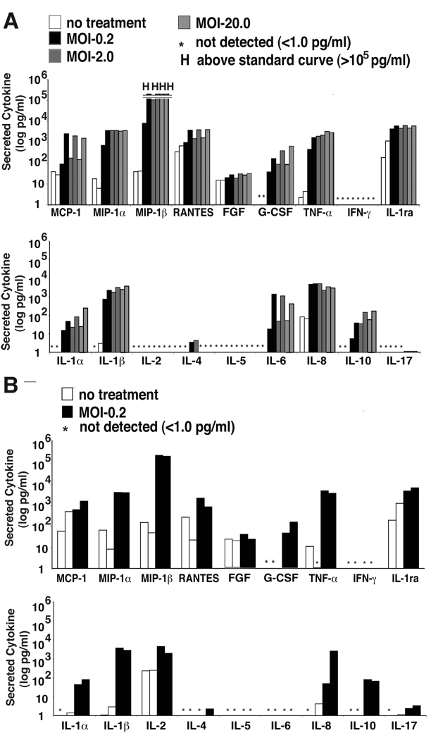
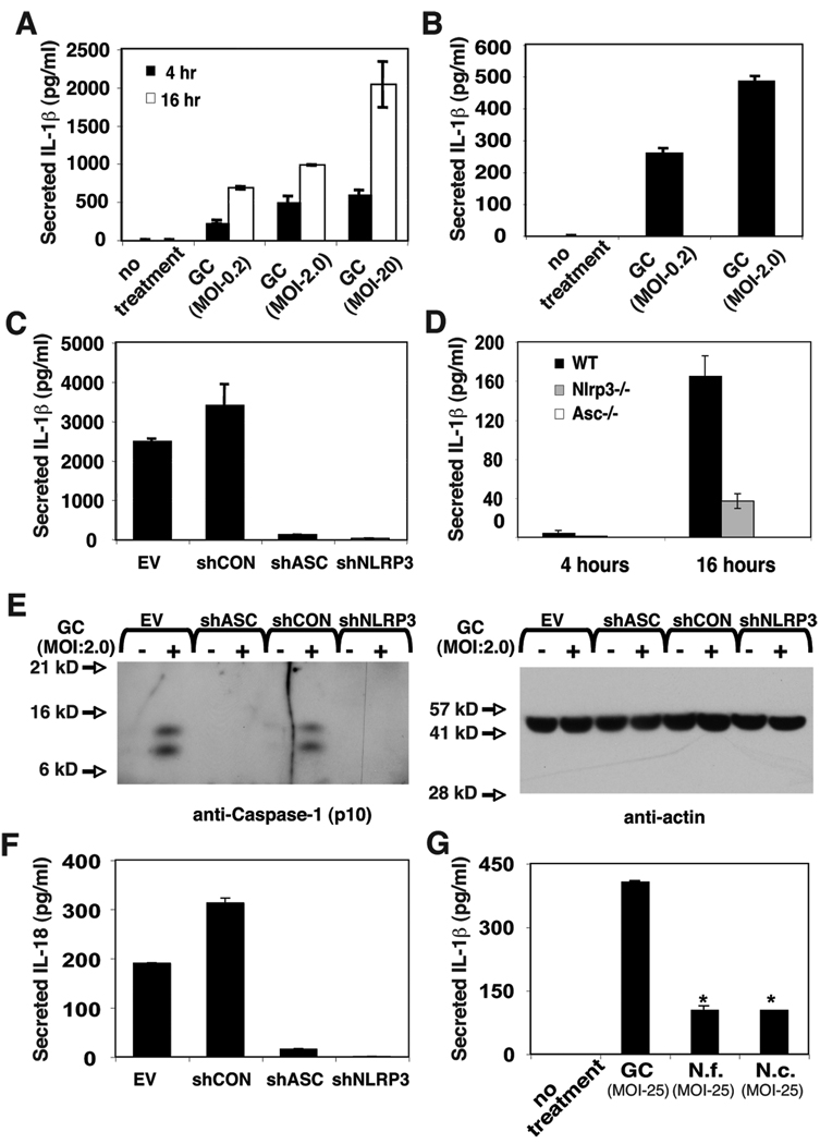
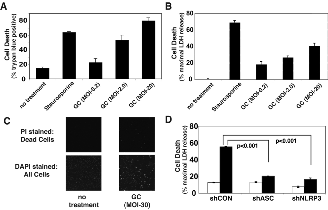
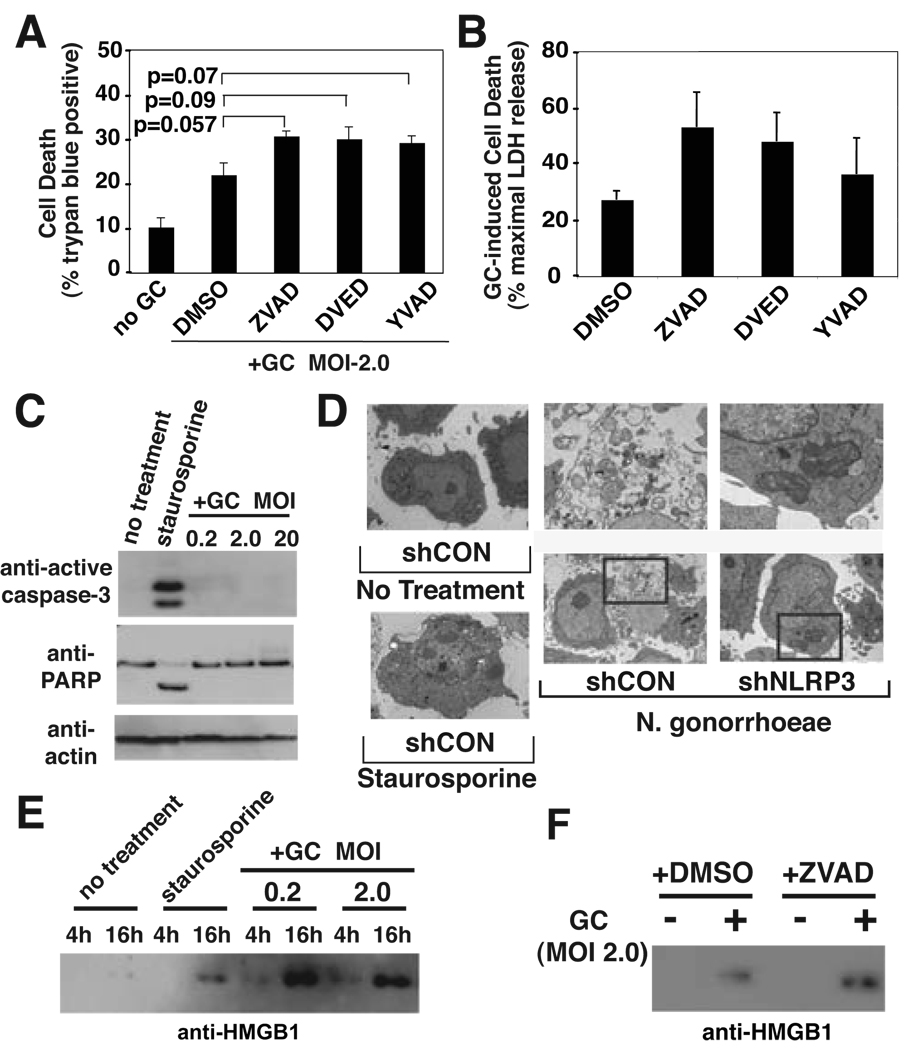
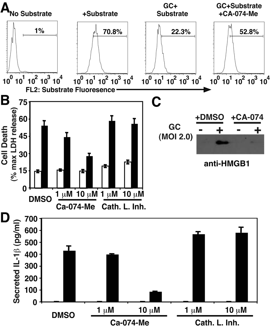
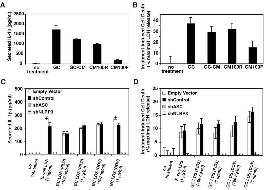
Neisseria gonorrhoeae is a common sexually transmitted pathogen that significantly impacts female fertility, neonatal health, and transmission of HIV worldwide. N. gonorrhoeae usually causes localized inflammation of the urethra and cervix by inducing production of IL-1beta and other inflammatory cytokines. Several NLR (nucleotide-binding domain, leucine-rich repeat) proteins are implicated in the formation of pro-IL-1beta-processing complexes called inflammasomes in response to pathogens. We demonstrate that NLRP3 (cryopyrin, NALP3) is the primary NLR required for IL-1beta/IL-18 secretion in response to N. gonorrhoeae in monocytes. We also show that N. gonorrhoeae infection promotes NLRP3-dependent monocytic cell death via pyronecrosis, a recently described pathway with morphological features of necrosis, including release of the strong inflammatory mediator HMBG1. Additionally, N. gonorrhoeae activates the cysteine protease cathepsin B as measured by the breakdown of a cathepsin B substrate. Inhibition of cathepsin B shows that this protease is an apical controlling step in the downstream activities of NLRP3 including IL-1beta production, pyronecrosis, and HMGB1 release. Nonpathogenic Neisseria strains (Neisseria cinerea and Neisseria flavescens) do not activate NLRP3 as robustly as N. gonorrhoeae. Conditioned medium from N. gonorrhoeae contains factors capable of initiating the NLRP3-mediated signaling events. Isolated N. gonorrhoeae lipooligosaccharide, a known virulence factor from this bacterium that is elaborated from the bacterium in the form of outer membrane blebs, activates both NLRP3-induced IL-1beta secretion and pyronecrosis. Our findings indicate that activation of NLRP3-mediated inflammatory response pathways is an important venue associated with host response and pathogenesis of N. gonorrhoeae.


Figures

Figure 1
Figure 1. Infection with N. gonorrhoeae induces production of chemotactic and inflammatory cytokines from primary monocytes and monocyte derived cell lines
A) THP-1 cells at 1×10⁶ cell/ml were incubated with N. gonorrhoeae at the indicated MOI as noted in the experimental methods. At 4 hours, extracellular bacteria were killed by addition of gentamicin. Supernatants were collected and cytokine production quantitated using multiplexed cytokine bead arrays. Each of two bars at each dose of infection represents an independent experiment. B) Primary monocytes were isolated from human blood as described in the Materials and Methods. The cells were seeded in plates at a density of 1.0 ×10⁶ cells/ ml and were infected with gonococcus at an MOI of 0.2. The samples were processed and cytokines measured as described in A. The * indicates cytokine levels below 1 pg/ml and “H” indicates cytokine levels were greater than the highest standard measured, 150,000 pg/ml.
Figure 2
Figure 2. Infection with N. gonorrhoeae induces caspase-1-dependent cytokine production by THP-1 cells and PBMC-derived monocytes
A) THP-1 cells at 1×10⁶ cell/ml were incubated with gonococci (GC) at the indicated MOI as noted in the experimental methods. At 4 hours, extracellular bacteria were killed by addition of gentamicin. Supernatants were collected after 4 hours (black bars) and 20 hours (white bars) and secreted IL-1β was measured by ELISA. B) Primary monocytes were isolated from PBMC by adherence. The cells were seeded at 1×10⁶ cell/ml and incubated with GC at the indicated MOI. At 4 hours, extracellular bacteria were killed by addition of gentamicin and supernatants were collected. C) THP-1-derived cell lines stabily transduced with shRNA expressing retrovirus were infected with GC at MOI of 2.0 and IL-1β production determined at 4 hours as described in A. The shRNA’s are directed to knock down expression as follows: shCON - negative control (scrambled sequence with base content equal to shASC); shASC – shRNA directed against Apoptotic Speck Containing-protein; shNLRP3 – shRNA directed against NLRP3. D) Bone marrow derived macrophages were isolated from C57/B6 mice which were either wild type (WT) or bearing genetic knock out of the genes encoding NLRP3 (Nlrp3−/−) or ASC (Asc −/−), cultured and infected with GC (MOI-0.2) as described in the materials and methods. At the indicated time points, culture supernatant was removed and assayed for the presence of IL-1β using ELISA. E) Immunoblot analysis for activated caspase-1 (P10 subunit) and control protein, actin, was performed on protein extracts from cellular infections described in (C) as described in the materials and methods. F) shRNA-expressing THP-1 cells were infected with GC (MOI-2.0) as described in (C) and secreted IL-18 was measured using ELISA. G) THP-1 cells were infected with pathogenic and commensal Neisseria species at the indicated MOI as described in A; N. gonorrhoeae (GC), Neisseria flavescens (N.f.), or Neisseria cinerea (N.c.). Secreted IL-1β was measured using ELISA. Experiments were performed in triplicate and results from representative experiments are shown. Error bars represent the standard error of the mean for duplicate or triplicate measurements of IL-1β.
Figure 3
Figure 3. N. gonorrhoeae-induces NLRP3-dependent cell death in THP-1 cells
A) THP-1 cells were infected with GC at the indicated MOI as described in Figure 2 or treated with staurosporine as a positive control for cell death. After 4 hours the cells were stained with trypan blue and the percentage of viable cells counted using the Nexcelom Cellometer Auto T4. B) Culture supernatants from infected cells were assayed for LDH released from injured or dead cells using a fluorometric assay. Levels of LDH above the background of LDH present in untreated cell culture supernatants are reported as a percent of the maximal LDH activity detected after detergent lysis. C) THP-1 cells were uninfected or infected with GC at an MOI of 0.2 for 20 hours. The cells with compromised membrane integrity were stained with the membrane impermeant dye, propidium iodide (left panels). All cells were subsequently stained with Hoechst 33342 to indicate total cell population (right panels). D) LDH released from cell lines expressing shRNA targeting the inflammasome components ASC or NLRP3 after a 4 hour exposure to GC at MOI of 2.0 was assayed. Results shown are representative of at least 3 independent experiments. Error bars are standard error of the mean for duplicate or triplicate measurements of cell death.
Figure 4
Figure 4. N. gonorrhoeae-induced cell death uses a necrotic mechanism
A) and B) THP-1 cells were pretreated with vehicle (DMSO), 20 µM pan-caspase inhibitor (AC-VAD-CHO), 20 µM caspase-3 inhibitor (AC-DVED-CHO), or 20 µM caspase-1 inhibitor (AC-YVAD-CHO) for 30 min, then infected with GC at MOI of 2.0 for 4 hours. Cell death was assayed by Trypan Blue exclusion (A) or LDH release (B). Error bars represent standard deviation of triplicate measurements. Representative experiments of at least three independent infections are shown. C) THP-1 cells were treated for 4 hours with either staurosporine or GC at the indicated MOI. Lysates from these cells were analyzed by SDS-PAGE and immunoblot directed against active caspase-3, PARP, or actin. D) THP-1 derived cell-lines expressing either a control hairpin RNA (shCON) or a hairpin RNA directed towards NLRP3 (shNLRP3) were untreated, treated with staurosporine, or infected with GC at an MOI of 2.0 as indicated. The cells were processed and examined by transmission electron microscopy as described in the Materials and Methods. Normal cellular morphology is demonstrated in the untreated cells (upper left panel), apoptotic morphology is demonstrated by a staurosporine-treated cell (lower left panel), a representative intact dying control cell with associated bacteria is shown in the lower middle panel and a NLRP3 knockdown cell with a large burden of internalized N. gonorrhoeae and no morphologic features of cell death is shown in the lower right panel. The upper middle and upper right panels show bacteria associated with these cells. In panel E and F, N. gonorrhoeae-induced HMGB1 release by THP-1 cells was analyzed by SDS-PAGE of the cell free culture supernatants and immunoblot against HMGB1. In E, Cells were treated for the indicated time period with either staurosporine or the indicated MOI of GC. In F, cells were treated with vehicle (DMSO) or pan caspase inhibitor as described in A and B, followed by exposure to GC for 4 hours at an MOI of 2.
Figure 5
Figure 5. N. gonorrhoeae-mediated Cathepsin B activation is required to mediate NLRP3-dependent IL-1β secretion and cell death
Cathepsin B activity was measured via degradation of a fluorescent substrate, Magic Red™ Cathepsin B substrate (Immunochemistry Technologie). Magic Red™ Cathepsin B substrate was added to THP-1 cells treated or not treated with GC. A cohort of GC-treated cells were also treated with 10 µM Ca-074-me, a specific inhibitor of Cathepsin B. Degradation of the Magic Red substrate was measured via Flow Cyometry on a BD FACSCalibur. Analysis of the data was accomplished with FloJo software with detection of the optimal Magic Red Substrate fluorescence emission in FL-2 on a logarithmic scale. A) Representative FACS plot of THP-1 cells with various treatment conditions. The percentage of Magic Red+ cells is indicated for each plot. A number in the third panel indicates increased Cathepsin B activity upon GC infection. B) THP-1 cells were treated with inhibitors of Cathepsin B, Cathepsin L, or DMSO vehicle at the indicated concentration for 15 min prior to infection with GC at MOI of 2.0. Cell death was assessed after 4 hours using release of LDH into the culture media as noted in the materials and methods.. C) THP-1 cells were pretreated with vehicle (DMSO) or 10 µM Cathepsin B inhibitor prior to infection with N. gonorrhoeae at MOI of 2.0. The cell culture supernatant from each condition was analyzed by SDS-PAGE and immunoblot with antibody directed to HMGB1. D) Cell culture supernatants from cells described in (B) were assayed for IL-1β using ELISA. In B and D, open bars indicate cells not treated with N. gonorrhoeae and closed bars indicate cells treated with N. gonorrhoeae. Error bars represent standard deviation. Experiments were performed in triplicate.
Figure 6
Figure 6. N. gonorrhoeae LOS induces NLRP3-dependent IL-1β secretion and pyronecrosis in THP-1 cells
A) and B) N. gonorrhoeae -conditioned media (GC-CM), N. gonorrhoeae-conditioned media 100kD retentate (CM100R) and filtrate (CM100F) were prepared as described in the materials and methods. THP-1 cells were incubated with live N. gonorrhoeae (GC) at an MOI of 1 or conditioned media preparations from and equivalent amount of bacteria for 4 hours and secreted IL-1β and cell death were assessed as previousl described. C) and D) THP-1-derived cell lines stably transduced with shRNA expressing retrovirus (described in Figure 2) were incubated with the indicated concentration of E. coli derived LPS or purified gonococcal LOS from strains PID2 or DOV (15, 35). After 4 hours, cell death was determined by measurement of LDH release into the media and secreted IL-1β was determined by ELISA. The shRNA’s are directed to knock down expression as follows: shCON - negative control (scrambled sequence with base content equal to shASC); shASC – shRNA directed against Apoptotic Speck Containing-protein; shNLRP3 – shRNA directed against NLRP3. LDH release and IL-1β secretion into the media that was not detectable is indicated by an asterisk (*). Representative experiments (of three) are shown, the bars indicate mean values of triplicate measurements with error bars representing the SEM.

References

    1. Tapsall JW. Antibiotic resistance in Neisseria gonorrhoeae. Clin Infect Dis. 2005;41 Suppl 4:S263–S268. - PubMed
    1. Fox KK, Thomas JC, Weiner DH, Davis RH, Sparling PF, Cohen MS. Longitudinal evaluation of serovar-specific immunity to Neisseria gonorrhoeae. Am J Epidemiol. 1999;149:353–358. - PubMed
    1. Pantelic M, Kim YJ, Bolland S, Chen I, Shively J, Chen T. Neisseria gonorrhoeae kills carcinoembryonic antigen-related cellular adhesion molecule 1 (CD66a)-expressing human B cells and inhibits antibody production. Infect Immun. 2005;73:4171–4179. - PMC - PubMed
    1. Boulton IC, Gray-Owen SD. Neisserial binding to CEACAM1 arrests the activation and proliferation of CD4+ T lymphocytes. Nat Immunol. 2002;3:229–236. - PubMed
    1. Ramsey KH, Schneider H, Kuschner RA, Trofa AF, Cross AS, Deal CD. Inflammatory cytokine response to experimental human infection with Neisseria gonorrhoeae. Ann N Y Acad Sci. 1994;730:322–325. - PubMed

Publication types

MeSH terms