

Nitrosative stress and inducible nitric oxide synthase expression in periventricular leukomalacia
- PMID: 19415311
- PMCID: PMC2909016
- DOI: 10.1007/s00401-009-0540-1
Nitrosative stress and inducible nitric oxide synthase expression in periventricular leukomalacia
Abstract
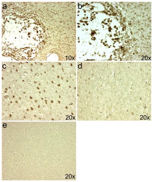
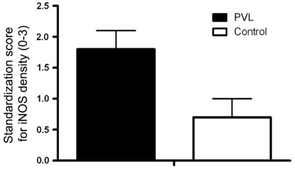
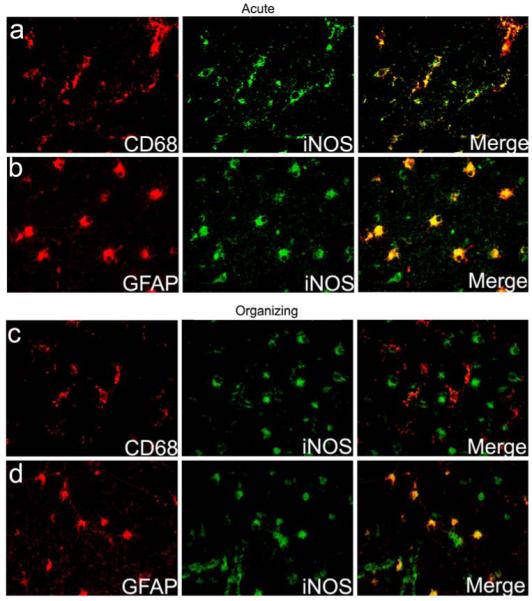
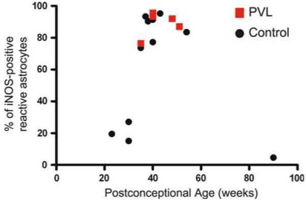
Periventricular leukomalacia (PVL) is a lesion of the immature cerebral white matter in the perinatal period and associated predominantly with prematurity and cerebral ischemia/reperfusion as well as inflammation due to maternofetal infection. It consists of focal necrosis in the periventricular region and diffuse gliosis with microglial activation and premyelinating oligodendrocyte (pre-OL) injury in the surrounding white matter. We previously showed nitrotyrosine in pre-OLs in PVL, suggesting involvement of nitrosative stress in this disorder. Here we hypothesize that inducible nitric oxide synthase (iNOS) expression is increased in PVL relative to controls. Using immunocytochemistry in human archival tissue, the density of iNOS-expressing cells was determined in the cerebral white matter of 15 PVL cases [29-51 postconceptional (PC) weeks] and 16 control cases (20-144 PC weeks). Using a standardization score of 0-3, the density of iNOS-positive cells was significantly increased in the diffuse component of PVL (score of 1.8 +/- 0.3) cases compared to controls (score of 0.7 +/- 0.3) (P = 0.01). Intense iNOS expression occurred in reactive astrocytes in acute through chronic stages and in activated microglia primarily in the acute stage, suggesting an early role for microglial iNOS in PVL's pathogenesis. This study supports an important role for iNOS-induced nitrosative stress in the reactive/inflammatory component of PVL.
Figures
Similar articles
-
12/15-lipoxygenase expression is increased in oligodendrocytes and microglia of periventricular leukomalacia.Dev Neurosci. 2013;35(2-3):140-54. doi: 10.1159/000350230. Epub 2013 Apr 20. Dev Neurosci. 2013. PMID: 23838566 Free PMC article.
-
Nitrosative and oxidative injury to premyelinating oligodendrocytes in periventricular leukomalacia.J Neuropathol Exp Neurol. 2003 May;62(5):441-50. doi: 10.1093/jnen/62.5.441. J Neuropathol Exp Neurol. 2003. PMID: 12769184
-
Interferon-gamma expression in periventricular leukomalacia in the human brain.Brain Pathol. 2004 Jul;14(3):265-74. doi: 10.1111/j.1750-3639.2004.tb00063.x. Brain Pathol. 2004. PMID: 15446581 Free PMC article.
-
Oxidative and nitrative injury in periventricular leukomalacia: a review.Brain Pathol. 2005 Jul;15(3):225-33. doi: 10.1111/j.1750-3639.2005.tb00525.x. Brain Pathol. 2005. PMID: 16196389 Free PMC article. Review.
-
Periventricular leukomalacia: overview and recent findings.Pediatr Dev Pathol. 2006 Jan-Feb;9(1):3-13. doi: 10.2350/06-01-0024.1. Epub 2006 Apr 4. Pediatr Dev Pathol. 2006. PMID: 16808630 Review.
Cited by
-
Oligodendroglial alterations and the role of microglia in white matter injury: relevance to schizophrenia.Dev Neurosci. 2013;35(2-3):102-29. doi: 10.1159/000346157. Epub 2013 Feb 27. Dev Neurosci. 2013. PMID: 23446060 Free PMC article.
-
Modeling the encephalopathy of prematurity in animals: the important role of translational research.Neurol Res Int. 2012;2012:295389. doi: 10.1155/2012/295389. Epub 2012 May 23. Neurol Res Int. 2012. PMID: 22685653 Free PMC article.
-
Infection-induced inflammation and cerebral injury in preterm infants.Lancet Infect Dis. 2014 Aug;14(8):751-762. doi: 10.1016/S1473-3099(14)70710-8. Epub 2014 May 28. Lancet Infect Dis. 2014. PMID: 24877996 Free PMC article. Review.
-
Current therapeutic strategies to mitigate the eNOS dysfunction in ischaemic stroke.Cell Mol Neurobiol. 2012 Apr;32(3):319-36. doi: 10.1007/s10571-011-9777-z. Epub 2011 Dec 25. Cell Mol Neurobiol. 2012. PMID: 22198555 Free PMC article. Review.
-
12/15-lipoxygenase expression is increased in oligodendrocytes and microglia of periventricular leukomalacia.Dev Neurosci. 2013;35(2-3):140-54. doi: 10.1159/000350230. Epub 2013 Apr 20. Dev Neurosci. 2013. PMID: 23838566 Free PMC article.
References
-
- Askalan R, Deveber G, Ho M, Ma J, Hawkins C. Astrocytic-inducible nitric oxide synthase in the ischemic developing human brain. Pediatr Res. 2006;60:687–692. doi:10.1203/01.pdr.0000246226.89215.a6. - PubMed
-
- Back SA, Luo NL, Mallinson RA, et al. Selective vulnerability of preterm white matter to oxidative damage defined by F2-isoprostanes. Ann Neurol. 2005;58:108–120. doi:10.1002/ana.20530. - PubMed
-
- Brown GC. Mechanisms of inflammatory neurodegeneration: iNOS and NADPH oxidase. Biochem Soc Trans. 2007;35:1119–1121. doi:10.1042/BST0351166. - PubMed
-
- Contestabile A. Roles of NMDA receptor activity and nitric oxide production in brain development. Brain Res Brain Res Rev. 2000;32:476–509. doi:10.1016/S0165-0173(00)00018-7. - PubMed
Publication types
MeSH terms
Substances
Grants and funding
LinkOut - more resources
Full Text Sources
