

Linking whole-slide microscope images with DICOM by using JPEG2000 interactive protocol
- PMID: 19415383
- PMCID: PMC2896636
- DOI: 10.1007/s10278-009-9200-1
Linking whole-slide microscope images with DICOM by using JPEG2000 interactive protocol
Abstract
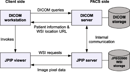
The use of digitized histopathologic specimens (also known as whole-slide images (WSIs)) in clinical medicine requires compatibility with the Digital Imaging and Communications in Medicine (DICOM) standard. Unfortunately, WSIs usually exceed DICOM image object size limit, making it impossible to store and exchange them in a straightforward way. Moreover, transmitting the entire DICOM image for viewing is ineffective for WSIs. With the JPEG2000 Interactive Protocol (JPIP), WSIs can be linked with DICOM by transmitting image data over an auxiliary connection, apart from patient data. In this study, we explored the feasibility of using JPIP to link JPEG2000 WSIs with a DICOM-based Picture Archiving and Communications System (PACS). We first modified an open-source DICOM library by adding support for JPIP as described in the existing DICOM Supplement 106. Second, the modified library was used as a basis for a software package (JVSdicom), which provides a proof-of-concept for a DICOM client-server system that can transmit patient data, conventional DICOM imagery (e.g., radiological), and JPIP-linked JPEG2000 WSIs. The software package consists of a compression application (JVSdicom Compressor) for producing DICOM-compatible JPEG2000 WSIs, a DICOM PACS server application (JVSdicom Server), and a DICOM PACS client application (JVSdicom Workstation). JVSdicom is available for free from our Web site ( http://jvsmicroscope.uta.fi/ ), which also features a public JVSdicom Server, containing example X-ray images and histopathology WSIs of breast cancer cases. The software developed indicates that JPEG2000 and JPIP provide a well-working solution for linking WSIs with DICOM, requiring only minor modifications to current DICOM standard specification.
Figures


References
-
- Digital Imaging and Communications in Medicine (DICOM). Available at ftp://medical.nema.org/dicom/2008/. Accessed 9 October 2008
Publication types
MeSH terms
LinkOut - more resources
Full Text Sources
Other Literature Sources
