

Local controlled delivery of anti-neoplastic RNAse to the brain
- PMID: 19415468
- PMCID: PMC3085080
- DOI: 10.1007/s11095-009-9893-3
Local controlled delivery of anti-neoplastic RNAse to the brain
Abstract
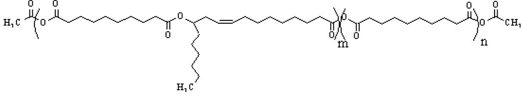
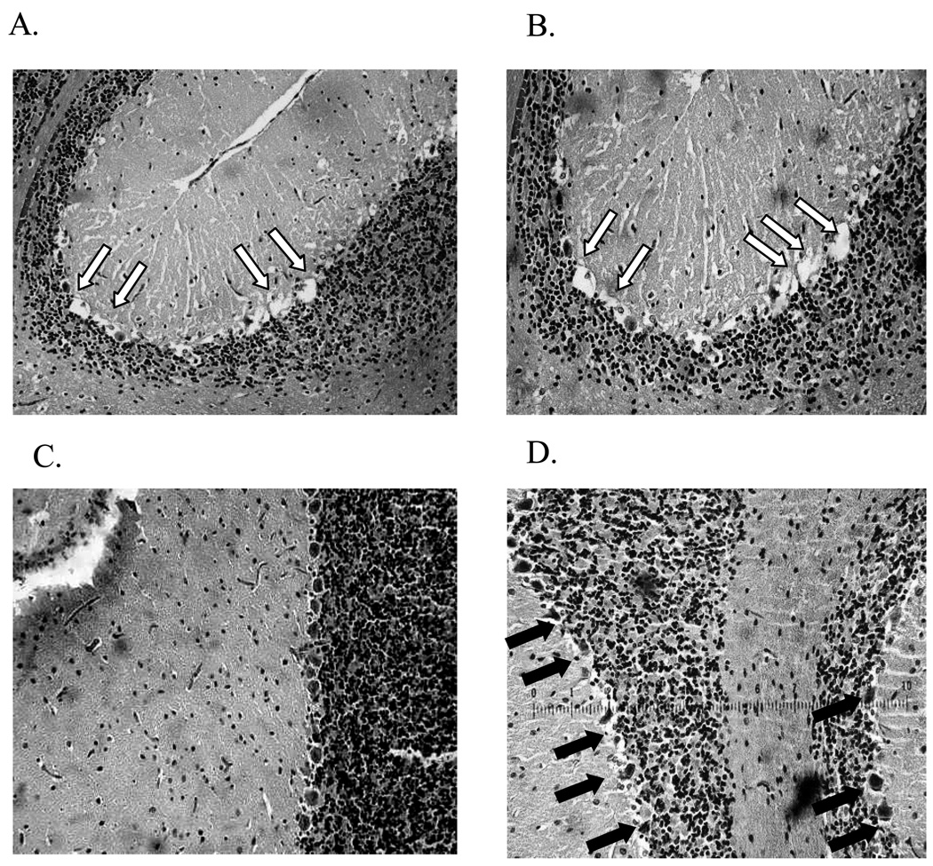
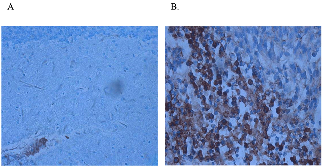
Purpose: Antineoplastic RNAse proteins, also known as Amphibinases, have been shown effective against various solid tumors but were found selectively neurotoxic to Purkinje cells in the cerebellum. This work describes the use of a waxy biodegradable poly(ricinoleic-co-sebacic acid) for the local controlled delivery of cytotoxic amphibinases in the parietal lobe of the brain in an attempt to overcome cerebellar neuronal toxicity while affecting glioma cells.
Methods: Amphibinase analogues were encapsulated in poly(ricinoleic-co-sebacic acid) formulations using mix-melt technology and loaded onto surgical foam. In-vitro release was monitored by BCA colorimetry and by RNAse specific bioactivity. The implants were inserted into rat brains bearing 9L glioma to assess toxicity and efficacy.
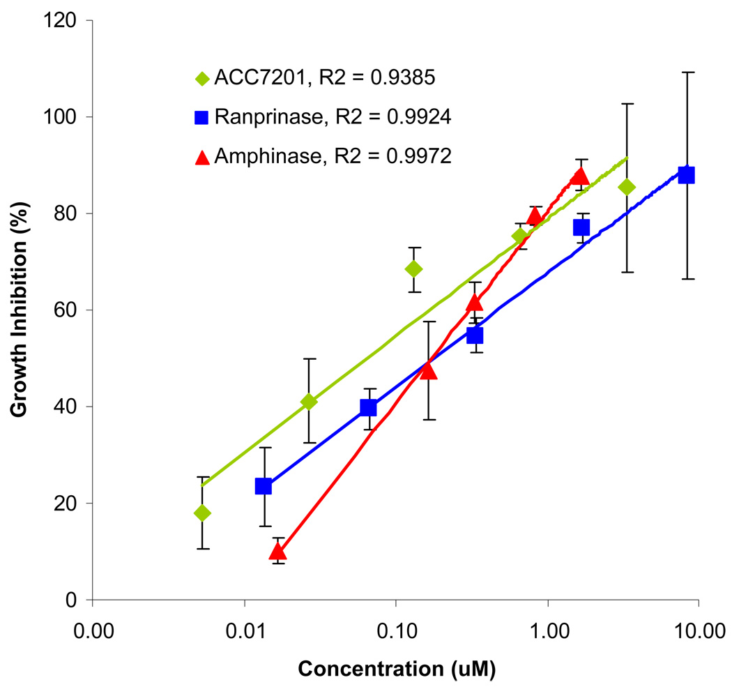
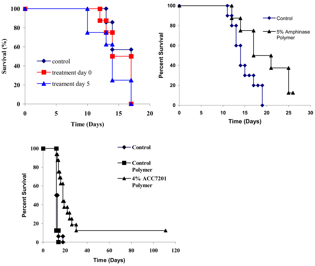
Results: The various formulations showed extended linear release for several weeks with minimal burst effect. Best in-vivo efficacy was obtained with ACC7201 containing implants, resulting in the extension of the median survival from 13 to 18 days with 13% long-term survivors.
Conclusion: Antineoplastic proteins were released from a p(SA-RA) polyanhydride implants in a controlled manner, providing efficacy against 9L glioma, while evading neurotoxicity in the cerebellum. The controlled release of Amphibinases forms the potential for a new therapy against brain tumors.
Figures
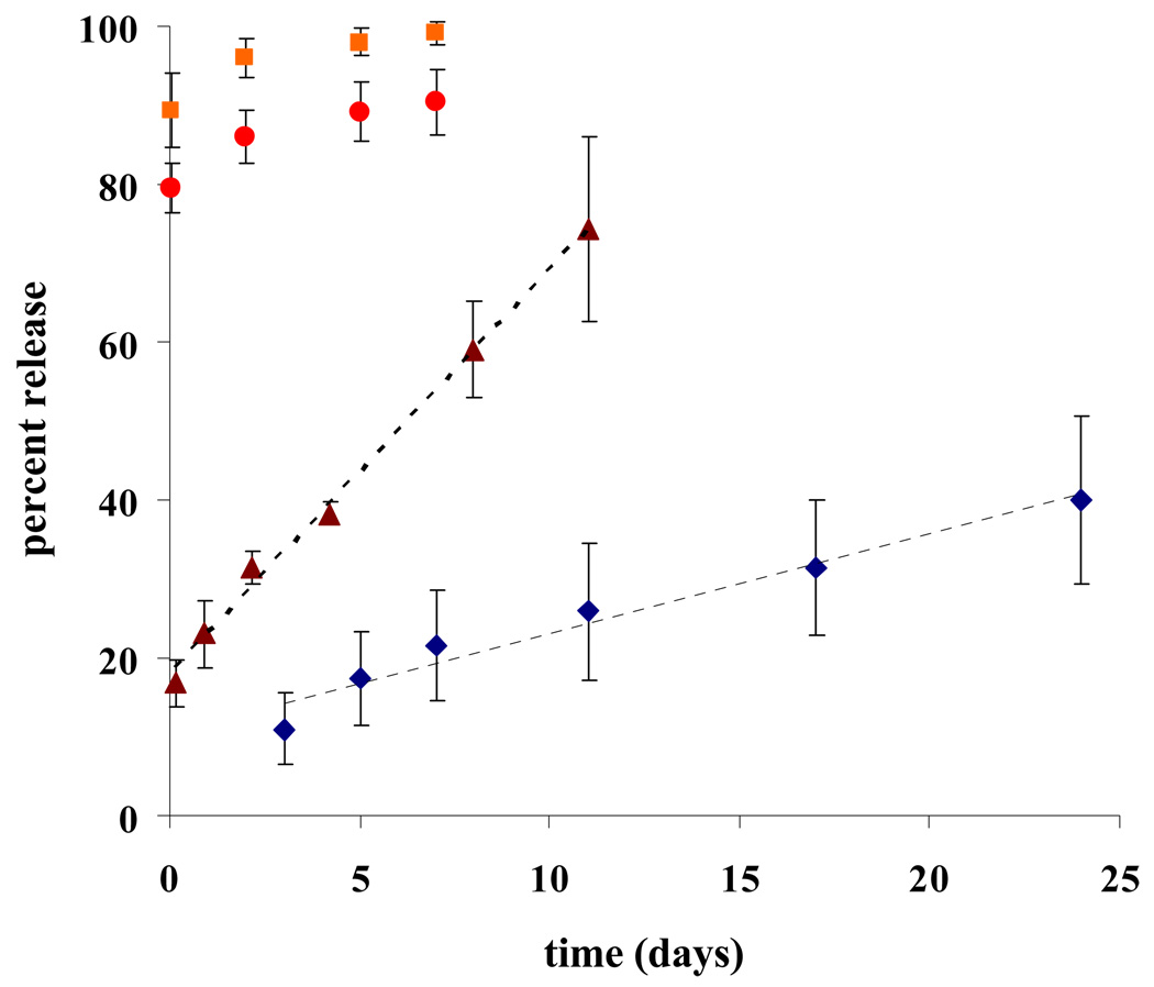
 2% Ranpirnase w/w polymer;
  Amphinase in ratio 4%,
  10%, and
  20% w/w polymer.
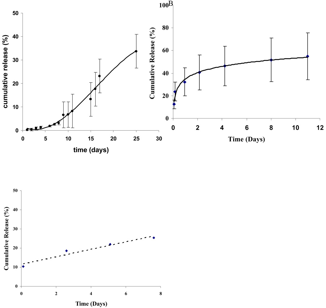
Similar articles
-
Lactacystin exhibits potent anti-tumor activity in an animal model of malignant glioma when administered via controlled-release polymers.J Neurooncol. 2006 May;77(3):225-32. doi: 10.1007/s11060-005-6937-3. J Neurooncol. 2006. PMID: 16609837 Free PMC article.
-
Effectiveness of controlled release of a cyclophosphamide derivative with polymers against rat gliomas.J Neurosurg. 1995 Mar;82(3):481-6. doi: 10.3171/jns.1995.82.3.0481. J Neurosurg. 1995. PMID: 7861228
-
Interstitial delivery of carboplatin via biodegradable polymers is effective against experimental glioma in the rat.Cancer Chemother Pharmacol. 1996;39(1-2):90-6. doi: 10.1007/s002800050542. Cancer Chemother Pharmacol. 1996. PMID: 8995504
-
Pharmacokinetics of the carmustine implant.Clin Pharmacokinet. 2002;41(6):403-19. doi: 10.2165/00003088-200241060-00002. Clin Pharmacokinet. 2002. PMID: 12074689 Review.
-
Blood flow and blood-to-tissue transport in 9L gliosarcomas: the role of the brain tumor model in drug delivery research.J Neurooncol. 1991 Dec;11(3):185-97. doi: 10.1007/BF00165526. J Neurooncol. 1991. PMID: 1823340 Review.
Cited by
-
Biohybrid polymer-antimicrobial peptide medium against Enterococcus faecalis.PLoS One. 2014 Oct 3;9(10):e109413. doi: 10.1371/journal.pone.0109413. eCollection 2014. PLoS One. 2014. PMID: 25279943 Free PMC article.
References
-
- Lesniak MS, Brem H. Targeted Therapy for Brain Tumours. Nat Rev Drug Discov. 2004;3:499–508. - PubMed
-
- Stupp R, Mason WP, van den Bent MJ, Weller M, Fisher B, Taphoorn MJ, Belanger K, Brandes AA, Marosi C, Bogdahn U, Curschmann J, Janzer RC, Ludwin SK, Gorlia T, Allgeier A, Lacombe D, Cairncross JG, Eisenhauer E, Mirimanoff RO. Radiotherapy plus concomitant and adjuvant temozolomide for glioblastoma. N Engl J Med. 2005;352(10):987–996. - PubMed
-
- Attenello FJ, Mukherjee D, Datoo G, McGirt MJ, Bohan E, Weingart JD, Olivi A, Quinones-Hinojosa A, Brem H. Use of Gliadel (BCNU) wafer in the surgical treatment of malignant glioma: a 10-year institutional experience. Ann Surg Oncol. 2008;15(10):2887–2893. - PubMed
-
- Lesniak MS. Novel advances in drug delivery to brain cancer. Technol Cancer Res Treat. 2005;4:417–428. - PubMed
-
- DiMeco F, Li KW, Tyler BM, Wolf AS, Brem H, Olivi A. Local Delivery of Mitoxantrone for the Treatment of Malignant Brain Tumors in Rats. Neurosurgery. 2002;97:1173–1178. - PubMed
Publication types
MeSH terms
Substances
Grants and funding
LinkOut - more resources
Full Text Sources
Other Literature Sources
Medical
