. 2009 May;27(5):1006-20.
doi: 10.1002/stem.30.

Identification of gastric cancer stem cells using the cell surface marker CD44

Affiliations

Identification of gastric cancer stem cells using the cell surface marker CD44

Shigeo Takaishi et al. Stem Cells. 2009 May.

Abstract

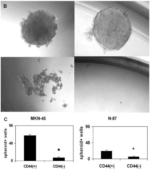
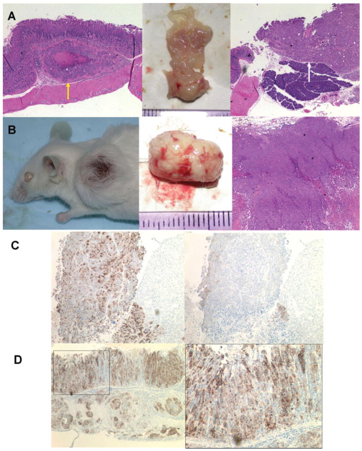
Cancer stem cells (CSCs) have been defined as a unique subpopulation in tumors that possess the ability to initiate tumor growth and sustain tumor self-renewal. Although the evidence has been provided to support the existence of CSCs in various solid tumors, the identity of gastric CSCs has not been reported. In this study, we have identified gastric cancer-initiating cells from a panel of human gastric cancer cell lines using cell surface marker CD44. Among six gastric cancer cell lines, three lines MKN-45, MKN-74, and NCI-N87 had a sizeable subpopulation of CD44(+) cells, and these cells showed spheroid colony formation in serum-free media in vitro as well as tumorigenic ability when injected into stomach and skin of severe combined immunodeficient (SCID) mice in vivo. The CD44(+) gastric cancer cells showed the stem cell properties of self-renewal and the ability to form differentiated progeny and gave rise to CD44(-) cells. CD44 knockdown by short hairpin RNA resulted in much reduced spheroid colony formation and smaller tumor production in SCID mice, and the CD44(-) populations had significantly reduced tumorigenic ability in vitro and in vivo. Other potential CSC markers, such as CD24, CD133, CD166, stage-specific embryonic antigen-1 (SSEA-1), and SSEA-4, or sorting for side population did not show any correlation with tumorigenicity in vitro or in vivo. The CD44(+) gastric cancer cells showed increased resistance for chemotherapy- or radiation-induced cell death. These results support the existence of gastric CSCs and may provide novel approaches to the diagnosis and treatment of gastric cancer.


Conflict of interest statement

Disclosure of Potential Conflicts of Interest The authors indicate no potential conflicts of interest.

Figures

Figure 1
Figure 1
Analysis of cell surface markers and spheroid colony formation assay. (A): Fluorescence-activated cell sorter (FACS) analysis of candidate surface markers for gastric cancer stem cells CD44 and CD24. Top left: Analysis of cell surface markers and spheroid colony formation assay. MKN-45; top right: MKN-74; bottom left: N-87; bottom right: MKN-28; horizontal axis: FITC-CD44; vertical axis: PE-CD24. (B): Spheroid colony formation of gastric cancer cell lines in serum-free media. Each human gastric cancer cell line was fractionated and inoculated by FACS sorting for CD44 positivity in ultra-low-attachment 96-well plates with serum-free medium (10 cells per plate) containing human epidermal growth factor (20 ng/ml) and basic fibroblast growth factor (10 ng/ml). After 3–4 weeks of culture, the number of spheroid colony-positive wells was counted. Top left: MKN-45; top right: N-87; bottom left: AGS; bottom right: KATO-III. MKN-45 and N-87 cells produced spheroid colonies, while AGS and KATO-III cells did not. (C): Spheroid colony-positive well numbers. Left: MKN-45; right: N-87. CD44-positive cells produced significantly higher numbers of spheroid colonies in low-attachment 96-well cell plates. *, p < .01. Abbreviations: FITC, fluorescein isothiocyanate; PE, phycoerythrin.
Figure 1
Figure 1
Analysis of cell surface markers and spheroid colony formation assay. (A): Fluorescence-activated cell sorter (FACS) analysis of candidate surface markers for gastric cancer stem cells CD44 and CD24. Top left: Analysis of cell surface markers and spheroid colony formation assay. MKN-45; top right: MKN-74; bottom left: N-87; bottom right: MKN-28; horizontal axis: FITC-CD44; vertical axis: PE-CD24. (B): Spheroid colony formation of gastric cancer cell lines in serum-free media. Each human gastric cancer cell line was fractionated and inoculated by FACS sorting for CD44 positivity in ultra-low-attachment 96-well plates with serum-free medium (10 cells per plate) containing human epidermal growth factor (20 ng/ml) and basic fibroblast growth factor (10 ng/ml). After 3–4 weeks of culture, the number of spheroid colony-positive wells was counted. Top left: MKN-45; top right: N-87; bottom left: AGS; bottom right: KATO-III. MKN-45 and N-87 cells produced spheroid colonies, while AGS and KATO-III cells did not. (C): Spheroid colony-positive well numbers. Left: MKN-45; right: N-87. CD44-positive cells produced significantly higher numbers of spheroid colonies in low-attachment 96-well cell plates. *, p < .01. Abbreviations: FITC, fluorescein isothiocyanate; PE, phycoerythrin.
Figure 2
Figure 2
Tumor formation of tumorigenic gastric cancer cell lines implanted in the stomach and skin of SCID mice. (A, B): CD44-positive fraction of MKN-74 cells implanted in the stomach and skin of SCID mice (30,000 cells per site) produced tumors in each site after 8–12 weeks. Peritoneal dissemination was observed in the mice injected with CD44(+) cells into the stomach, and some of these mice also showed tumors invaded into pancreas or liver. (A): Stomach with tumor. Yellow arrow indicates tumor in submucosal area, and white arrow indicates tumor invasion into pancreas. (B): Skin tumor. H&E images: original magnification, ×40. (C): Immunohistochemistry of tumors produced by CD44-positive MKN-45 cells implanted in the skin of SCID mice; left: immunostaining with anti-human-specific epithelial membrane antigen (EMA) antibody; right: immunostaining with anti-human-specific CD44 antibody. CD44-positive cells in skin tumor were mainly detected at the marginal area of tumor. Most of the cells were CD44 negative. (D): Immunohistochemistry of tumors produced by CD44-positive MKN-45 cells implanted in the stomach of SCID mice; left and right: both immunostaining with anti-human-specific EMA antibody. CD44-positive MKN-45 cells produced not only submucosal tumor but also human gastric glands in the SCID mouse stomach. Left: original magnification, ×60; right: higher magnification image (original magnification, ×200) of the rectangular area in the left.
Figure 3
Figure 3
Chemoresistance and radioresistance of CD44-positive and -negative fractions of tumorigenic human gastric cell lines MKN-45 and MKN-74. (A): FACS-sorted CD44-positive and -negative MKN-74 cells in 12-well plates (10,000 cells per well) were treated with 5-FU (10 mM), etoposide (VP-16; 200 μM), or without drugs for 8 days (media with respective drug were changed to fresh one after 4 days), and then living cell numbers were counted using Cell Counting Kit-8. Y-axis data show optical absorbance of reagent WST-8 at a wavelength of 450 nm, which represent the number of survived cells. **, p < .01. (B): Survived cells treated with 5-FU after 8 days. Left: CD44 positive; right: CD44 negative. (C): FACS-sorted CD44-positive and -negative MKN-45 cells in six-well plates (50,000 cells per well) were irradiated with 3 or 6 Gy, respectively. The number of living cells was counted using Cell Counting Kit-8 after 6 days (medium with 10% FBS was changed to fresh one after 3 days). Y-axis data show optical absorbance of reagent WST-8 at a wavelength of 450 nm, which represent the number of survived cells. *, p < .05; **, p < .01. (D): Survived cells at 6 days after irradiation. Left: CD44 positive; right: CD44 negative. Abbreviation: 5-FU, 5-fluorouracil.
Figure 3
Figure 3
Chemoresistance and radioresistance of CD44-positive and -negative fractions of tumorigenic human gastric cell lines MKN-45 and MKN-74. (A): FACS-sorted CD44-positive and -negative MKN-74 cells in 12-well plates (10,000 cells per well) were treated with 5-FU (10 mM), etoposide (VP-16; 200 μM), or without drugs for 8 days (media with respective drug were changed to fresh one after 4 days), and then living cell numbers were counted using Cell Counting Kit-8. Y-axis data show optical absorbance of reagent WST-8 at a wavelength of 450 nm, which represent the number of survived cells. **, p < .01. (B): Survived cells treated with 5-FU after 8 days. Left: CD44 positive; right: CD44 negative. (C): FACS-sorted CD44-positive and -negative MKN-45 cells in six-well plates (50,000 cells per well) were irradiated with 3 or 6 Gy, respectively. The number of living cells was counted using Cell Counting Kit-8 after 6 days (medium with 10% FBS was changed to fresh one after 3 days). Y-axis data show optical absorbance of reagent WST-8 at a wavelength of 450 nm, which represent the number of survived cells. *, p < .05; **, p < .01. (D): Survived cells at 6 days after irradiation. Left: CD44 positive; right: CD44 negative. Abbreviation: 5-FU, 5-fluorouracil.
Figure 4
Figure 4
CD44 knockdown by lentivirus-mediated shRNA and the significant reduction of the tumorigenic ability of gastric cancer stem cells. (A): Fluorescence-activated cell sorter analysis of CD44 knockdown in MKN-45 cells by lentivirus-mediated human CD44-specific shRNA. Left: scramble shRNA as control; right: human CD44-specific shRNA. CD44 expression in MKN-45 cells was successfully knocked down. CD44 expression in MKN-74 cells was also downregulated to a similar level. (B): Spheroid colony formation of CD44 downregulated MKN-45 and MKN-74 cells. After 4 weeks of culture, CD44 shRNA-infected cells produced significantly lower number of colonies than scramble shRNA virus-infected cells. (C): Skin tumors produced by CD44 downregulated MKN-45 cells. CD44 shRNA-infected MKN-45 cells as well as scramble shRNA virus-infected cells were injected under the skin of SCID mice, respectively, and after 4 weeks, although all mice had tumors, CD44 shRNA virus-infected cells produced much smaller tumors than scramble shRNA virus-infected cells. CD44 downregulated MKN-74 cells also showed similar results. (D): Spheroid colony formation of high CD44-expressing and low CD44-expressing populations in MKN-45 and MKN-74 cells. After 4 weeks of culture, high CD44-expressing cells produced significantly larger number of colonies than low CD44-expressing cells. (E): Skin tumors produced by high CD44-expressing MKN-45 cells. High CD44-expressing as well as low CD44-expressing populations in MKN-45 cells were injected under the skin of SCID mice, respectively, and after 4 weeks, high CD44-expressing cells produced much larger tumors than low CD44-expressing cells. High CD44-expressing MKN-74 cells also showed similar results. Abbreviations: FITC, fluorescein isothiocyanate; PE, phycoerythrin; shRNA, short hairpin RNA.
Figure 4
Figure 4
CD44 knockdown by lentivirus-mediated shRNA and the significant reduction of the tumorigenic ability of gastric cancer stem cells. (A): Fluorescence-activated cell sorter analysis of CD44 knockdown in MKN-45 cells by lentivirus-mediated human CD44-specific shRNA. Left: scramble shRNA as control; right: human CD44-specific shRNA. CD44 expression in MKN-45 cells was successfully knocked down. CD44 expression in MKN-74 cells was also downregulated to a similar level. (B): Spheroid colony formation of CD44 downregulated MKN-45 and MKN-74 cells. After 4 weeks of culture, CD44 shRNA-infected cells produced significantly lower number of colonies than scramble shRNA virus-infected cells. (C): Skin tumors produced by CD44 downregulated MKN-45 cells. CD44 shRNA-infected MKN-45 cells as well as scramble shRNA virus-infected cells were injected under the skin of SCID mice, respectively, and after 4 weeks, although all mice had tumors, CD44 shRNA virus-infected cells produced much smaller tumors than scramble shRNA virus-infected cells. CD44 downregulated MKN-74 cells also showed similar results. (D): Spheroid colony formation of high CD44-expressing and low CD44-expressing populations in MKN-45 and MKN-74 cells. After 4 weeks of culture, high CD44-expressing cells produced significantly larger number of colonies than low CD44-expressing cells. (E): Skin tumors produced by high CD44-expressing MKN-45 cells. High CD44-expressing as well as low CD44-expressing populations in MKN-45 cells were injected under the skin of SCID mice, respectively, and after 4 weeks, high CD44-expressing cells produced much larger tumors than low CD44-expressing cells. High CD44-expressing MKN-74 cells also showed similar results. Abbreviations: FITC, fluorescein isothiocyanate; PE, phycoerythrin; shRNA, short hairpin RNA.
Figure 5
Figure 5
CD44 expression in mouse and human gastric cancer tissues. (A, B): Immunostaining with anti-CD44 antibody of hypergastrinemia (insulin-gastrin) mouse with Helicobacter felis infection (7 months postinfection). (C, D): Immunostaining with anti-CD44 antibody of human gastric adenocarcinoma (moderately differentiated type). Original magnification: (A), ×60; (B), ×200; (C), ×40; (D), ×200. Scale bar = (A, B), 200 μm; (C, D), 50 μm. Many gastric cancer cells, especially invasive front area, were strongly CD44 positive for both mouse and human gastric cancer tissues. In addition, some parts of immune cells as well as stromal cells were CD44 positive.

References

    1. Fox JG, Wang TC. Inflammation, atrophy, and gastric cancer. J Clin Invest. 2007;117:60–69. - PMC - PubMed
    1. Jemal A, Siegel R, Ward E, et al. Cancer statistics, 2008. CA Cancer J Clin. 2008;58:71–96. - PubMed
    1. Takaishi S, Okumura T, Wang TC. Gastric cancer stem cells. J Clin Oncol. 2008;26:2876–2882. - PMC - PubMed
    1. Houghton J, Morozov A, Smirnova I, et al. Stem cells and cancer. Semin Cancer Biol. 2007;17:191–203. - PubMed
    1. Lapidot T, Sirard C, Vormoor J, et al. A cell initiating human acute myeloid leukaemia after transplantation into SCID mice. Nature. 1994;367:645–648. - PubMed

Publication types