Comparative Study
. 2009 Jun 19;4(6):401-8.
doi: 10.1021/cb900082q.

Comparative assessment of large-scale proteomic studies of apoptotic proteolysis

Affiliations
Comparative Study

Comparative assessment of large-scale proteomic studies of apoptotic proteolysis

Gabriel M Simon et al. ACS Chem Biol. .

Abstract

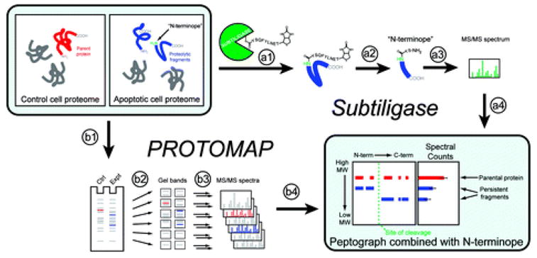
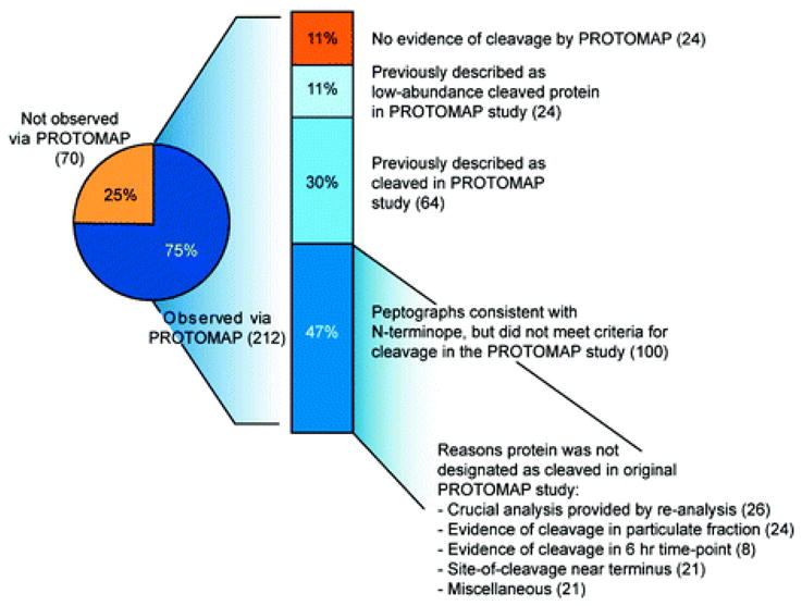
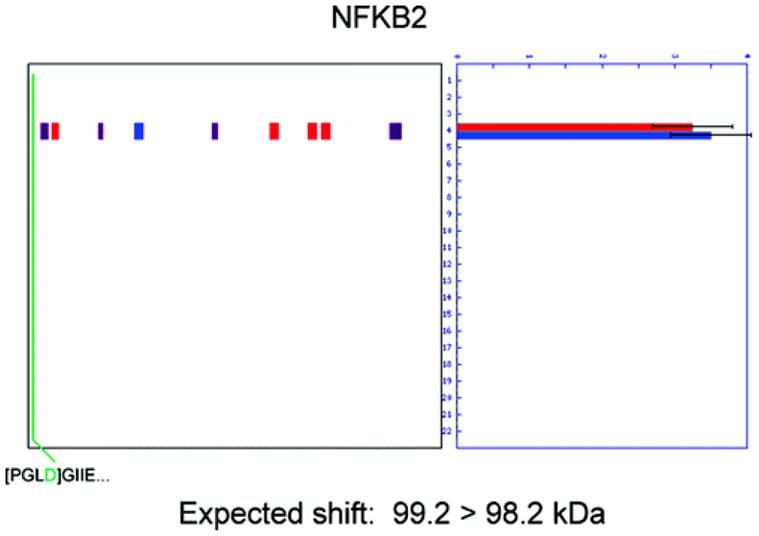
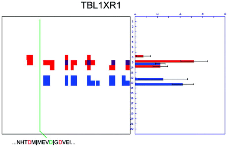
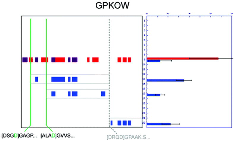
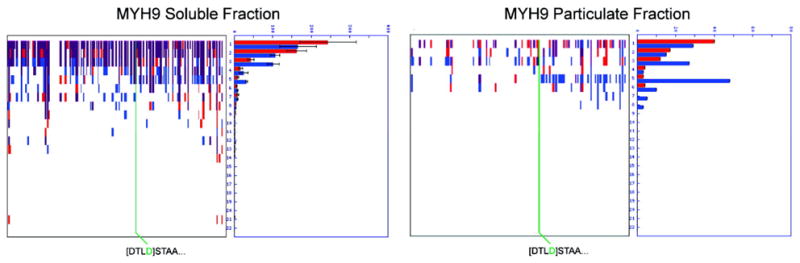
Two proteomic methods were recently introduced to globally map proteolytic cleavage events in biological systems, one that characterized proteolyzed proteins by differential gel migration (PROTOMAP) and the other by enzymatic tagging and enrichment of the nascent N-terminal peptides generated by proteolysis (Subtiligase). Both technologies were applied to apoptosis, and each uncovered hundreds of novel proteolytic events. An initial survey, however, revealed only minimal overlap in the two data sets. In this article, we perform an in-depth comparative analysis of the PROTOMAP and Subtiligase results that assimilates the complementary information acquired by each method. This analysis uncovered substantial agreement between the PROTOMAP and Subtiligase data sets, which in integrated form yield a highly enriched portrait of the proteome-wide impact of proteolysis in apoptosis. We discuss the respective strengths of each proteomic method and the potential for these technologies to expand the scope and sensitivity of large-scale studies of proteolysis in biological systems.


Figures

Figure 1
Figure 1
Overview of Subtiligase and PROTOMAP methods. Comparison of healthy and apoptotic cell proteomes was accomplished using two complementary techniques. The Subtiligase method utilizes an engineered enzyme (a1) “subtiligase” that covalently reacts with a custom biotinylated peptide ester containing a TEV protease cleavage site. Subtiligase then selectively transfers this biotinylated peptide to free amines on the N-termini of proteins. (a2) Proteins are digested with trypsin, and “N-terminopes”, peptides corresponding to the N-termini of proteins, are purified with avidin affinity chromatography and elution with TEV protease. N-Terminopes are then (a3) sequenced using LC-MS/MS, and internally located N-terminopes that are found in apoptotic but not control proteomes are considered to be direct evidence of proteolytic cleavage and can be (a4) combined with the topographical information provided by PROTOMAP data. The PROTOMAP approach begins with (b1) separation of control and apoptotic proteins in distinct lanes of a 1D SDS–PAGE gel. (b2) Each lane is then sliced into evenly sized bands and (b3) proteins in each band are in-gel digested with trypsin and sequenced via LC-MS/MS. The resulting (b4) peptides are bioinformatically integrated into a “peptograph”, which plots peptides from control and apoptotic samples in red and blue, respectively, according to their position in the primary sequence of each protein from left to right (N- to C-terminus) and their position in the gel, from top to bottom (high to low molecular weight), thereby revealing changes in gel migration and topography such as would be expected upon proteolysis. The combination of peptographs with N-terminopes provides a near-complete description of each proteolytic event.
Figure 2
Figure 2
Analysis of the overlap between the Subtiligase and PROTOMAP data sets. The pie chart shows that 75% of the proteins detected as cleaved with the Subtiligase approach generated interpretable peptographs in the PROTOMAP study. Of these 212 proteins, 88% showed patterns that were consistent with the cleavage event predicted by the N-terminope (see Supplementary Table 1 for a direct comparison). Eighty-eight proteins were included in one of the two supplemental tables from the original PROTOMAP study describing cleaved proteins, whereas an additional 100 proteins were not designated as “cleaved” in this study for a variety of reasons: many proteins only displayed evidence of cleavage in the particulate fraction or a later time point (24 and 8, respectively). Additionally, re-analysis of the data with advanced statistical techniques identified peptographs that provide clear evidence of cleavage for many (26) low-abundance proteins and persistent fragments. See Supplementary Table 1 for the complete integration of the Subtiligase and PROTOMAP data sets.
Figure 3
Figure 3
Evidence of cleavage for low-abundance proteins. RNA polymerase II-associated protein 3 (RPAP3) was detected with only 23 total spectral counts, below the original threshold of 30 spectral counts required to make confident assignments by PROTOMAP. However, at least three cleavage events are evident that are consistent with the two N-terminopes detected by the Subtiligase approach.
Figure 4
Figure 4
Evidence of cleavage found in the particulate fraction. B-Cell receptor-associated protein 31 (BAP31) was not included in the original PROTOMAP study because of insufficient abundance in the soluble fraction; however, inspection of the peptograph from the particulate fraction reveals clear evidence of cleavage that is consistent with the N-terminope detected in the Subtiligase study.
Figure 5
Figure 5
Cleavage events that are too near the termini of proteins to produce shifts in gel migration. The N-terminopes for several proteins are found so close to one terminus that no shift in gel migration is expected. In the case of NFKB2, the N-terminope detected with Subtiligase indicates a caspase cleavage event occurred at D10 that is predicted to cause a change in molecular weight from 99.2 to 98.2 kDa. No shift in gel migration is seen by PROTOMAP; however, these results can be said to be in agreement because no gel shift would be expected for such a small change in protein molecular weight.
Figure 6
Figure 6
Subtiligase can reveal sites of cleavage that are otherwise difficult to detect. Proteolysis of TBL1XR1 is clearly evident from the peptograph for this protein; however, a precise site of cleavage was not detected by PROTOMAP. An N-terminope was detected that identifies the scissile residue as MEVD152, thereby underscoring the value of peptide enrichment achieved with the Subtiligase strategy.
Figure 7
Figure 7
PROTOMAP and Subtiligase provide complementary and synergistic information content-mapping the topographical impact of multiple cleavage events. The often complex nature of proteolytic cleavage is exemplified by the GPKOW protein, in which three persistent fragments are evident from the peptograph (outlined in gray). Only two N-terminopes were detected (shown in green), but a third cleavage event (occurring at DRQD341) is known from the literature (12) and can also be inferred from the topography of persistent fragments detected by PROTOMAP. It is also clear from the migration rates and topography that the N-terminal portion of GPKOW has already been cleaved from the other two (internal) persistent fragments. Such a complete description of a complex proteolytic event probably could not be obtained without combining gel-based and peptide-centric approaches.
Figure 8
Figure 8
PROTOMAP and Subtiligase provide complementary and synergistic information content-mapping low-magnitude cleavage events in abundant proteins. In this example, it is clear from the peptograph of MYH9 in the soluble fraction that the magnitude of cleavage is small and obscured by the bleeding of this abundant protein into lower molecular weight regions of the gel. An N-terminope was nonetheless detected in the middle of the protein by the Subtiligase method, thereby confirming proteolysis (and persistent fragments corresponding to this site of cleavage can be observed in the particulate fraction; right peptograph). The peptographs generated by PROTOMAP confirm this protelytic event as minor in magnitude with the majority of the parent protein remaining intact.

Similar articles

Cited by

References

    1. Danial NN, Korsmeyer SJ. Cell death: critical control points. Cell. 2004;116:205–219. - PubMed
    1. Van Damme P, Martens L, Van Damme J, Hugelier K, Staes A, Vandekerckhove J, Gevaert K. Caspase-specific and nonspecific in vivo protein processing during Fas-induced apoptosis. Nat Methods. 2005;2:771–777. - PubMed
    1. Lee AY, Park BC, Jang M, Cho S, Lee DH, Lee SC, Myung PK, Park SG. Identification of caspase-3 degradome by two-dimensional gel electrophoresis and matrix-assisted laser desorption/ionization-time of flight analysis. Proteomics. 2004;4:3429–3436. - PubMed
    1. Anke Rickers EB, Markus Y Mapara, Albrecht Otto, Bernd Drken, Kurt Bommert. Inhibition of CPP32 blocks surface IgM-mediated apoptosis and D4-GDI cleavage in human BL60 Burkitt lymphoma cells. Eur J Immunol. 1998;28:296–304. - PubMed
    1. Brockstedt E, Rickers A, Kostka S, Laubersheimer A, Dorken B, Wittmann-Liebold B, Bommert K, Otto A. Identification of apoptosis-associated proteins in a human Burkitt lymphoma cell line. Cleavage of heterogeneous nuclear ribonucleoprotein A1 by caspase 3. J Biol Chem. 1998;273:28057–28064. - PubMed

Publication types