Review
. 2009 Aug;11(8):2025-42.
doi: 10.1089/ars.2009.2495.

Gene and cell therapy for heart failure

Affiliations
Review

Gene and cell therapy for heart failure

Ebo D de Muinck. Antioxid Redox Signal. 2009 Aug.

Abstract

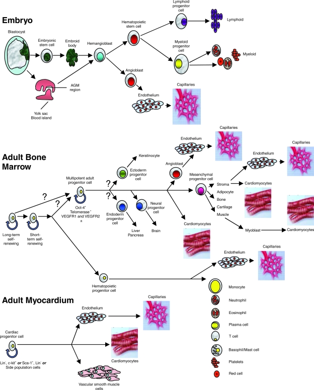
Cardiac gene and cell therapy have both entered clinical trials aimed at ameliorating ventricular dysfunction in patients with chronic congestive heart failure. The transduction of myocardial cells with viral constructs encoding a specific cardiomyocyte Ca(2+) pump in the sarcoplasmic reticulum (SR), SRCa(2+)-ATPase has been shown to correct deficient Ca(2+) handling in cardiomyocytes and improvements in contractility in preclinical studies, thus leading to the first clinical trial of gene therapy for heart failure. In cell therapy, it is not clear whether beneficial effects are cell-type specific and how improvements in contractility are brought about. Despite these uncertainties, a number of clinical trials are under way, supported by safety and efficacy data from trials of cell therapy in the setting of myocardial infarction. Safety concerns for gene therapy center on inflammatory and immune responses triggered by viral constructs, and for cell therapy with myoblast cells, the major concern is increased incidence of ventricular arrhythmia after cell transplantation. Principles and mechanisms of action of gene and cell therapy for heart failure are discussed, together with the potential influence of reactive oxygen species on the efficacy of these treatments and the status of myocardial-delivery techniques for viral constructs and cells.


Figures

FIG. 1.
FIG. 1.
Hemangioblasts can be derived from blastocyst-derived embryonic stem cells, and in vivo, they arise from the yolk sac or AGM region of the early embryo. With appropriate stimulation in vitro, hemangioblasts develop into hematopoietic progenitor cells that can give rise to all cell lines of the lymphoid and hematopoietic system, or they can develop into angioblasts that form endothelial cells and capillaries. In adult bone marrow, self-renewing cells differentiate into hemangioblasts that generate all cells of hematopoietic lineage, but cells of monocyte lineage can also differentiate into endothelial cells. The origin of the multipotent adult progenitor cell is unknown, but its progeny forms all mesenchymal tissues, and it can differentiate into cardiomyocytes and endothelial cells. Skeletal muscle is of mesenchymal lineage and can dedifferentiate into a myoblast phenotype, and then can differentiate into a cell with cardiomyocyte features. In adult myocardium, several multipotent cells have been identified by immunohistochemistry and fluorescence-activated cell sorting, and by sorting for side population cells. These cells have been shown to differentiate into endothelial cells and capillaries, cardiomyocytes, and vascular smooth muscle cells. (Reproduced with permission of Nature Publishing Group from ref. . © 2006.)
FIG. 2.
FIG. 2.
Microsphere retention and distribution in normal hearts and in hearts with 1- and 2-week-old MI. The retention is expressed as percentage of the total microspheres injected. Values <0.5% are shown as 0. (A) Moving from left to right in these hearts, the anterior wall is shown on the left side, followed by the septum, posterior wall, and lateral wall. (Reproduced with permission of John Wiley and Sons, Inc. from ref. . © 2006.) (B) When comparing endo- and epicardial distribution, in healthy hearts, significantly more retention of microspheres is found in the epicardium after retrograde infusion; this relation is reversed in 7-day-old MI, but in 2-week-old MI characterized by a transmural scar, retrograde infusion deposits more spheres in the epicardium than does transendocardial injection (Reproduced with permission of John Wiley and Sons, Inc. from ref. . © 2006.)
FIG. 2.
FIG. 2.
Microsphere retention and distribution in normal hearts and in hearts with 1- and 2-week-old MI. The retention is expressed as percentage of the total microspheres injected. Values <0.5% are shown as 0. (A) Moving from left to right in these hearts, the anterior wall is shown on the left side, followed by the septum, posterior wall, and lateral wall. (Reproduced with permission of John Wiley and Sons, Inc. from ref. . © 2006.) (B) When comparing endo- and epicardial distribution, in healthy hearts, significantly more retention of microspheres is found in the epicardium after retrograde infusion; this relation is reversed in 7-day-old MI, but in 2-week-old MI characterized by a transmural scar, retrograde infusion deposits more spheres in the epicardium than does transendocardial injection (Reproduced with permission of John Wiley and Sons, Inc. from ref. . © 2006.)
FIG. 3.
FIG. 3.
Vascular smooth muscle cells labeled ex vivo with iron particles (Feridex i.v.; Bayer Healthcare, Leverkusen, Germany) and injected retrogradely into the anterior interventricular vein in a porcine model of myocardial infarction, 1 week after induction of myocardial infarction by a 45-min balloon occlusion in the midsegment of the left anterior descending coronary artery. T2*-weighted magnetic resonance imaging shows a dark shadow indicating accumulation of the cells at the border of the infarct zone (arrow). After Prussian blue staining, iron-labeled cells are visible on 4-μm sections from the myocardium, and the fact that the iron label is retained within the cytoplasm of the labeled cells can be interpreted as indicating that these cells are still intact and viable (lower panel). (Reproduced with permission of John Wiley and Sons, Inc. from ref. . © 2004.)

References

    1. Abdel-Latif A. Bolli R. Tleyjeh IM. Montori VM. Perin EC. Hornung CA. Zuba-Surma EK. Al-Mallah M. Dawn B. Adult bone marrow-derived cells for cardiac repair: a systematic review and meta-analysis. Arch Intern Med. 2007;167:989–997. - PubMed
    1. Agbulut O. Vandervelde S. Al Attar N. Larghero J. Ghostine S. Leobon B. Robidel E. Borsani P. Le Lorc'h M. Bissery A. Chomienne C. Bruneval P. Marolleau JP. Vilquin JT. Hagège A. Samuel JL. Menasché P. Comparison of human skeletal myoblasts and bone marrow-derived CD133+ progenitors for the repair of infarcted myocardium. J Am Coll Cardiol. 2004;44:458–463. - PubMed
    1. American Heart Association. Heart disease and stroke statistics update. Dallas, TX: American Heart Association; 2008.
    1. Anilkumar N. Sirker A. Shah AM. Redox sensitive signaling pathways in cardiac remodeling, hypertrophy and failure. Front Biosci. 2009;14:3168–3187. - PubMed
    1. Asahara T. Murohara T. Sullivan A. Silver M. van der Zee R. Li T. Witzenbichler B. Schatteman G. Isner JM. Isolation of putative progenitor endothelial cells for angiogenesis. Science. 1997;275:964–967. - PubMed