

Review
doi: 10.1146/annurev.biophys.050708.133728.
Imaging transcription in living cells
Affiliations
- PMID: 19416065
- PMCID: PMC3166783
- DOI: 10.1146/annurev.biophys.050708.133728
Item in Clipboard
Review
Imaging transcription in living cells
Annu Rev Biophys.
2009.
Abstract
The advent of new technologies for the imaging of living cells has made it possible to determine the properties of transcription, the kinetics of polymerase movement, the association of transcription factors, and the progression of the polymerase on the gene. We report here the current state of the field and the progress necessary to achieve a more complete understanding of the various steps in transcription. Our Consortium is dedicated to developing and implementing the technology to further this understanding.
Figures
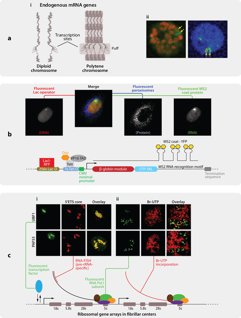
Three models for visualizing transcription in vivo. (a) Drosophila polytene chromosome. (i) Diagram of diploid chromosome and polytene chromosome. Active transcription site of heat shock gene hsp70 loci at 87A and 87C exhibits “chromosome puffs” (yellow arrows). (ii) (Left) hsp70 loci are enriched with Pol II (red: Hoechst33342 stain; green: EGFP-Rpb3). (Reproduced with permission from Reference .) (Right) Upon heat shock, EGFP-Rpb3 exhibits a strong doublet at 87A and 87C loci that can be recognized in polytene nucleus (arrows). Image in pseudocolor. (b) An artificial reporter gene was stably inserted as multiple copies into a single locus in the genome of U2OS cells. The locus of integration was tracked with the use of a Lac-repressor-fused RFP. The MS2 stem loop structures on the RNA, which bind stably to MS2 coat proteins, fused with YFP, allowing the investigator to localize single molecules of mRNA in vivo (78).(c) Ribosomal RNA transcription occurring inside fibrillar centers in CMT3 cells. (i) PAF53, a Pol I subunit, and UBF1, a rDNA transcription factor, colocalize with nascent rRNA in fibrillar centers (green: PAF53-GFP or UBF1-GFP; red: fluorescent in situ hybridization probe directed to rRNA 5’ ETS core). (ii) GFP-tagged PAF53 and UBF1 accumulate in active rRNA transcription sites (green: PAF53-GFP or UBF1-GFP; red: Bromo-UTP stain) (Adapted with permission from Reference 24). Abbreviations: EGFP, enhanced green fluorescent protein; ETS, external transcribed spacer; Pol I (II), polymerase I (II)
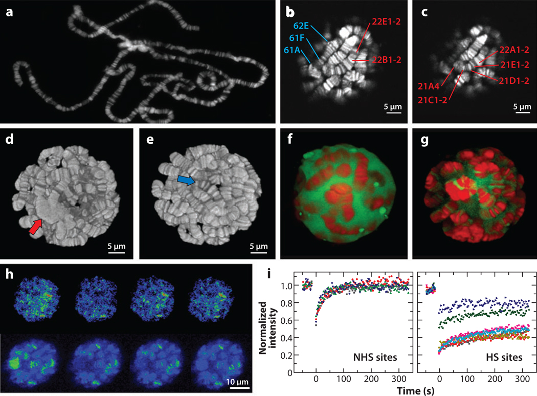
Imaging transcription kinetics at Drosophila polytene chromosomes.(a) An image of Drosophila polytene chromosomes spread onto a glass slide and stained by Hoechst33342. (b, c) Optical sections of polytene chromosomes in a live salivary gland stained with Hoechst33342. The z-distance is 0.5 µm. Labels identify specific bands on chromosome arms 2L (red) and 3L (blue). (d, e) Three dimensional reconstructions of a polytene nucleus. Red and blue arrows indicate the centromeric region and telomere, respectively. Panels b–e are adapted with permission from Reference . (f, g) HSF expression and localization in salivary glands at NHS (f) and after HS (g). Shown are the three-dimensional reconstructions of the two-photon optical sections of polytene nuclei expressing HSF-EGFP (green) stained with Hoechst33342 (red). (h, i) FRAP of HSF-EGFP at endogenous gene loci in salivary glands. (h) Intensity images of a nucleus of a heat-shocked salivary gland in pseudocolor during FRAP. (i) FRAP curves at NHS and HS sites. (Adapted with permission from References and 96). Abbreviations: EGFP, enhanced green fluorescent protein; FRAP, fluorescence recovery after photobleaching; HS, heat shock; HSF, heat shock factor; NHS, non-heat shock.
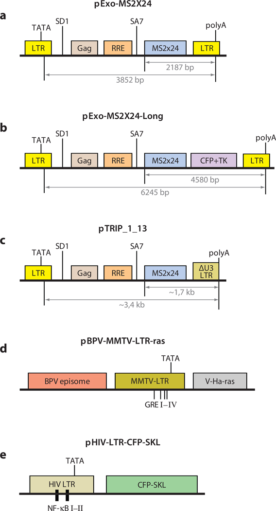
Diagrams of vectors containing viral sequences used to produce tandem arrays for real-time live cell imaging.(a) Vector used by Molle and colleagues to determine the residency times of Tat and Cdk9 on the HIV-1-LTR-driven transgene (57). (b, c) Modifications of the vector presented in panel a used in Reference to measure transcription elongation rate. (d) MMTV-LTR containing glucocorticoid receptor (GR) binding sites developed by McNally and colleagues was used to determine various kinetic parameters of gene activation by hormones (45, 54, 58, 80). Similarly, the vector presented in panel e has been used to study NF-κB interaction with its binding sites (9). The original vector designations and references in which they were first described are given. The kinetic parameters of transcription were obtained on tandem arrays in each of these vectors. Systems are discussed in the text. Diagrams are not drawn to scale. (For a compilation of other existing gene arrays, see Reference .) Abbreviations: BPV: bovine papilloma virus; Gag: retroviral gene coding for internal structural proteins; GRE: glucocorticoid-responsive element; NF-κB: nuclear factor κB; RRE: Rev-responsive element; SA7: splice acceptor 7; SD1: splicedonor 1; SKL: Ser-Lys-Leu peroxisome-targeting peptide; v-Ha-ras: Harvey viral ras.
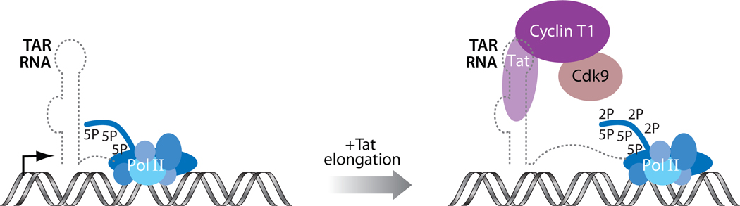
The role of the TAR:Tat:P-TEFb complex in HIV transcription. RNA polymerase II (Pol II) is stalled soon after initiation of transcription of the HIV provirus. The viral protein Tat recruits host cellular P-TEFb to the nascent stem-bulge-loop leader RNA, TAR (trans-activation responsive). The elongation factor P-TEFb composed of the cyclin T and the kinase Cdk9 phosphorylates the C-terminal domain repeats of the large subunit of Pol II at the Ser2 positions to stimulate processive elongation.
References
-
- Abramowitz M, Stegun IA. Handbook of Mathematical Functions. New York: Dover; 1965.
-
- Akhtar A, Gasser SM. The nuclear envelope and transcriptional control. Nat. Rev. Genet. 2007;8:507–517. - PubMed
-
- Beach DL, Salmon ED, Bloom K. Localization and anchoring of mRNA in budding yeast. Curr. Biol. 1999;9:569–578. - PubMed
Publication types
MeSH terms
Substances
Grants and funding
LinkOut - more resources
Full Text Sources
