

Matrix metalloproteinase induction by relaxin causes cartilage matrix degradation in target synovial joints
- PMID: 19416213
- PMCID: PMC2757096
- DOI: 10.1111/j.1749-6632.2009.03830.x
Matrix metalloproteinase induction by relaxin causes cartilage matrix degradation in target synovial joints
Abstract
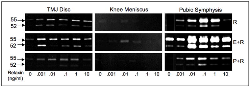
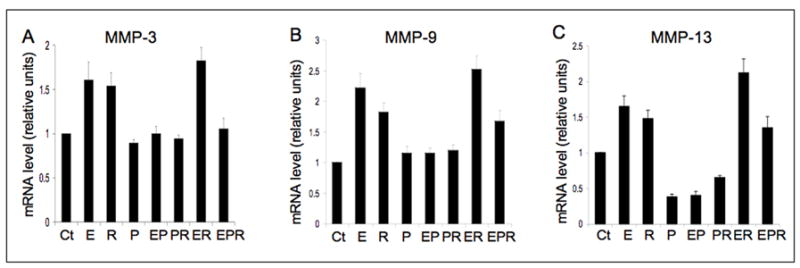
Our long-term goal is to understand the mechanisms by which relaxin and estrogen potentially contribute to joint diseases, particularly those afflicting the fibrocartilaginous temporomandibular joint (TMJ). Previously, we showed that relaxin produces a dose-dependent induction of tissue-degrading enzymes of the matrix metalloproteinase (MMP) family, specifically MMP-1 (collagenase-1), MMP-3 (stromelysin-1), MMP-9 (92-kDa gelatinase), and MMP-13 (collagenase-3) in cell isolates and tissue explants from TMJ fibrocartilage. The induction of these MMPs is accompanied by loss of collagen and glycosaminoglycans (GAGs), which was blocked by a pan-MMP inhibitor. We also found the targeted in vivo loss of collagen and GAGs in TMJ discs of ovariectomized rabbits treated with beta-estradiol, relaxin, or both hormones together. Progesterone attenuated the induction of MMPs and matrix loss by relaxin and estrogen. The modulation of matrix composition in TMJ fibrocartilage by these hormones was similar to that observed in the pubic symphysis and differed from that of the knee meniscus. The two target tissues showing the greatest modulation of MMPs and matrix loss, namely, the TMJ disc and pubic symphysis, had similar expression profiles of the estrogen receptors alpha and beta, relaxin-1 receptor (RXFP1, LGR7), and insulin-like peptide 3 receptor (RXFP2, LGR8) and these profiles differed from those in cells from the knee meniscus. These findings suggest a novel model for targeted tissue turnover of cartilage of specific joints through hormone-mediated induction of select MMPs.
Figures
Similar articles
-
Relaxin's induction of metalloproteinases is associated with the loss of collagen and glycosaminoglycans in synovial joint fibrocartilaginous explants.Arthritis Res Ther. 2005;7(1):R1-11. doi: 10.1186/ar1451. Epub 2004 Oct 29. Arthritis Res Ther. 2005. PMID: 15642129 Free PMC article.
-
Relaxin and beta-estradiol modulate targeted matrix degradation in specific synovial joint fibrocartilages: progesterone prevents matrix loss.Arthritis Res Ther. 2006;8(4):R98. doi: 10.1186/ar1978. Arthritis Res Ther. 2006. PMID: 16784544 Free PMC article.
-
Targeted induction of collagenase and stromelysin by relaxin in unprimed and beta-estradiol-primed diarthrodial joint fibrocartilaginous cells but not in synoviocytes.Lab Invest. 1998 Aug;78(8):925-38. Lab Invest. 1998. PMID: 9714180
-
Matrix metalloproteinases: role in arthritis.Front Biosci. 2006 Jan 1;11:529-43. doi: 10.2741/1817. Front Biosci. 2006. PMID: 16146751 Review.
-
Histochemistry for studying structure and function of the articular disc of the human temporomandibular joint.Eur J Histochem. 2012 Feb 29;56(1):e11. doi: 10.4081/ejh.2012.e11. Eur J Histochem. 2012. PMID: 22472889 Free PMC article. Review.
Cited by
-
Late onset corneal ectasia after LASIK surgery.Saudi J Ophthalmol. 2011 Jul;25(3):225-30. doi: 10.1016/j.sjopt.2011.05.003. Epub 2011 Dec 5. Saudi J Ophthalmol. 2011. PMID: 23960929 Free PMC article.
-
VEGF promotes cartilage angiogenesis by phospho-ERK1/2 activation of Dll4 signaling in temporomandibular joint osteoarthritis caused by chronic sleep disturbance in Wistar rats.Oncotarget. 2017 Mar 14;8(11):17849-17861. doi: 10.18632/oncotarget.14874. Oncotarget. 2017. PMID: 28147322 Free PMC article.
-
Endogenous estrogen exacerbates UV-induced inflammation and photoaging in mice.J Invest Dermatol. 2014 Aug;134(8):2290-2293. doi: 10.1038/jid.2014.160. Epub 2014 Mar 27. J Invest Dermatol. 2014. PMID: 24675752 No abstract available.
-
Relationship Between Estrogen and Idiopathic Mandibular Condylar Resorption: A Systematic Literature Review.Medicina (Kaunas). 2025 Jan 23;61(2):201. doi: 10.3390/medicina61020201. Medicina (Kaunas). 2025. PMID: 40005318 Free PMC article.
-
Level of Disability after Total Hip Replacement in Patients with Some COMT Gene Polymorphism.J Clin Med. 2023 Dec 13;12(24):7652. doi: 10.3390/jcm12247652. J Clin Med. 2023. PMID: 38137720 Free PMC article.
References
-
- Felson DT, Nevitt MC. The effects of estrogen on osteoarthritis. Curr Opin Rheumatol. 1998;10:269–72. - PubMed
-
- Von Korff M, et al. An epidemiologic comparison of pain complaints. Pain. 1988;32:173–83. - PubMed
-
- Warren MP, Fried JL. Temporomandibular disorders and hormones in women. Cells Tissues Organs. 2001;169:187–92. - PubMed
-
- Lipton JA, Ship JA, Larach-Robinson D. Estimated prevalence and distribution of reported orofacial pain in the United States. J Am Dent Assoc. 1993;124:115–21. - PubMed
-
- Milam SB, et al. Sexual dimorphism in the distribution of estrogen receptors in the temporomandibular joint complex of the baboon. Oral Surg Oral Med Oral Pathol. 1987;64:527–32. - PubMed
Publication types
MeSH terms
Substances
Grants and funding
LinkOut - more resources
Full Text Sources
Miscellaneous
