. 2009 Jul;11(7):1114-27.
doi: 10.1111/j.1462-5822.2009.01315.x. Epub 2009 Mar 12.

Peptidoglycan induces loss of a nuclear peptidoglycan recognition protein during host tissue development in a beneficial animal-bacterial symbiosis

Affiliations

Peptidoglycan induces loss of a nuclear peptidoglycan recognition protein during host tissue development in a beneficial animal-bacterial symbiosis

Joshua V Troll et al. Cell Microbiol. 2009 Jul.

Abstract

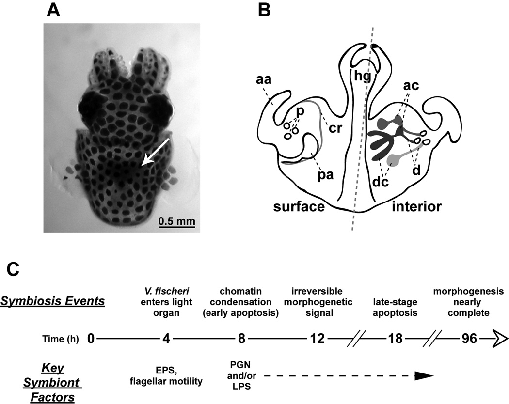
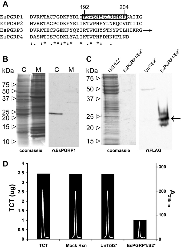
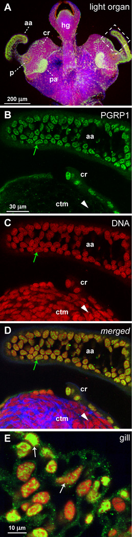
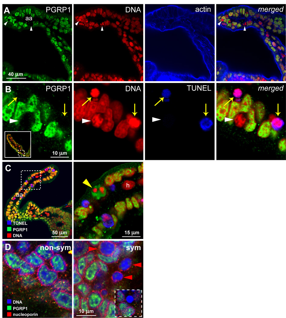
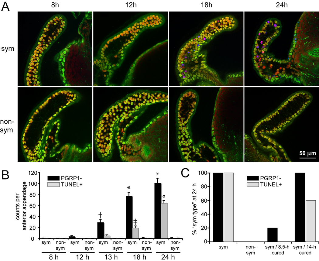
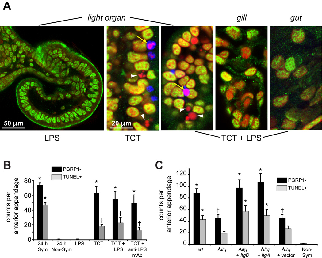
Peptidoglycan recognition proteins (PGRPs) are mediators of innate immunity and recently have been implicated in developmental regulation. To explore the interplay between these two roles, we characterized a PGRP in the host squid Euprymna scolopes (EsPGRP1) during colonization by the mutualistic bacterium Vibrio fischeri. Previous research on the squid-vibrio symbiosis had shown that, upon colonization of deep epithelium-lined crypts of the host light organ, symbiont-derived peptidoglycan monomers induce apoptosis-mediated regression of remote epithelial fields involved in the inoculation process. In this study, immunofluorescence microscopy revealed that EsPGRP1 localizes to the nuclei of epithelial cells, and symbiont colonization induces the loss of EsPGRP1 from apoptotic nuclei. The loss of nuclear EsPGRP1 occurred prior to DNA cleavage and breakdown of the nuclear membrane, but followed chromatin condensation, suggesting that it occurs during late-stage apoptosis. Experiments with purified peptidoglycan monomers and with V. fischeri mutants defective in peptidoglycan-monomer release provided evidence that these molecules trigger nuclear loss of EsPGRP1 and apoptosis. The demonstration of a nuclear PGRP is unprecedented, and the dynamics of EsPGRP1 during apoptosis provide a striking example of a connection between microbial recognition and developmental responses in the establishment of symbiosis.


Figures

Figure 1
Figure 1. Events in the early symbiosis of the squid/vibrio association
A) A dorsal view of a hatchling E. scolopes. E. scolopes harvests V. fischeri cells from ambient seawater within hours of hatching and maintains a population of the symbiont in a specialized ‘light organ’. The light organ appears as a dark region in the center of the body cavity (white arrow). B) A diagram depicting the ventral view of the hatchling light organ; left of the dashed line surface features are shown, right of the dashed line interior structures are displayed. Ciliated epithelial fields specific to the surface of the juvenile light organ promote colonization by the symbiont. These ciliated epithelial fields that undergo morphogenesis include the anterior appendage (aa), the posterior appendage (pa), and the ciliated ridge (cr). C) A timeline illustrating relevant characteristics of the early symbiosis. V. fischeri cells aggregate near the ciliated epithelial fields and enter the light organ around 4 to 6 hours post hatching. Within hours of successful colonization of the deep crypts of the light organ, the symbionts deliver an irreversible signal that triggers an extensive developmentally-programmed tissue destruction (morphogenesis) of the light organ into a mature form lacking the ciliated epithelial fields. ac, antechamber; d, ducts; dc, deep crypts; EPS, exopolysaccharide; hg, hindgut; LPS, lipopolysaccharide; p, pores; and PGN, peptidoglycan.
Figure 2
Figure 2. Biochemical characteristics of the EsPGRP1 protein
A) An alignment of the EsPGRP C-termini (arrow indicates additional C-terminal sequence for EsPGRP3). Boxed area highlights antigen sequence used for anti-EsPGRP1 antibody generation. *, identical residues; :, conserved substitutions; ., semi-conserved substitutions. B) A representative immunoblot of E. scolopes tissue. The anti-EsPGRP1 antibody reacts with a major band in the aqueous soluble fraction at ~24 kDa. C, cytoplasmic (aqueous) fraction; M, membrane (SDS soluble) fraction. C) Anti-FLAG immunoblot and companion gel of 24 µg crude protein extract from untransfected Drosophila S2* cells and 256 ng of an EsPGRP1-enriched fraction of S2* cells transfected with an EsPGRP1-FLAG expression construct. The arrow indicates an anti-FLAG reactive band at the expected molecular weight for EsPGRP1. D) An EsPGRP1 enriched EsPGRP1/S2* fraction degraded TCT in vitro, but a protein extract of untransfected S2* cells had no effect on TCT levels. Bars indicate mass of TCT (left axis) in each reaction, white peaks inside bars are the TCT peaks following treatment as the absorbance at 215nm on a reverse-phase HPLC chromatograph (right axis). UnT/S2*, untransfected S2* cell extract +; EsPGRP1/S2*, EsPGRP1-enriched fraction of S2* cells transfected with pJT28; mock rxn, TCT incubated in reaction buffer for 3 h; TCT, 7 µL 1mM TCT loaded directly onto HPLC column.
Figure 3
Figure 3. Localization of EsPGRP1 in host tissue
Confocal immunocytochemistry of hatchling and 24-h non-symbiotic E. scolopes localized EsPGRP1 to epithelial nuclei. A) A low magnification image of the light organ. Anti-EsPGRP1 antibodies intensely labeled the ciliated epithelial fields on the lateral surfaces of the light organ. B–D) Representative higher magnification images displaying the anterior appendage and a portion of the more medial ciliated field (dashed box in A). The nuclei of these epithelial cells (aa and cr) labeled strongly with the anti-EsPGRP1 antibody (green arrows), whereas non-epithelial nuclei of the underlying tissues (ctm) appeared devoid of EsPGRP1 (white arrowheads). E) Epithelia across the body contain nuclear EsPGRP1. A representative image taken from gill tissue is shown, note variations of staining intensity in the nuclei (white arrows). aa, anterior appendage; cr, ciliated ridge; ctm, connective tissue matrix; hg, hindgut; p, pores; pa, posterior appendage. EsPGRP1, green; DNA, red; and actin cytoskeleton, blue.
Figure 4
Figure 4. Loss of EsPGRP1 during progression through apoptosis
Confocal microscopy of symbiotic light organs revealed nuclear loss of EsPGRP1 and entry into late-stage apoptosis in anterior appendage epithelial nuclei. A) Micrographs of an optical section of the anterior appendage; individual channels on the left, merged image on the right. Colonization of the light organ by V. fischeri for 24 h induces a subset of nuclei in the ciliated epithelial fields to lose EsPGRP1 staining (e.g., white arrowheads). B) High magnification micrographs of epithelial cells from the anterior appendage. TUNEL staining of cleaved DNA only occurs in nuclei that have lost EsPGRP1 staining (yellow arrows), but not all EsPGRP1-negative nuclei are TUNEL positive (white arrowhead). Dashed box in inset indicates area of the anterior appendage shown in high magnification. C) Micrograph of an anterior appendage. Dashed box indicates area of high magnification in right panel that displays a transient alteration of intranuclear (based on size and shape) localization of EsPGRP1 staining after chromatin condensation but prior to the loss of EsPGRP1-signal from the nucleus (yellow arrowhead) as seen in A. Note also that hemocytes (h), which have migrated to the light organ blood sinus, fail to stain with EsPGRP1. Color palette is the same as in B: EsPGRP1, green; DNA, Red; and TUNEL, Blue. D) High magnification micrographs of anterior appendage epithelial cells from non-symbiotic and symbiotic animals. Labeling of the nucleoporins (red) with the monoclonal Ab 414 indicates that loss of EsPGRP1 from the nucleus (blue) occurs prior to the dissolution of the nuclear membrane (red arrowheads). Inset in sym panel exhibits an EsPGRP1-negative nucleus with complete breakdown of the nuclear envelope.
Figure 5
Figure 5. The timing of EsPGRP1 nuclear loss
EsPGRP1 loss occurs before entry into late-stage apoptosis and is triggered by the irreversible morphogenesis signal. A) Representative confocal micrographs from a timecourse of 8–24 h symbiotic and non-symbiotic hatchlings. EsPGRP1, green; DNA, red; and TUNEL, blue. B) Direct-count estimates of EsPGRP1-negative nuclei (black bars) and TUNEL-positive nuclei (grey bars) per anterior appendage (n=15). Statistical analysis included ANOVA with Tukey’s pairwise comparisons to identify statistically significant differences between time points (group error rate p ≤ 0.05). Symbols represent groupings that are statistically different from other groups. Error bars represent standard error. C) A qualitative comparison of light organs from a “curing experiment.” Symbiotic hatchlings were treated at 8.5 h or 14 h with 20 µg/ml chloramphenicol and scored for EsPGRP1-negative and TUNEL-positive nuclei in the anterior appendage at 24 h. The 95% confidence intervals (CI) for EsPGRP1-negative and TUNEL-positive nuclei were determined for 24-h symbiotic animals and the values for each animal from the two cured and the non-symbiotic groups were compared to the 24-h sym CI. If the number of EsPGRP1-negative or TUNEL-positive nuclei in a given anterior appendage fell within the 24-h sym CI, then that animal was considered “sym-type” for that category (protein localization or cell-death).
Figure 6
Figure 6. TCT induction of EsPGRP1 loss and late-stage apoptosis
A) Representative confocal micrographs of E. scolopes epithelia. Hatchling E. scolopes were exposed to 10 ng/ml LPS and/or 10 µM TCT and assayed for EsPGRP1-negative and TUNEL-positive nuclei at 24 h. TCT induces EsPGRP1-negative (white arrowheads) and TUNEL-positive (yellow arrows) nuclei. EsPGRP1, green; DNA, red; TUNEL, blue. B) Direct-count estimates of EsPGRP-negative and TUNEL-positive nuclei in TCT/LPS treated light organs (n = 10). Symbols represent groupings that are statistically different from other groups. C) Direct-count estimates of EsPGRP1-negative and TUNEL-positive nuclei in light organs colonized by V. fischeri mutants defective in TCT secretion. Δltg represents mutant strain DMA388, which is defective for three lytic transglycosylases (ΔltgA, Δ ltgD, ltgY::erm). Complementation strains carry indicated gene on the plasmid pVSV107 (Dunn et al., 2006). Symbols represent groupings that are statistically different from non-symbiotic light organs († represents an intermediate level of EsPGRP1 loss that is different from both sym and non-sym). For both B and C, Statistical analysis included ANOVA with Tukey’s pairwise comparisons to identify statistically significant differences between time points (group error rate p ≤ 0.05). Error bars represent standard error.

Similar articles

Cited by

References

    1. Adin DM, Engle JT, Goldman WE, McFall-Ngai MJ, Stabb EV. Mutations in ampG and lytic transglycosylase genes affect the net release of peptidoglycan monomers from Vibrio fischeri. J Bacteriol. 2008 - PMC - PubMed
    1. Anselme C, Perez-Brocal V, Vallier A, Vincent-Monegat C, Charif D, Latorre A, et al. Identification of the weevil immune genes and their expression in the bacteriome tissue. BMC Biol. 2008;6:43. - PMC - PubMed
    1. Anselme C, Vallier A, Balmand S, Fauvarque MO, Heddi A. Host PGRP gene expression and bacterial release in endosymbiosis of the weevil Sitophilus zeamais. Appl Environ Microbiol. 2006;72:6766–6772. - PMC - PubMed
    1. Apicella MA, Griffiss JM, Schneider H. Isolation and characterization of lipopolysaccharides, lipooligosaccharides, and lipid A. Methods Enzymol. 1994;235:242–252. - PubMed
    1. Beg AA, Baltimore D. An essential role for NF-κB in preventing TNF-alpha -induced cell death. Science. 1996;274:782–784. - PubMed

Publication types

MeSH terms

LinkOut - more resources