. 2009 Jun;24(3):190-6.
doi: 10.1111/j.1399-302X.2008.00494.x.

Expression of receptor activator of nuclear factor-kappaB ligand by B cells in response to oral bacteria

Affiliations

Expression of receptor activator of nuclear factor-kappaB ligand by B cells in response to oral bacteria

X Han et al. Oral Microbiol Immunol. 2009 Jun.

Abstract

Introduction: We investigated receptor activator of nuclear factor-kappaB ligand (RANKL) expression by B lymphocytes during early and late aspects of the immune response to Aggregatibacter actinomycetemcomitans, a gram-negative, anaerobic bacterium associated with aggressive periodontal disease.

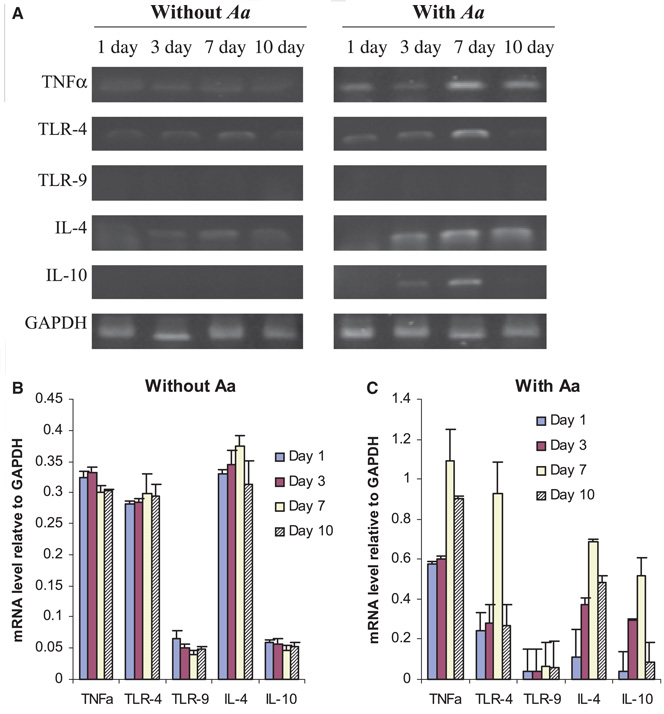
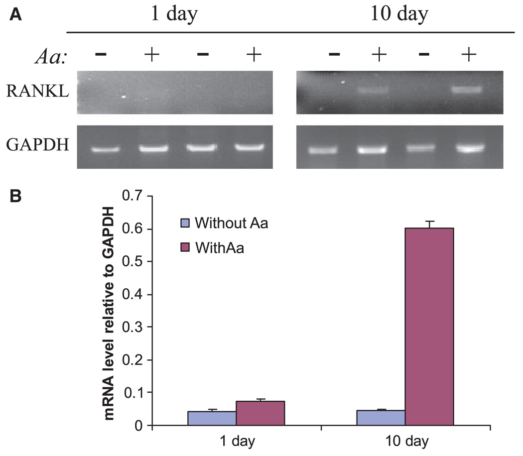
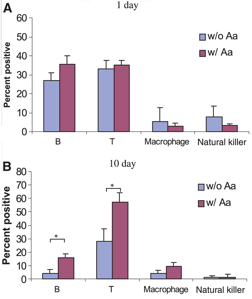
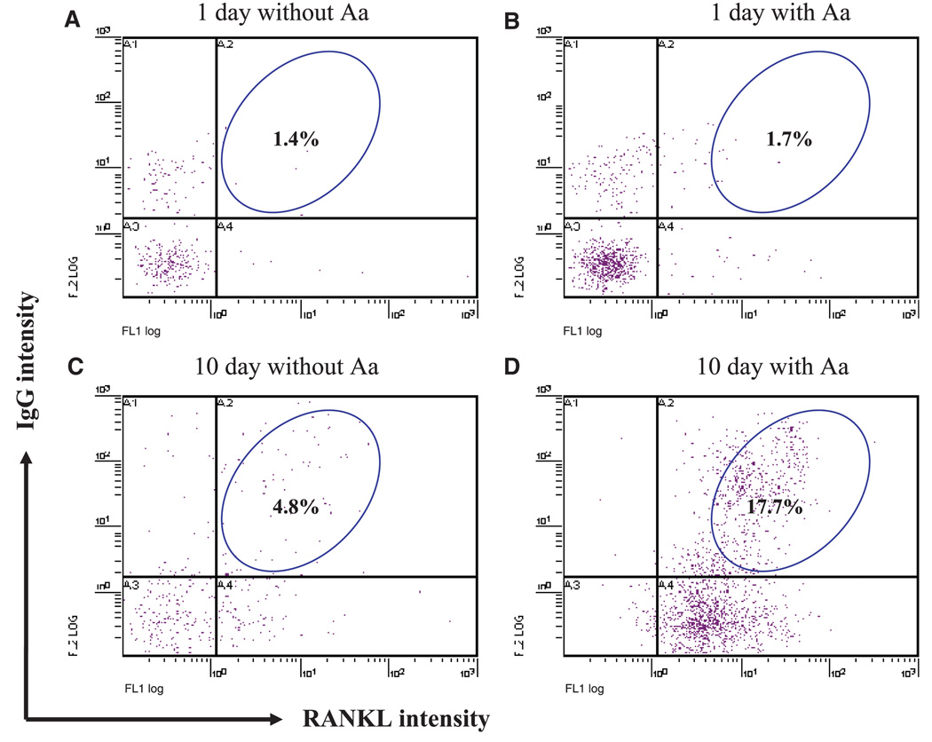
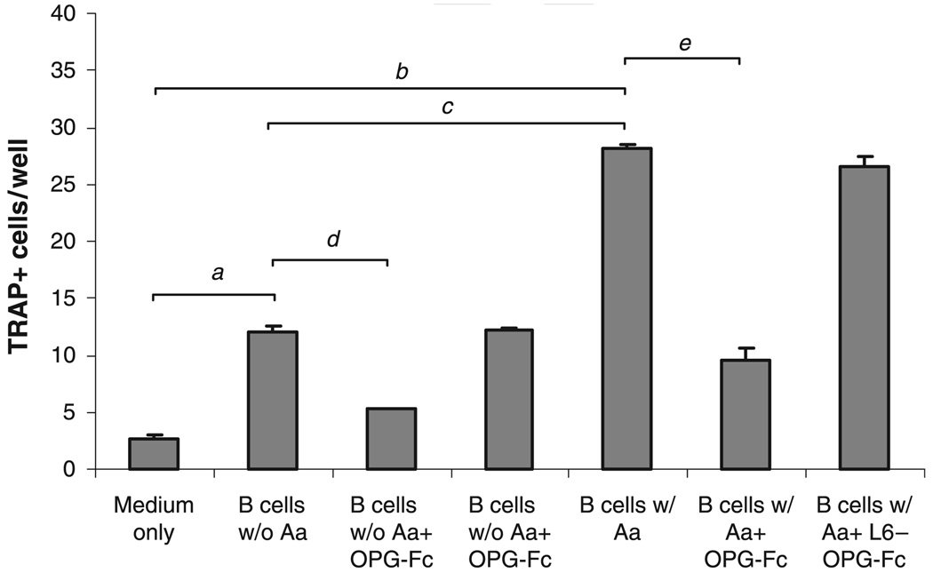
Methods: Expression of messenger RNA transcripts (tumor necrosis factor-alpha, Toll-like receptors 4 and 9, interleukins 4 and 10, and RANKL) involved in early (1-day) and late (10-day) responses in cultured rat splenocytes was examined by reverse transcription-polymerase chain reaction (RT-PCR). The immune cell distribution (T, B, and natural killer cells and macrophages) in cultured rat splenocytes and RANKL expression in B cells were determined by flow cytometric analyses. B-cell capacity for induction of osteoclast differentiation was evaluated by coculture with RAW 264.7 cells followed by a tartrate-resistant acid phosphatase (TRAP) activity assay.

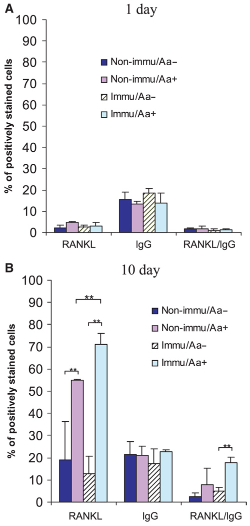
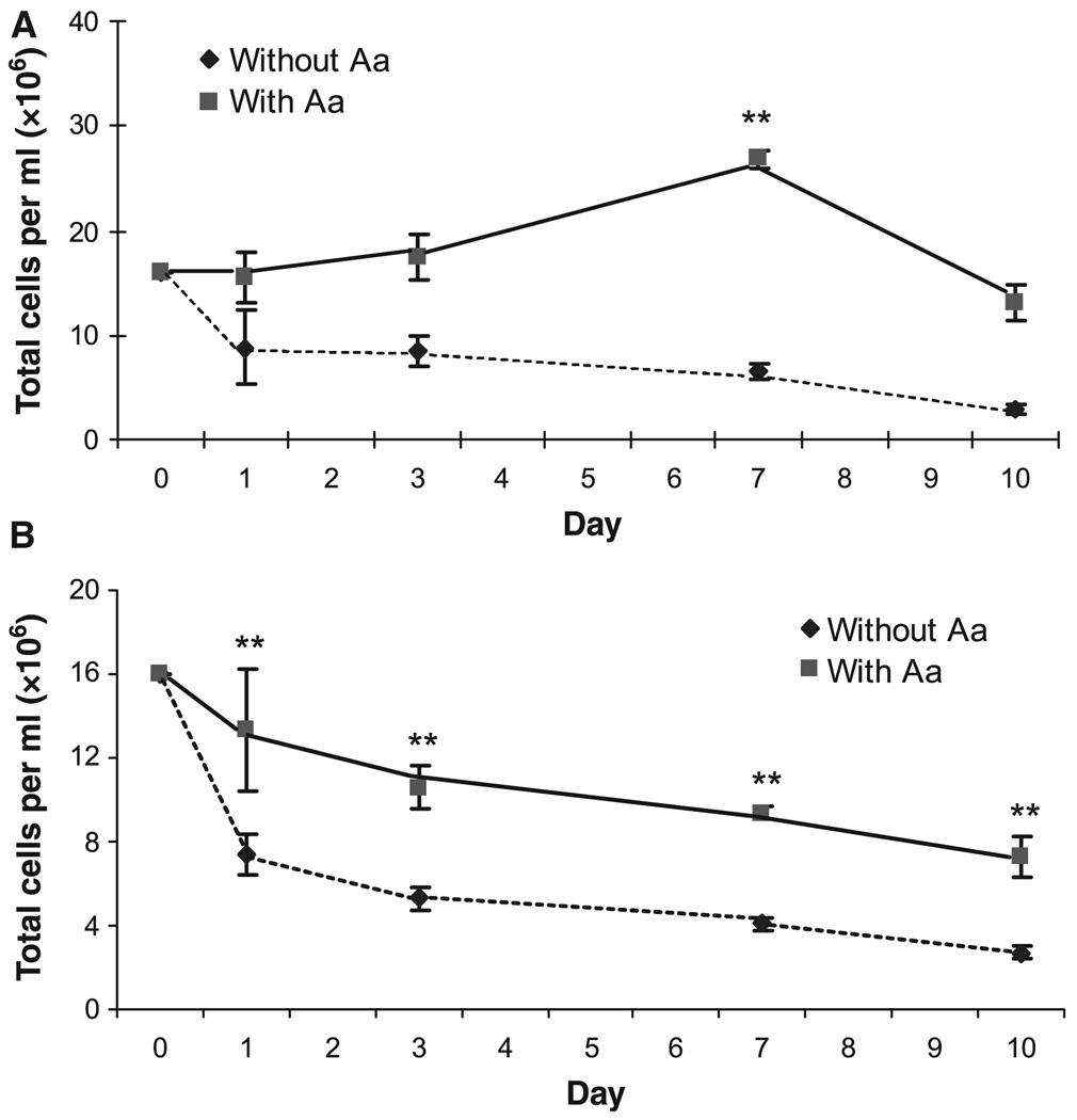
Results: The expression levels of interleukins 4 and 10 in cultured cells were not changed in the presence of A. actinomycetemcomitans until cultured for 3 days, and peaked after 7 days. After culture for 10 days, the percentages of B and T cells, the overall RANKL messenger RNA transcripts, and the percentage of RANKL-expressing immunoglobulin G-positive cells were significantly increased in the presence of A. actinomycetemcomitans. These increases were considerably greater in cells isolated from A. actinomycetemcomitans-immunized animals than from non-immunized animals. RAW 264.7 cells demonstrated significantly increased TRAP activity when cocultured with B cells from A. actinomycetemcomitans-immunized animals. The addition of human osteoprotegerin-Fc to the culture significantly diminished such increases.

Conclusion: This study suggests that B-lymphocyte involvement in the immune response to A. actinomycetemcomitans through upregulation of RANKL expression potentially contribute to bone resorption in periodontal disease.


Figures

Fig. 1
Fig. 1
Expression of molecular markers of innate and/or adaptive response in cultured splenocytes from naïve rats. Splenocytes from non-immunized rats were cultured for the times indicated and total RNA was extracted from cultured cells. Single-strand complementary DNA (cDNA) was synthesized from 0.1 µg total RNA by reverse transcription, and the specific cDNAs for tumor necrosis factor-α (TNF-α), Toll-like receptors 4 and 9 (TLR-4, TLR-9), interleukins 4 and 10 (IL-4, IL-10), and glyceraldehyde 3-phosphate dehydrogenase (GAPDH) were amplified by polymerase chain reaction (PCR). (A) PCR fragments were resolved on a 1% agarose gel and stained with ethidium bromide. (B, C). Signal intensity was quantified by densitometry and data are presented as mean ± SE of three independent repeats and the respective transcript levels are expressed as ratios relative to GAPDH; Aggregatibacter actinomycetemcomitans (Aa).
Fig. 2
Fig. 2
Receptor activator of nuclear factor-κB ligand (RANKL) messenger RNA expression by reverse transcription–polymerase chain reaction (RT-PCR) in cultured splenocytes from naïve rats. Splenocytes from non-immunized rats were cultured for 1 day or 10 days in the presence or absence of Aggregatibacter actinomycetemcomitans (Aa). After indicated times cultured splenocytes were collected and total RNA was extracted. Single-strand complementary DNA (cDNA) was synthesized from 0.1 µg total RNA by reverse transcription, and the specific cDNAs for RANKL and glyceraldehyde 3-phosphate dehydrogenase (GAPDH) were amplified by PCR. (A) PCR fragments were resolved on a 1% agarose gel and stained with ethidium bromide. (B) Signal intensity was quantified by densitometry and data are presented as mean ± SE of three independent duplicates and the respective transcript levels are expressed as ratios relative to GAPDH.
Fig. 3
Fig. 3
Cell type distribution of cultured splenocytes from non-immunized rats. Non-immune splenocytes were cultured for (A) 1 day or (B) 10 days in the presence or absence of Aggregatibacter actinomycetemcomitans (Aa). Cells were stained with the mouse anti-rat antibodies against different surface makers: OX33 for B cells, R73 for T cells, ED1 for macrophages, and NK-RP1 for natural killer cells. The cells were then stained with fluorescein isothiocyanate-conjugated rat anti-mouse immunoglobulin G. The percentage of each cell type in cultured splenocytes was determined by flow cytometry. At least 20,000 cells were counted for each sample (n = 4, mean ± SD, *P < 0.05, t-test).
Fig. 4
Fig. 4
Receptor activator of nuclear factor-κB ligand (RANKL) expression in immunoglobulin G-positive (IgG+) cells. Splenocytes from non-immunized rats were cultured for 1 day or 10 days in the presence or absence of Aggregatibacter actinomycetemcomitans (Aa). Cells were stained with rabbit anti-rat IgG followed by phycoerythrin-conjugated goat anti-rabbit IgG for the detection of IgG+ cells. Cells were also stained with human osteoprotegerin (OPG) -Fc (a fusion protein kindly provided by Dr Colin Dunstan from Amgen Inc., Thousand Oaks, CA) or the irrelavant human peptide L6-Fc (as controls), followed by fluorescein isothiocyanate-conjugated goat anti-human IgG for the detection of RANKL-expressing cells. At least 20,000 cells were counted for each sample.
Fig. 5
Fig. 5
Receptor activator of nuclear factor-κB ligand (RANKL) -expressing immunoglobulin G-positive (IgG+) cells in immunized vs. non-immunized animals. The same experiments as described in Fig. 3 were repeated on animals immunized intraperitoneally with Aggregatibacter actinomycetemcomitans (Aa) or with phosphate-buffered saline (as non-immunized controls). Splenocytes from these rats were cultured for (A) 1 day or (B) 10 days in the presence or absence of A. actinomycetemcomitans. Cells were then double-stained for the detection of RANKL-expressing IgG+ cells. At least 20,000 cells were counted for each sample (n = 4, mean ± SD, **P < 0.01, t-test).
Fig. 6
Fig. 6
Cell viability after exposure to Aggregatibacter actinomycetemcomitans. Splenocytes were cultured for the indicated time in the presence or absence of A. actinomycetemcomitans (Aa). Cultured cells were then harvested and stained with 40 µg/ml acridine orange and 100 µg/ml ethidium bromide, and visualized by fluorescence microscopy using an inverted microscope. Each experiment was performed in triplicates and the results were presented as mean ± SD (**P < 0.01, t-test).
Fig. 7
Fig. 7
Induction of osteoclast differentiation by purified B cells. Splenocytes from immunized animals (n = 3) were cultured in the presence or absence of formalin-fixed Aggregatibacter actinomycetemcomitans as described in the Materials and methods. B cells were purified from cultured splenocytes by fluorescence-activated cell sorting (FACS). RAW 264.7 cells were cocultured with purified B cells for 3 days. For some experiments, cocultures were performed in the presence or absence of human osteoprotegerin (OPG)-Fc at a concentration of 1 µg/ml. An irrelevant fusion protein, L6-Fc, was used as control. For tartrate-resistant acid phosphatase (TRAP) staining, cells were stained using a Leukocyte Acid Phosphatase kit (Sigma) and multinucleated TRAP-positive cells were counted. Each experiment was performed in triplicate and the results were presented as mean ± SE (a,b,d,eP < 0.01, cP < 0.001, t-test).

References

    1. Taubman MA, Yoshie H, Ebersole JL, Smith DJ, Olson CL. Host response in experimental periodontal disease. J Dent Res. 1984;63:455–460. - PubMed
    1. Stoufi ED, Taubman MA, Ebersole JL, Smith DJ, Stashenko PP. Phenotypic analyses of mononuclear cells recovered from healthy and diseased human periodontal tissues. J Clin Immunol. 1987;7:235–245. - PubMed
    1. Seymour GJ, Powell RN, Cole KL, et al. Experimental gingivitis in humans. A histochemical and immunological characterization of the lymphoid cell subpopulations. J Periodontal Res. 1983;18:375–385. - PubMed
    1. Malberg K, Molle A, Streuer D, Gangler P. Determination of lymphocyte populations and subpopulations extracted from chronically inflamed human periodontal tissues. J Clin Periodontol. 1992;19:155–158. - PubMed
    1. Tew J, Engel D, Mangan D. Polyclonal B-cell activation in periodontitis. J Periodontal Res. 1989;24:225–241. - PubMed

Publication types

MeSH terms

LinkOut - more resources