. 2009 May 19;106(20):8344-9.
doi: 10.1073/pnas.0812808106. Epub 2009 May 4.

The iron-sulfur clusters of dehydratases are primary intracellular targets of copper toxicity

Affiliations

The iron-sulfur clusters of dehydratases are primary intracellular targets of copper toxicity

Lee Macomber et al. Proc Natl Acad Sci U S A. .

Abstract

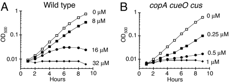
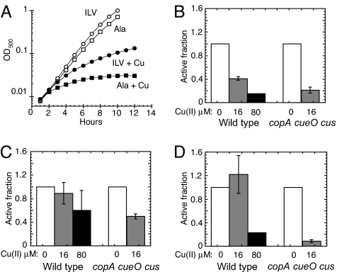
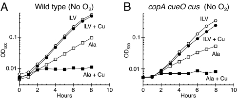
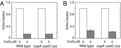
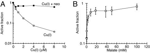
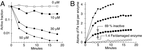
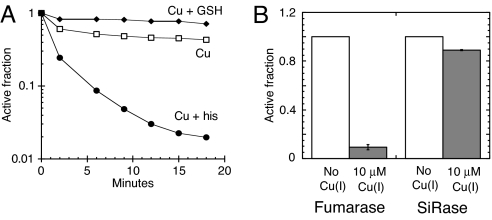
Excess copper is poisonous to all forms of life, and copper overloading is responsible for several human pathologic processes. The primary mechanisms of toxicity are unknown. In this study, mutants of Escherichia coli that lack copper homeostatic systems (copA cueO cus) were used to identify intracellular targets and to test the hypothesis that toxicity involves the action of reactive oxygen species. Low micromolar levels of copper were sufficient to inhibit the growth of both WT and mutant strains. The addition of branched-chain amino acids restored growth, indicating that copper blocks their biosynthesis. Indeed, copper treatment rapidly inactivated isopropylmalate dehydratase, an iron-sulfur cluster enzyme in this pathway. Other enzymes in this iron-sulfur dehydratase family were similarly affected. Inactivation did not require oxygen, in vivo or with purified enzyme. Damage occurred concomitant with the displacement of iron atoms from the solvent-exposed cluster, suggesting that Cu(I) damages these proteins by liganding to the coordinating sulfur atoms. Copper efflux by dedicated export systems, chelation by glutathione, and cluster repair by assembly systems all enhance the resistance of cells to this metal.


Conflict of interest statement

The authors declare no conflict of interest.

Figures

Fig. 1.
Fig. 1.
Copper is toxic to aerobic cells at micromolar doses. E. coli cultures were grown at 37 °C in aerobic glucose medium, and CuSO4 was added. (A) W3110 (wild type) cultures were challenged with 0 μM (open squares), 8 μM (closed squares), 16 μM (closed circles), or 32 μM CuSO4 (closed diamonds). (B) LEM33 (copA cueO cusCFBA) cultures were challenged with 0 μM (open squares), 0.25 μM (closed squares), 0.5 μM (closed circles), or 1 μM CuSO4 (closed diamonds). The data are representative of 3 independent experiments.
Fig. 2.
Fig. 2.
Copper damages iron-sulfur-cluster dehydratases. (A) LEM33 (copA cueO cusCFBA) was grown at 37 °C in aerobic glucose medium with 1.5 mM alanine (Ala) (squares) or 0.5 mM each of isoleucine (I), leucine (L), and valine (V) (circles), and CuSO4 was added to 0 μM (open symbols) or 10 μM (closed symbols). The data are a representative of 3 independent experiments. (B-D) W3110 (WT) and LEM33 (copA cueO cusCFBA) were grown aerobically to an OD500 of ≈0.1, then challenged with 0 μM (open bars), 16 μM (gray bars), or 80 μM CuSO4 (black bars) for 30 min. (B and C) Cells were grown in glucose/alanine, and IPMI (B) and fumarase (C) activities were measured. (D) Cells were grown aerobically in gluconate medium supplemented with 1.5 mM alanine, and 6-phosphogluconate activity was measured. (B-D) Data are the average of 3 independent experiments, and the error bars represent SD. (B) WT cells exposed to 80 μM Cu had IPMI activities below the detection limit (<15%).
Fig. 3.
Fig. 3.
Copper inhibits branched-chain biosynthesis even in the absence of oxygen. (A and B) E. coli cultures were grown anaerobically in glucose medium supplemented with either 1.5 mM alanine (ala) (squares) or 0.5 mM each of isoleucine (I), leucine (L), and valine (V) (circles), and they were then challenged with 0 μM (open symbols) or 2 μM CuSO4 (closed symbols). (A) W3110 (WT). (B) LEM33 (copA cueO cusCFBA). The data are representative of 3 independent experiments.
Fig. 4.
Fig. 4.
Copper damages iron-sulfur cluster dehydratases in the absence of oxygen. (A and B) W3110 (WT) and LEM33 (copA cueO cusCFBA) cultures were grown anaerobically in glucose/alanine medium to an OD500 of ≈0.1, and then challenged with 0 μM (open bars) or 4 μM CuSO4 (gray bars) for 30 min. (A) IPMI activity: copper-treated cells had IPMI activities below the detection limit (<15%). (B) Fumarase activity: the data are the average of 3 independent experiments, and the error bars represent SD.
Fig. 5.
Fig. 5.
Fumarase A is damaged by Cu(I) and protected by substrate. (A) Purified fumarase A (0.1 μM) was anaerobically challenged in 50 mM Tris-HCl, pH 7.6, with Cu(I) for 3 min at 27 °C either without a chelator (open squares) or in the presence of 100 μM neocuproine (neo) (closed squares). The data are a representative of 3 independent experiments. (B) Fumarase A was exposed to 3 μM Cu(I) for 3 min at 27 °C in the presence of the indicated concentrations of malate, and final activity was determined. The data are normalized to the starting activity. The data are the average of 3 independent experiments, and the error bars represent the SD.
Fig. 6.
Fig. 6.
Cu(I) damages the iron-sulfur cluster of fumarase A. (A and B) Fumarase A (4 μM) was challenged anaerobically at 27 °C with 0 μM (open squares), 10 μM (closed squares), 30 μM (closed circles), or 50 μM (closed diamonds) Cu(I) for 3 min. (A) Fumarase activity after challenge with Cu(I). (B) Time course of iron release from fumarase during copper challenge in A. The data are representative of 3 independent experiments.
Fig. 7.
Fig. 7.
The ability of Cu(I) to damage iron-sulfur clusters is modulated by copper-binding metabolites and requires that clusters be solvent-exposed. (A) Fumarase A (4 μM) was anaerobically challenged with 10 μM Cu(I) at 27 °C without supplementation (open squares) or in the presence of 100 μM histidine (his) (closed circles) or 100 μM reduced glutathione (GSH) (closed diamonds). The data are a representative of 3 independent experiments. (B) Fumarase A (0.1 μM) was mixed with sulfite reductase (SiRase), and the enzyme mixture was then challenged anaerobically with 0 μM (open bars) or 10 μM Cu(I) (gray bars) for 3 min at 27 °C. The data are the average of 3 independent experiments, and the error bars represent SD.

References

    1. Muller T, Feichtinger H, Berger H, Muller W. Endemic Tyrolean infantile cirrhosis: an ecogenetic disorder. Lancet. 1996;347:877–880. - PubMed
    1. Tanner MS, et al. Increased hepatic copper concentration in Indian childhood cirrhosis. Lancet. 1979;8128:1203–1205. - PubMed
    1. Bull PC, Thomas GR, Rommens JM, Forbes JR, Cox DW. The Wilson disease gene is a putative copper transporting P-type ATPase similar to the Menkes gene. Nat Genet. 1993;5:327–337. - PubMed
    1. Shah AB, et al. Identification and analysis of mutations in the Wilson disease gene (ATP7B): population frequencies, genotype-phenotype correlation, and functional analyses. Am J Hum Genet. 1997;61:317–328. - PMC - PubMed
    1. Tanzi RE, et al. The Wilson disease gene is a copper transporting ATPase with homology to the Menkes disease gene. Nat Genet. 1993;5:344–350. - PubMed

Publication types

MeSH terms