. 2009 May 19;106(20):8192-7.
doi: 10.1073/pnas.0811919106. Epub 2009 May 4.

Catalytically-active complex of HIV-1 integrase with a viral DNA substrate binds anti-integrase drugs

Affiliations

Catalytically-active complex of HIV-1 integrase with a viral DNA substrate binds anti-integrase drugs

Akram Alian et al. Proc Natl Acad Sci U S A. .

Abstract

HIV-1 integration into the host cell genome is a multistep process catalyzed by the virally-encoded integrase (IN) protein. In view of the difficulty of obtaining a stable DNA-bound IN at high concentration as required for structure determination, we selected IN-DNA complexes that form disulfide linkages between 5'-thiolated DNA and several single mutations to cysteine around the catalytic site of IN. Mild reducing conditions allowed for selection of the most thermodynamically-stable disulfide-linked species. The most stable complexes induce tetramer formation of IN, as happens during the physiological integration reaction, and are able to catalyze the strand transfer step of retroviral integration. One of these complexes also binds strand-transfer inhibitors of HIV antiviral drugs, making it uniquely valuable among the mutants of this set for understanding portions of the integration reaction. This novel complex may help define substrate interactions and delineate the mechanism of action of known integration inhibitors.


Conflict of interest statement

The authors declare no conflict of interest.

Figures

Fig. 1.
Fig. 1.
INP–DNA complex formation. (A) Ribbon structure of IN CCD, residues 52–210 (1BI4) (16), showing residues chosen for cysteine mutation: cluster 1 (orange) and cluster 2 (magenta). Catalytic triad residues D64, D116, and E152 are shown in red. C65, K159, and the proposed cut site of trypsin (R187) are shown in yellow, blue, and cyan, respectively. (B) DNA substrate for cross-linking is modeled on the HIV-1 3′-long terminal repeat (LTR). The DNA shown is an 18/20 oligonucleotide duplex. Thiol modification chemistry is indicated with 2-nitro-5- thiobenzoate (TNB) moiety and a linker of 6 carbons attached to the 5′-phosphate of the terminal adenine; the penultimate 3′-CA dinucleotide is underlined. (C) Nonreducing SDS/PAGE of INPK160C as native protein (lane 2) and after complexing with DNA of 2 different lengths: 9/11-mer and 18/20-mer (upper bands in lanes 3 and 4). The apparent molecular mass of the tethered DNA, as observed on the gel, is half (≈7 kDa for 18/20) of that of its double helix (≈14 kDa). Molecular mass standards (kDa) are provided to the left. (D) Relative IN–DNA complex formation by the different cysteine mutants shown as the percentage of cross-linked IN–DNA complex. INQ contains the original C65 (INC56S/W131D/F185D/C280S). All other IN mutants are based on INP, which contains an additional C65S mutation. Error bars represent SEM of 5 independent experiments. Bars are colored in accordance to residue color in A.
Fig. 2.
Fig. 2.
Oligomeric state of INP–DNA complex. MALLS-normalized refractive index (RI) (peaks) and molecular masses (lines) of SEC purified full-length INPY143C (thin line) and INPY143C–DNA complex (thick line) at 80 μM protein concentration are shown.
Fig. 3.
Fig. 3.
Impact of DNA substrate on IN–DNA complex formation. (A) IN–DNA complex formation using 3′-processed (18/20) and unprocessed DNA (20/20) for full-length INP constructs, each containing the indicated cysteine mutation. Thiol groups are indicated as TNB. (B) Nonreducing SDS/PAGE (4–12%), stained with Coomassie blue, of cross-linking analysis of full-length INPY143C and INPY143C–DNA complex without homo-bifunctional cross-linkers (lanes 2 and 6), and in the presence of homo-bifunctional cross-linkers of 2 different spacer arm lengths, 6.4 Å (lanes 3 and 7) and 11.4 Å (lanes 4 and 8). Lanes 1 and 5 are molecular standards. M: monomer; C: IN–DNA complex; D: dimer. Molecular masses (kDa) were determined by electrophoretic mobility. #: masses are the mean of 3 independent experiments. (C) Calculated molecular masses (kDa) of full length INP and INP–DNA. Asterisks indicate calculated masses of INP–DNA as expected to show on SDS/PAGE (≈7 kDa for each DNA as observed on gels). Masses in parentheses are as observed on the gel in B. Mono: monomer (IN); Di: dimer (IN2 or IN2–DNA); Tet: tetramer (IN4 or [IN2–DNA]2); Oct: octamer (IN8 or [IN2–DNA]4).
Fig. 4.
Fig. 4.
DNA tethering overcomes the IN NTD requirement for strand transfer. Single-end strand transfer activity of CCD + CTD untethered INPK160C/52–288 (◇, K160C) and INPY143C/52–288 (◆, Y143C), and tethered complexes of INPK160C/52–288–DNA (○, K160C/DNA) and INPY143C/52–288–DNA (●, Y143C/DNA). Error bars represent SEM of 3 independent experiments. RLU: relative luminescence units.
Fig. 5.
Fig. 5.
Inhibitor binding to INP–DNA complexes. Binding of GS-9160 to cross-linked INPK160C/52–288–DNA (diamonds, K160C/DNA) and INPY143C/52–288–DNA complexes (circles, Y143C/DNA) at 2 different immobilization concentrations of INP–DNA complexes: 500 nM (open symbols) and 1,500 nM (filled symbols). Error bars represent SEM of triplicates.
Fig. 6.
Fig. 6.
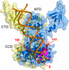
DNA binding by IN dimer. A model of IN dimer (1 protomer in blue and 1 in yellow) bound to DNA (orange ribbon). The 6C-thiol linker (≈9-Å length) is attached to the indicated 5′ end of the DNA. Dashed lines represent distances between the 5′ end and K160 (Cα in red sphere, 15Å) or Y143 (Cα in red sphere, 23Å). CCD flexible loop spanning Y143 (residues 139–150) is colored red. The inhibitor (L-870810, yellow sticks) and the definition of inhibitor binding pocket (magenta surface) have been described (30). Model of viral DNA bound to IN dimer has been described (15), and coordinates were generously obtained from M. Kvaratskhelia (Ohio State University, Columbus, OH). Loop coordinates were obtained from the CCD structure (1BI4) (16). The figure was made with Pymol (DeLano Scientific).

References

    1. Kukolj G, Skalka AM. Enhanced and coordinated processing of synapsed viral DNA ends by retroviral integrases in vitro. Genes Dev. 1995;9:2556–2567. - PubMed
    1. Brown PO. In: Retroviruses. Coffin JM, Hughes SH, Varmus HE, editors. Cold Spring Harbor, NY: Cold Spring Harbor Laboratory; 1997. pp. 161–203. - PubMed
    1. Deprez E, et al. Oligomeric states of the HIV-1 integrase as measured by time-resolved fluorescence anisotropy. Biochemistry. 2000;39:9275–9284. - PubMed
    1. Faure A, et al. HIV-1 integrase cross-linked oligomers are active in vitro. Nucleic Acids Res. 2005;33:977–986. - PMC - PubMed
    1. Engelman A, Bushman FD, Craigie R. Identification of discrete functional domains of HIV-1 integrase and their organization within an active multimeric complex. EMBO J. 1993;12:3269–3275. - PMC - PubMed

Publication types

LinkOut - more resources