. 2009 May 12;106(19):7798-803.
doi: 10.1073/pnas.0903188106. Epub 2009 Apr 27.

7SK snRNP/P-TEFb couples transcription elongation with alternative splicing and is essential for vertebrate development

Affiliations

7SK snRNP/P-TEFb couples transcription elongation with alternative splicing and is essential for vertebrate development

Matjaz Barboric et al. Proc Natl Acad Sci U S A. .

Abstract

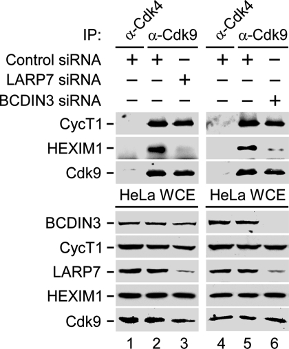
Eukaryotic gene expression is commonly controlled at the level of RNA polymerase II (RNAPII) pausing subsequent to transcription initiation. Transcription elongation is stimulated by the positive transcription elongation factor b (P-TEFb) kinase, which is suppressed within the 7SK small nuclear ribonucleoprotein (7SK snRNP). However, the biogenesis and functional significance of 7SK snRNP remain poorly understood. Here, we report that LARP7, BCDIN3, and the noncoding 7SK small nuclear RNA (7SK) are vital for the formation and stability of a cell stress-resistant core 7SK snRNP. Our functional studies demonstrate that 7SK snRNP is not only critical for controlling transcription elongation, but also for regulating alternative splicing of pre-mRNAs. Using a transient expression splicing assay, we find that 7SK snRNP disintegration promotes inclusion of an alternative exon via the increased occupancy of P-TEFb, Ser2-phosphorylated (Ser2-P) RNAPII, and the splicing factor SF2/ASF at the minigene. Importantly, knockdown of larp7 or bcdin3 orthologues in zebrafish embryos destabilizes 7SK and causes severe developmental defects and aberrant splicing of analyzed transcripts. These findings reveal a key role for P-TEFb in coupling transcription elongation with alternative splicing, and suggest that maintaining core 7SK snRNP is essential for vertebrate development.


Conflict of interest statement

The authors declare no conflict of interest.

Figures

Fig. 1.
Fig. 1.
Identification of core 7SK snRNP and elucidation of its assembly. (A and B) WCEs of 293, F.L7.293, or F.B3.293 cells were subjected to IP with α-FLAG. Levels of F.LARP7, F.BCDIN3, and their associating factors in WCEs (Left) and immunoprecipitates (Right) were detected by RT-PCR and Western blotting, respectively. WCEs or cells before lysis were incubated (+) or not (−) with RNase A or ActD, respectively. (C) GST.LARP7 chimeras were incubated with WCEs of 293 or F.B3.293 cells (Top). WCEs were incubated (+) or not (−) with RNase A. Levels of bound F.BCDIN3 proteins (Middle) and those of F.BCDIN3 and GST chimeras present in the binding reactions (Bottom) were analyzed by Western blotting. (D) Levels of indicated proteins in WCEs or α-FLAG immunoprecipitations of 293 or F.L7.293 cells are shown, which were analyzed by Western blotting. WCEs were incubated (+) or not (−) with RNase A.
Fig. 2.
Fig. 2.
Depletion of LARP7 or BCDIN3 releases HEXIM1 from P-TEFb. WCEs of HeLa cells that expressed the indicated siRNAs were subjected to IP. Levels of Cdk9-CycT1 and HEXIM1 proteins in immunoprecipitates (Middle) and the protein components of 7SK snRNP in WCEs (Bottom) were detected by Western blotting.
Fig. 3.
Fig. 3.
Depletion of LARP7 or BCDIN3 enhances the inclusion of EDA in P-TEFb-dependent manner. (A) Schematic of the pSVED-A Tot minigene. Arrow represents the transcription start site from the α-globin promoter. The third exon of the 3 α-globin exons (white rectangles) contains 3 fibronectin exons (black rectangles, exon number 32–34) with the introns (solid lines). Dashed lines represent the 2 possible mature EDA and ΔEDA mRNAs that include or lack the second fibronectin exon named EDA, respectively. The psv5′j/psv3′j primer pair used for RT-PCR analysis is indicated. (B and C) Levels of EDA and ΔEDA mRNAs were detected by RT-PCR by using total RNA samples isolated from HeLa cells that expressed siRNAs and pSVED-A Tot as indicated above the agarose gels.
Fig. 4.
Fig. 4.
P-TEFb controls alternative splicing via Ser2-P RNAPII and SF2/ASF. (A and B) Levels of EDA and ΔEDA mRNAs were detected by RT-PCR by using total RNA samples isolated from HeLa cells that expressed siRNAs and pSVED-A Tot as indicated above the agarose gels. Cells were treated (+) or not (−) with 60 nM of flavopiridol for 24 h before the isolation of total RNA. (C) Schematic of the pSVED-A Tot minigene used in the ChIP-qPCR. Positions of the promoter (Pr) and exonic (Ex) regions of the minigene that were amplified with qPCR are indicated. (D–F) ChIP-qPCR analysis of the pSVED-A Tot minigene in HeLa cells treated with control (white bars), BCDIN3 (black bars), or SF2/ASF (gray bars) siRNAs. Chromatin was immunoprecipitated with the depicted antibodies. Results for 3 ChIP-qPCR assays are shown ± SEM and are shown as percent (%) of input binding.
Fig. 5.
Fig. 5.
Knockdown of larp7 and bcdin3 in zebrafish caused developmental defects, decreased 7SK levels, and aberrant splicing. (A and B) RNA in situ hybridizations of whole-mount zebrafish embryos by using larp7 and bcdin3 antisense (1, 2) and sense probes (3, 4) at the indicated time points. Black arrowheads in panels 1 and 2 indicate larp7 and bcdin3 expressions in blastomeres and their enriched expressions in developing brain/head, respectively. (C) Zebrafish embryos injected with either larp7 MO or bcdin3 MO show developmental defects at 24 hpf compared with embryo injected with a control MO. Black arrowheads indicate the developing brain/head and trunk/tail. (D) Relative quantities of 7SK levels were determined by RT-qPCR at the indicated time points below the graph. The error bars represent the mean ± SD. (E) Splicing pattern analysis for the 2 genes indicated on the left at different time points as shown above the agarose gels. Levels of splicing variants were determined by RT-PCR by using the samples as in D and gene-specific primers.

References

    1. Core LJ, Lis JT. Transcription regulation through promoter-proximal pausing of RNA polymerase II. Science. 2008;319:1791–1792. - PMC - PubMed
    1. Zhou Q, Yik JH. The Yin and Yang of P-TEFb regulation: Implications for human immunodeficiency virus gene expression and global control of cell growth and differentiation. Microbiol Mol Biol Rev. 2006;70:646–659. - PMC - PubMed
    1. Shim EY, Walker AK, Shi Y, Blackwell TK. CDK-9/cyclin T (P-TEFb) is required in two postinitiation pathways for transcription in the C. elegans embryo. Genes Dev. 2002;16:2135–2146. - PMC - PubMed
    1. Ni Z, Schwartz BE, Werner J, Suarez JR, Lis JT. Coordination of transcription, RNA processing, and surveillance by P-TEFb kinase on heat shock genes. Mol Cell. 2004;13:55–65. - PubMed
    1. Ahn SH, Kim M, Buratowski S. Phosphorylation of serine 2 within the RNA polymerase II C-terminal domain couples transcription and 3′ end processing. Mol Cell. 2004;13:67–76. - PubMed

Publication types

Substances

LinkOut - more resources