. 2009 May 19;106(20):8290-5.
doi: 10.1073/pnas.0903196106. Epub 2009 Apr 30.

MR1 antigen presentation to mucosal-associated invariant T cells was highly conserved in evolution

Affiliations

MR1 antigen presentation to mucosal-associated invariant T cells was highly conserved in evolution

Shouxiong Huang et al. Proc Natl Acad Sci U S A. .

Abstract

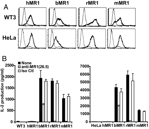
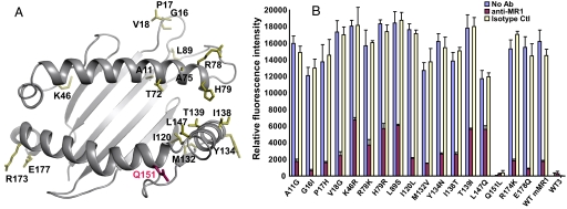
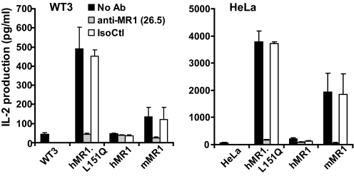
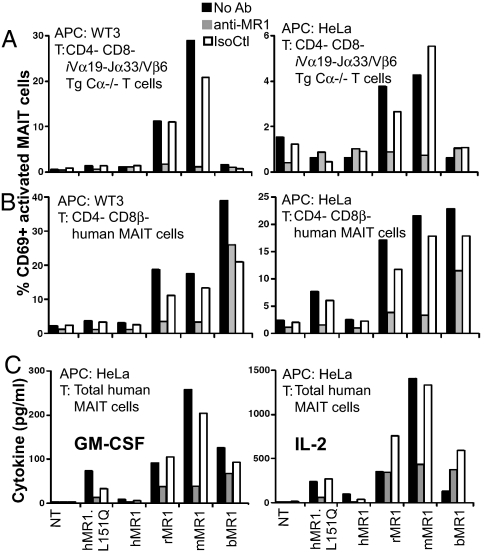
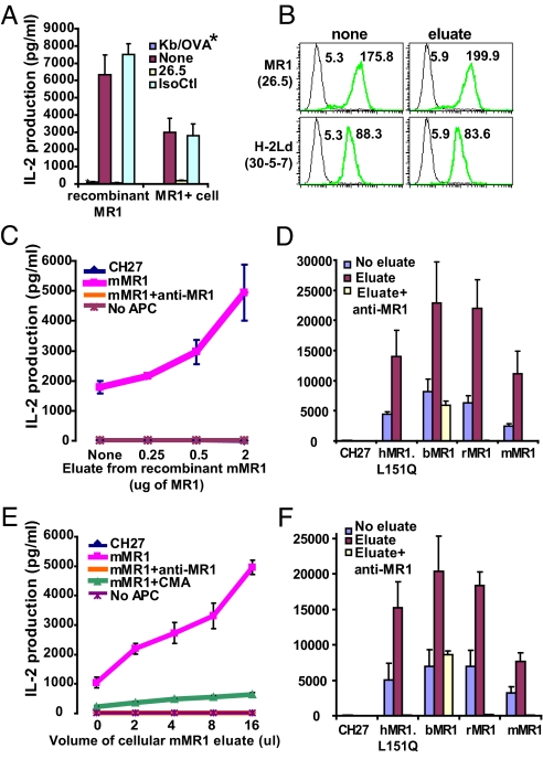
Several nonclassical major histocompatibilty antigens (class Ib molecules) have emerged as key players in the early immune response to pathogens or stress. Class Ib molecules activate subsets of T cells that mount effector responses before the adaptive immune system, and thus are called innate T cells. MR1 is a novel class Ib molecule with properties highly suggestive of its regulation of mucosal immunity. The Mr1 gene is evolutionarily conserved, is non-Mhc linked, and controls the development of mucosal-associated invariant T (MAIT) cells. MAIT cells preferentially reside in the gut, and their development is dependent on commensal microbiota. Although these properties suggest that MAIT cells function as innate T cells in the mucosa, this has been difficult to test, due to the (i) paucity of MAIT cells that display MR1-specific activation in vitro and (ii) lack of knowledge of whether or not MR1 presents antigen. Here we show that both mouse and human MAIT cells display a high level of cross-reactivity on mammalian MR1 orthologs, but with differences consistent with limited ligand discrimination. Furthermore, acid eluates from recombinant or cellular MR1 proteins enhance MAIT cell activation in an MR1-specific and cross-species manner. Our findings demonstrate that the presentation pathway of MR1 to MAIT cells is highly evolutionarily conserved.


Conflict of interest statement

The authors declare no conflict of interest.

Figures

Fig. 1.
Fig. 1.
Activation of mouse MAIT cells by bovine, rat, and mouse MR1 proteins. (A) Human, bovine, rat, and mouse MR1 were expressed on both mouse (WT3) and human (HeLa) cell lines using a retroviral transduction system. The surface expression was evaluated by FACS using anti-MR1 antibody 26.5 (coarse lines) compared with negative controls (thin lines) using the secondary antibody only (WT3) or the nontransduced HeLa cells. Similar results were seen with human 293T and mouse CH27 cells. (B) The mouse MAIT cell hybridoma 6C2 could be activated by bovine, rat, or mouse MR1 proteins in a manner blocked or inhibited by anti-mouse MR1 antibody 26.5.
Fig. 2.
Fig. 2.
The inability of human MR1 to activate mouse MAIT cell activation is mapped to the residue L151. (A) A mouse MR1 threading model was generated with the template of H-2Kb (1VAC) using the PHYRE Web server (32). The variable residues in the murine MR1 α1/α2 domains that were mutated to their human MR1 counterparts are shown with colored side chains using the PyMOL program. (B) The mMR1 mutants expressed on WT3 cells were tested for their ability to activate mouse MAIT cell hybridoma. Of the 17 mutants tested, only the mutation of Q151L [shown in magenta in (A)] resulted in decreased mouse MAIT cell activation.
Fig. 3.
Fig. 3.
Human-to-mouse reversal mutation at residues L151 restores the ability of hMR1 to activate mouse MAIT cells. The mutation of Leu-151 in hMR1 to Gln of mMR1 rescued the activation of mouse MAIT cells by hMR1 and was blocked by anti-MR1 antibody 26.5. Irrelevant isotype-matched antibody 34–2-12 was used as an isotype control (IsoCtl).
Fig. 4.
Fig. 4.
Primary quasi-monoclonal murine and polyclonal human MAIT cell are differentially activated on coculture with xenogeneic MR1 transfectants. TCRβ+/Vβ6+/CD4/CD8α (double-negative) transgenic iVα19-Jα33/Vβ6/Cα−/− mouse (A), TCRγδ/3C10+/CD3+/CD4/CD8β/CD161+ (double-negative and CD8αα) (B), and unseparated human MAIT T cells (C) were cultured with the indicated transduced cell lines for 16 h (A and B) or 48 h (C). Both mouse and human MAIT cells were activated when cocultured on mouse and rat MR1 overexpressed on WT3 and HeLa. Human MAIT cells also were activated by hMR1.L151Q and bovine MR1. Interactions were specifically blocked by the anti-MR1 26.5 antibody. NT, nontransduced cells; IsoCtl, isotype control antibody.
Fig. 5.
Fig. 5.
MR1 ligand isolated from recombinant MR1 enhances mouse MAIT cell activation by MR1 orthologous proteins. (A) The MAIT cell hybridoma (6C2) was activated by plate-bound recombinant MR1 protein compared with activation by the MR1-expressing cell. The plate-bound recombinant H-2Kb/OVA (*) complex was used as a negative control. (B) Surface staining of MR1 and H-2Ld molecules on mMR1–overexpressing CH27 cells after incubation with the acid eluate from recombinant MR1. The numbers indicate the mean fluorescent intensity. The green lines indicate the staining with anti-MR1 or Ld antibodies; the black lines indicate isotype controls. (C) Acid-eluted components (<10 kd) from recombinant mMR1 activated mouse MAIT cells in an MR1-dependent manner. (D) MAIT cell activation by MR1 protein from different species with addition of the acid eluate from recombinant MR1. (E) Acid-eluted components (<10 kd) from immunoprecipitated cellular mMR1 expressed in/on CH27 cells activated mouse MAIT cells in an MR1-dependent manner, and this activation was blocked by proton (H+) ATPase inhibitor CMA. (F) MAIT cell activation by MR1 protein from different species with addition of the acid eluate from cellular mMR1 protein. IsoCtl, isotype control antibody.

References

    1. Medzhitov R. Toll-like receptors and innate immunity. Nat Rev Immunol. 2001;1:135–145. - PubMed
    1. Matzinger P. The danger model: A renewed sense of self. Science. 2002;296:301–305. - PubMed
    1. Trinchieri G, Sher A. Cooperation of Toll-like receptor signals in innate immune defence. Nat Rev Immunol. 2007;7:179–190. - PubMed
    1. Bendelac A, Bonneville M, Kearney JF. Autoreactivity by design: Innate B and T lymphocytes. Nat Rev Immunol. 2001;1:177–186. - PubMed
    1. Colmone A, Wang CR. H2–M3–restricted T cell response to infection. Microbes Infect. 2006;8:2277–2283. - PubMed

Publication types

Substances

Associated data