. 2009 May 12;106(19):8043-8.
doi: 10.1073/pnas.0900358106. Epub 2009 Apr 29.

The organic cation transporter-3 is a pivotal modulator of neurodegeneration in the nigrostriatal dopaminergic pathway

Affiliations

The organic cation transporter-3 is a pivotal modulator of neurodegeneration in the nigrostriatal dopaminergic pathway

Mei Cui et al. Proc Natl Acad Sci U S A. .

Abstract

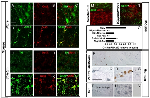
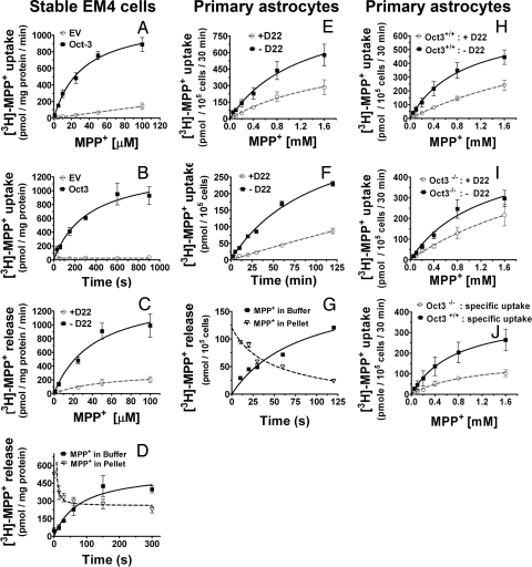
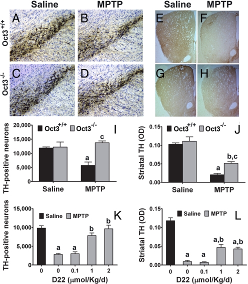
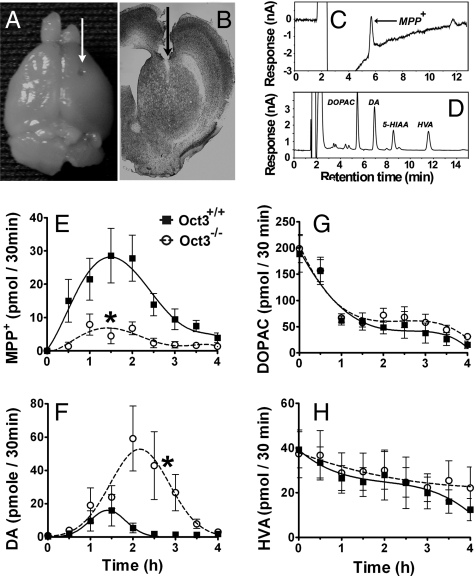
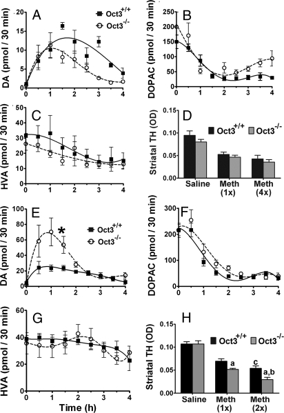
Toxic organic cations can damage nigrostriatal dopaminergic pathways as seen in most parkinsonian syndromes and in some cases of illicit drug exposure. Here, we show that the organic cation transporter 3 (Oct3) is expressed in nondopaminergic cells adjacent to both the soma and terminals of midbrain dopaminergic neurons. We hypothesized that Oct3 contributes to the dopaminergic damage by bidirectionally regulating the local bioavailability of toxic species. Consistent with this view, Oct3 deletion and pharmacological inhibition hampers the release of the toxic organic cation 1-methyl-4-phenylpyridinium from astrocytes and protects against 1-methyl-4-phenyl-1,2,3,6-tetrahydropyridine-induced dopaminergic neurodegeneration in mice. Furthermore, Oct3 deletion impairs the removal of the excess extracellular dopamine induced by methamphetamine and enhances striatal dopaminergic terminal damage caused by this psychostimulant. These results may have far-reaching implications for our understanding of the mechanism of cell death in a wide range of neurodegenerative diseases and may open new avenues for neuroprotective intervention.


Conflict of interest statement

The authors declare no conflict of interest.

Figures

Fig. 1.
Fig. 1.
Selective expression of Oct3 in the brain. Coronal mouse brain sections (A–N) were immunolabeled with antibodies against Oct3 in combination with either anti-TH (dopaminergic marker), anti-GFAP (astrocytic marker), or anti-MAP2 (neuronal marker). Oct3 colocalized with GFAP in the nigrostriatal region (D–F and J–L) but not with astrocytes in other regions (M and N). Although not detectable in dopaminergic structures (A–C and G–I), Oct3 immunoreactivity was detectable in other neurons (K, M, and N). The expression of Oct3 was further confirmed in cells captured by laser capture microdissection followed by quantitative real time RT-PCR analysis (O; BD, below detection). Primarily based on specific cellular markers, at least 800–1,000 cells of each type were captured. In postmortem human samples (P–V), OCT3 (blue–gray appearance) was robustly expressed in cells with morphology resembling that of nondopaminergic neurons (Q, T, and U) and astrocyte-like cells (S) but not in dopaminergic neurons (brown pigment of neuromelanin; P and R). As a negative control, Oct3 antibody was preabsorbed with Oct3 peptide and incubated with an adjacent cerebellar section (V). [Scale bars: 20 μm (A–N); 400 μm (P and T); and 100 μm (Q–S, U, and V)].
Fig. 2.
Fig. 2.
Bidirectional transport of MPP+. (A–D) EM4 cells stably overexpressing rat Oct3 or empty vector (EV), astrocytes from new born C57BL/6 mice (E–G), astrocytes from Oct3+/+ and Oct3−/− mice (H–J), were assessed for their transport activities of [3H]-MPP+. As described in the SI Materials and Methods, briefly, for the uptake studies (A, B, E, F, and H–J), cells were incubated with 10 nM [3H]-MPP+ and various concentrations of unlabeled MPP+, in the presence or absence of 5 μM D22 (a potent Oct3 inhibitor). Cell pellets were collected and radioactivity was counted using a scintillation counter. Specific uptake (in J) represents transport activity in the absence of D22 minus that of the group with D22. For the release studies (C, D, G), cells were preloaded for 20 min with 10 nM [3H]-MPP+ (plus 100 μM MPP+), washed, and then incubated at 37 °C for different time points in the assay buffer with or without 5 μM D22. Radioactivity released into the buffer and remaining in cells was counted using a scintillation counter. Oct3-mediated transport was time and concentration dependent and was blocked by D22 and Oct3 deficiency. Data represent mean ± SEM from 3–5 independent experiments with n = 4 per experiment.
Fig. 3.
Fig. 3.
Inhibition/ablation of Oct3 protected against MPTP neurotoxicity. Oct3−/− mice and their wild-type littermates Oct3+/+ (A–J), and C57BL/6 mice (K and L) infused s.c. with either saline or varying doses of D22, were injected with MPTP or saline. The loss of TH-positive neurons in the nigra (A–D and I) was completely prevented in the Oct3−/− mice (I) and by D22 (K, in a dose-dependent manner). Damage to the striatal density of TH-positive fibers (E–H, J, and L) was also attenuated in these animals. (I and J) (a) P < 0.01 compared to the Oct3+/+ saline group; (b) P < 0.01 compared to the Oct3−/− saline group; (c) P < 0.05 compared to the Oct3+/+ MPTP group, analyzed by 2-way ANOVA with treatments crossed with genotypes (panel I: genotype: F1,15 = 8.70, P = 0.01; treatment: F1,15 = 2.99, P = 0.10; panel J genotype: F1,15: = 6.60, P = 0.021; treatment: F1,15 = 67.07, P < 0.001) followed by the Newman-Keuls posthoc test. (K and L) (a) P < 0.01 compared to the control saline group; (b) P < 0.05 compared to the MPTP group without D22, analyzed by 1-way ANOVA followed by the Newman-Keuls post hoc test (panel K: F4,13 = 30.13, P < 0.001; panel L: F4,14 = 55.37, P < 0.001). Data represent mean ± SEM from 3–5 animals per group.
Fig. 4.
Fig. 4.
Oct3 ablation altered extracellular levels of MPP+ and DA in MPTP injected mice. Oct3−/− and their Oct3+/+ littermates were stereotaxically implanted with microdialysis probes into the right striatum (A), as verified in coronal striatal sections stained with thionin (B). Dialysates were collected every 30 min for 1 h before MPTP injection (30 mg/kg, i.p.) for baseline measurements (pooled for 0 time point) and for an additional 4 h after the injection, followed by HPLC analyses for the levels of MPP+ (E), DA (F), and its metabolites (G and H). Representative HPLC chromatograms for these measurements at 90 min after MPTP injection were illustrated in C and D. Data represent mean ± SEM, n = 9 WT and 11 KO. Areas under the curve were generated using GraphPad Prism followed by a 2-tailed t test. *, P < 0.05 compared to the Oct3+/+ group.
Fig. 5.
Fig. 5.
Oct3 ablation increased extracellular levels of DA and striatal neurotoxicity in methamphetamine injected mice. Striatal microdialysis dialysates were collected every 30 min for 1 h before methamphetamine injections (A–C: 5 mg/kg i.p., E–G: 30 mg/kg s.c.) for baseline measurements (pooled for 0 time point) and for an additional 4 h after the injection, followed by HPLC analyses for DA (A and E) and its metabolite levels (B, C, F, and G). Data represent mean ± SEM, n = 4–5 per genotype. Areas under the curve were generated using GraphPad Prism followed by a 2-tailed t test. *, P < 0.05 compared to the Oct3+/+ group. In other separate studies, animals were injected with either 5 mg/kg i.p. (single or every 2 h for 4 injections, D) or 30 mg/kg s.c. (single or 2 injections 4 h apart, H) and processed for striatal dopaminergic terminal density (D and H). Data represent mean ± SEM, n = 3–5 per group (D), n = 6–9 per group (H), (a) P < 0.05 compared to the respective Oct3+/+ methamphetamine group, (b) P < 0.01 compared to the Oct3−/− methamphetamine 1 injection group, (c) P < 0.05 compared to the Oct3+/+ methamphetamine 1 injection group, analyzed by 2-way ANOVA with treatments crossed with genotypes (genotype: F1,38 = 11.19, P = 0.002; treatment: F2,38 = 78.86, P < 0.001) followed by the Newman-Keuls post hoc test.

Similar articles

Cited by

References

    1. Dauer W, Przedborski S. Parkinson's disease: Mechanisms and models. Neuron. 2003;39:889–909. - PubMed
    1. Miller GW, Gainetdinov RR, Levey AI, Caron MG. Dopamine transporters and neuronal injury. Trends Pharmacol Sci. 1999;20:424–429. - PubMed
    1. Nagai M, et al. Astrocytes expressing ALS-linked mutated SOD1 release factors selectively toxic to motor neurons. Nat Neurosci. 2007;10:615–622. - PMC - PubMed
    1. Custer SK, et al. Bergmann glia expression of polyglutamine-expanded ataxin-7 produces neurodegeneration by impairing glutamate transport. Nat Neurosci. 2006;9:1302–1311. - PubMed
    1. Shin JY, et al. Expression of mutant huntingtin in glial cells contributes to neuronal excitotoxicity. J Cell Biol. 2005;171:1001–1012. - PMC - PubMed

Publication types

Substances