. 2009 May 5;106(18):7426-31.
doi: 10.1073/pnas.0812191106. Epub 2009 Apr 22.

Identification and structural basis of the reaction catalyzed by CYP121, an essential cytochrome P450 in Mycobacterium tuberculosis

Affiliations

Identification and structural basis of the reaction catalyzed by CYP121, an essential cytochrome P450 in Mycobacterium tuberculosis

Pascal Belin et al. Proc Natl Acad Sci U S A. .

Abstract

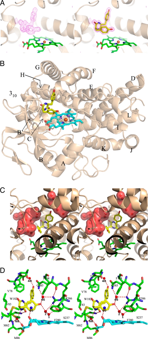
The gene encoding the cytochrome P450 CYP121 is essential for Mycobacterium tuberculosis. However, the CYP121 catalytic activity remains unknown. Here, we show that the cyclodipeptide cyclo(l-Tyr-l-Tyr) (cYY) binds to CYP121, and is efficiently converted into a single major product in a CYP121 activity assay containing spinach ferredoxin and ferredoxin reductase. NMR spectroscopy analysis of the reaction product shows that CYP121 catalyzes the formation of an intramolecular C-C bond between 2 tyrosyl carbon atoms of cYY resulting in a novel chemical entity. The X-ray structure of cYY-bound CYP121, solved at high resolution (1.4 A), reveals one cYY molecule with full occupancy in the large active site cavity. One cYY tyrosyl approaches the heme and establishes a specific H-bonding network with Ser-237, Gln-385, Arg-386, and 3 water molecules, including the sixth iron ligand. These observations are consistent with low temperature EPR spectra of cYY-bound CYP121 showing a change in the heme environment with the persistence of the sixth heme iron ligand. As the carbon atoms involved in the final C-C coupling are located 5.4 A apart according to the CYP121-cYY complex crystal structure, we propose that C-C coupling is concomitant with substrate tyrosyl movements. This study provides insight into the catalytic activity, mechanism, and biological function of CYP121. Also, it provides clues for rational design of putative CYP121 substrate-based antimycobacterial agents.


Conflict of interest statement

The authors declare no conflict of interest.

Figures

Fig. 1.
Fig. 1.
Spectral titration of CYP121 with cYY. The spectral changes induced on titration of CYP121 (2 μM) with cYY (0–463 μM) are shown. Spectra corresponding to 0 and 463 μM cYY are in black. Arrows indicate directions of change in spectra upon addition of cyclodipeptide. The chemical structure of cYY is shown with the diketopiperazine (DKP) ring in bold.
Fig. 2.
Fig. 2.
EPR features of the LS ferric heme of cYY-bound CYP121. 9-GHz EPR spectra of substrate-free CYP121 (black) and CYP121 after addition of a 2-fold excess of cYY (blue) were recorded at 20 K under non-saturating conditions using a microwave power of 0.05 mW and a modulation amplitude of 16 G.
Fig. 3.
Fig. 3.
cYY conversion by CYP121. CYP121, an electron transport chain, NADPH and cYY were incubated at 30 °C for various times (0, 1, 3, 10, and 30 min) then acidified and analyzed by LC-MS. Chromatograms were monitored at 214 nm and are shown for elution times from 10 to 45 min. NADPH and cYY were identified by comparison with standards. The chemical structure of P1 is shown.
Fig. 4.
Fig. 4.
Crystal structure of cYY-bound CYP121. (A) Fo-Fc electron density map contoured at 4σ level (pink) was calculated after 20 cycles of refinement upon removal of the ligand. The overall structure of CYP121 is in cartoon mode colored in wheat, the heme and Cys-345 are represented as sticks and colored in green, with oxygen, nitrogen and sulfur in red, blue and pale yellow, respectively. (Left) Representation of the active site after removal of cYY. (Right) The refined cYY is represented as sticks and colored in yellow (carbon atoms), red (oxygen atoms), and blue (nitrogen atoms). (B) The complex is shown in cartoon representation, with the heme and cYY represented as sticks. The CYP121 backbone is in wheat. The heme is in cyan with the iron in orange, and the cYY is in yellow, with oxygen atoms in red and nitrogen atoms in blue. The helices are labeled according to the previously published CYP121 crystal structure (10). (C) A stereoview of the active site highlighting the water-filled pockets surrounding the cYY. CYP121 is shown in cartoon representation in wheat. The heme and cYY are represented as sticks. The heme is in cyan with the iron in orange, and the cYY is in yellow, with oxygen atoms in red and nitrogen atoms in blue. Water molecules are represented as red spheres and water-filled pockets are shown in surface mode in red. (D) A stereoview in stick representation of the active site of CYP121 in complex with cYY. Cyclodipeptide is shown in yellow, the heme is in cyan with the iron represented as an orange sphere, CYP121 residues are in green and oxygen and nitrogen atoms are in red and blue, respectively. Water molecules are represented as red spheres and the cation presumed to interact with cYY is shown as a blue sphere. Probable H-bonds between CYP121, cyclodipeptide and non-bonded molecules are indicated as dotted lines. The numbering of non-bonded molecules is according to the pdb file.
Fig. 5.
Fig. 5.
Proposed mechanism for the intramolecular C-C coupling reaction of cYY by CYP121. For simplification, the DKP ring is represented as a sphere.

References

    1. Corbett EL, et al. The growing burden of tuberculosis: Global trends and interactions with the HIV epidemic. Arch Intern Med. 2003;163:1009–1021. - PubMed
    1. Cole ST, et al. Deciphering the biology of Mycobacterium tuberculosis from the complete genome sequence. Nature. 1998;393:537–544. - PubMed
    1. Isin EM, Guengerich FP. Complex reactions catalyzed by cytochrome P450 enzymes. Biochim Biophys Acta. 2007;1770:314–329. - PubMed
    1. McLean KJ, et al. The preponderance of P450s in the Mycobacterium tuberculosis genome. Trends Microbiol. 2006;14:220–228. - PubMed
    1. Ahmad Z, Sharma S, Khuller GK. Azole antifungals as novel chemotherapeutic agents against murine tuberculosis. FEMS Microbiol Lett. 2006;261:181–186. - PubMed

Publication types

MeSH terms

LinkOut - more resources