. 2009 Jul 3;284(27):18033-46.
doi: 10.1074/jbc.M109.006999. Epub 2009 May 5.

Functional pre- mRNA trans-splicing of coactivator CoAA and corepressor RBM4 during stem/progenitor cell differentiation

Affiliations

Functional pre- mRNA trans-splicing of coactivator CoAA and corepressor RBM4 during stem/progenitor cell differentiation

Yang S Brooks et al. J Biol Chem. .

Abstract

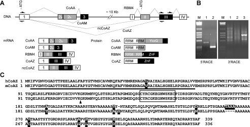
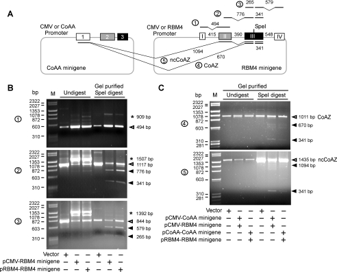
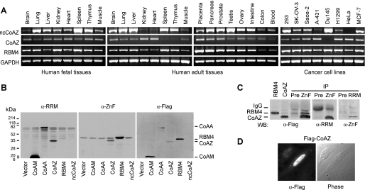
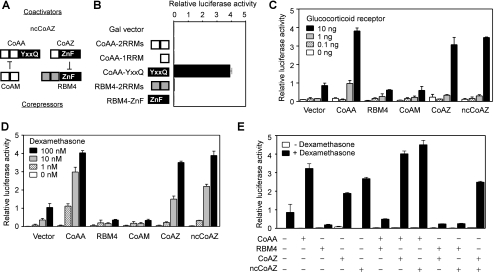
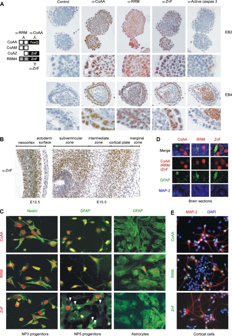
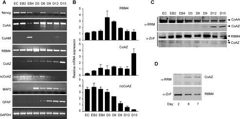
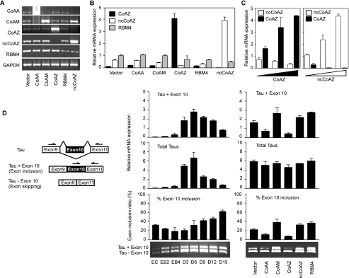
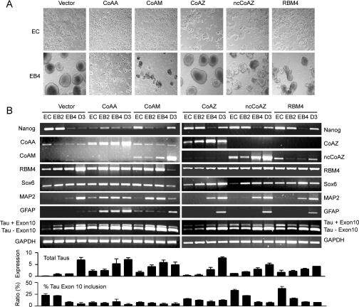
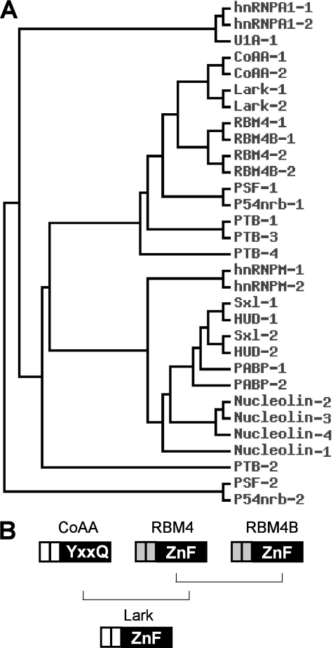
Alternative splicing yields functionally distinctive gene products, and their balance plays critical roles in cell differentiation and development. We have previously shown that tumor-associated enhancer loss in coactivator gene CoAA leads to its altered alternative splicing. Here we identified two intergenic splicing variants, a zinc finger-containing coactivator CoAZ and a non-coding transcript ncCoAZ, between CoAA and its downstream corepressor gene RBM4. During stem/progenitor cell neural differentiation, we found that the switched alternative splicing and trans-splicing between CoAA and RBM4 transcripts result in lineage-specific expression of wild type CoAA, RBM4, and their variants. Stable expression of CoAA, RBM4, or their variants prevents the switch and disrupts the embryoid body formation. In addition, CoAA and RBM4 counter-regulate the target gene Tau at exon 10, and their splicing activities are subjected to the control by each splice variant. Further phylogenetic analysis showed that mammalian CoAA and RBM4 genes share common ancestry with the Drosophila melanogaster gene Lark, which is known to regulate early development and circadian rhythms. Thus, the trans-splicing between CoAA and RBM4 transcripts may represent a required regulation preserved during evolution. Our results demonstrate that a linked splicing control of transcriptional coactivator and corepressor is involved in stem/progenitor cell differentiation. The alternative splicing imbalance of CoAA and RBM4, because of loss of their common enhancer in cancer, may deregulate stem/progenitor cell differentiation.


Figures

FIGURE 1.
FIGURE 1.
Identification of CoAZ and ncCoAZ as trans-splicing variants of CoAA and RBM4. A, schematic representation of CoAA and RBM4 genes in which introns are shown as lines and exons are boxes with splicing events depicted (not to scale). CoAA exons are numbered as 1–3 and RBM4 exons as I-IV. Translation start sites are indicated with ATG. The position of primers for 5′- and 3′-RACE of CoAZ are shown as open and filled arrows, respectively. The mRNA transcripts of CoAA and RBM4 and their corresponding protein products are shown with each region correlated with open, shaded, and filled boxes. RRM, CoAA activation domain (YXXQ), and RBM4 zinc finger-containing C terminus (ZnF) are indicated. Deposited accession numbers at NCBI GenBankTM are human CoAZ, EU287938; human ncCoAZ, EU287939; mouse CoAZ, EU287940; and mouse ncCoAZ, EU287941. B, 5′- and 3′-RACE analyses of CoAZ. Number indicates sequential PCR steps. C, aligned primary sequences of human and mouse CoAZ. The RRM domain is underlined, and the zinc finger is boxed. Non-identical amino acids between human and mouse are shaded. The arrow indicates the trans-splicing in-frame fusion site.
FIGURE 2.
FIGURE 2.
CoAZ and ncCoAZ are trans-splicing products of CoAA and RBM4 genes. A, CoAA and RBM4 minigenes with shortened intron sequences were constructed in individual plasmids under the control of either CMV or their native promoter. A SpeI restriction site was inserted into the third exon of RBM4 to distinguish minigene products from the endogenous transcripts. Digested PCR fragments and introns are depicted as lines with the number of base pairs indicated. B, 293 cells were transfected with vector alone or RBM4 minigenes (200 ng) with either CMV or RBM4 promoter as indicated. The RBM4 minigenes were analyzed for individual intron splicing capacity by RT-PCR. Gel-purified spliced transcripts (open arrow) were SpeI digested (filled arrows). Unspliced intron-containing transcripts are indicated by asterisks. Numbers indicate nucleotide base pairs. Each PCR reaction labeled by circled numbers was depicted in A. C, 293 cells were cotransfected with CoAA (100 ng) and RBM4 (100 ng) minigenes with either CMV or their native promoters as indicated. RT-PCR analyses depicted in A were followed by SpeI digestion using gel-purified PCR products for both CoAZ and ncCoAZ. The presence of digested fragments (filled arrows) indicates the trans-splicing events of the minigenes. Non-digestible fragments (open arrows) were derived from endogenous transcripts.
FIGURE 3.
FIGURE 3.
Expression of CoAZ, ncCoAZ, and RBM4. A, endogenous mRNA expression of CoAZ, ncCoAZ, and RBM4 was analyzed by PCR using normalized first strand cDNA panels from human adult and fetal tissues as well as cancer cell lines (Clontech). GAPDH was a control. B, Western blot analyses of CoAZ, ncCoAZ, and RBM4. 293 cells were transfected with FLAG-tagged CoAA, CoAM, CoAZ, RBM4, and ncCoAZ. Nuclear extracts were immunoblotted with anti-RRM, which recognizes the RRM domains of CoAA, CoAM, and CoAZ, with anti-ZnF, which recognizes the zinc-finger domain of RBM4 and CoAZ, and with anti-FLAG. ncCoAZ did not yield protein product. C, co-immunoprecipitation (IP) using overexpressed CoAZ and RBM4 in 293 cells. Individually expressed CoAZ or RBM4 were used as position markers. Preimmune sera were used as controls in co-immunoprecipitation. WB, Western blot. D, nuclear localization of FLAG-tagged CoAZ detected in transfected cells by immunofluorescence staining using anti-FLAG antibody.
FIGURE 4.
FIGURE 4.
CoAZ and ncCoAZ stimulate transcription and promote CoAA activity. A, domain structures of CoAA, CoAM, CoAZ, and RBM4 proteins are depicted with open boxes as RRMs and filled boxes as the YXXQ or the ZnF domain. CoAM inhibits CoAA via RRMs, and CoAZ inhibits RBM4 via zinc finger domain. B, each depicted CoAA and RBM4 domain was fused to a Gal4 DNA binding domain for testing transcriptional activity. CV-1 cells were transiently transfected with Gal4-fusion proteins and a luciferase reporter containing five Gal4-binding sites. C, full-length CoAA, CoAM, RBM4, CoAZ, or ncCoAZ was tested for transcriptional activity using a MMTV-luciferase reporter in CV-1 cells. Each clone (200 ng) was cotransfected with a MMTV-luciferase reporter (100 ng) and an increasing amount of glucocorticoid receptor (0, 0.1, 1, 10 ng) which binds to the MMTV promoter. Cells were induced by glucocorticoid receptor ligand dexamethasone (100 nm) overnight, and the luciferase activity was measured by Dynex luminometer. D, similar transfections as in C except using 10 ng of glucocorticoid receptor and an increasing amount of dexamethasone (0, 1, 10, 100 nm). E, cells were cotransfected as indicated in the presence or the absence of dexamethasone (100 nm). Luciferase activities shown are the means of triplicate transfections ± S.D.
FIGURE 5.
FIGURE 5.
Endogenous expression of CoAA, RBM4, and CoAZ during neural differentiation. A, the diagram at the left indicates antibodies recognizing each variant. ES cell-derived embryoid bodies at 2 days (EB2) and 4 days (EB4) of differentiation stage were paraffin-embedded, sectioned, and stained with affinity-purified anti-CoAA (CoAA only), anti-RRM (CoAA, CoAM, and CoAZ), anti-ZnF (RBM4 and CoAZ), and anti-active caspase-3 (cleaved caspase-3) antibodies and counterstained with hematoxylin. Enlarged views are shown below. B, immunohistochemistry analyses of mouse embryonic brain at gestation stages of E12.5 and E15.5. The sagittal sections were stained with anti-ZnF antibody (1:200). C, immunofluorescence staining of ES cell-derived neural progenitors after 3 days (NP3) or 5 days (NP5) of culture using anti-CoAA, anti-RRM, and anti-ZnF antibodies co-stained with the neural progenitor marker nestin or astrocyte cell marker GFAP (left and middle panels). White arrows indicate negatively stained cells. Double staining of isolated astrocytes from brain of P3 neonates using GAFP (right panel). D, mouse brain sections from P3 neonates were stained with anti-CoAA, anti-RRM, and anti-ZnF antibodies and co-stained with GFAP and MAP-2. E, double staining in MAP-2 positive neurons or MAP-2 negative astrocytes from isolated rat brain cortical cells at E18.5 after 6 days of culture in vitro. Slides are counter-stained with 4′,6-diamidino-2-phenylindole.
FIGURE 6.
FIGURE 6.
Alternative splicing switch between CoAZ and ncCoAZ during P19 embryonal carcinoma cell differentiation. A, undifferentiated P19 cells (EC) were induced by 500 nm retinoic acid in suspension culture up to 4 days to form embryoid bodies (EB2–EB4). Cells were further differentiated for an additional 15 days in the absence of retinoic acid (D3–D15). Total RNA was isolated and normalized at each stage and analyzed using gene-specific primers by RT-PCR as indicated. GAPDH was control. B, quantitative real-time PCR analysis of RBM4, CoAZ, and ncCoAZ at each stage using clone-specific primers. C, immunoblot analyses of CoAZ and RBM4 using anti-RRM and anti-ZnF antibodies during P19 cell differentiation. The asterisk indicates a nonspecific band. D, immunoblot analyses of CoAZ and RBM4 in isolated primary rat brain cortical cells after 2, 6, and 7 days of in vitro culture as indicated.
FIGURE 7.
FIGURE 7.
CoAA, RBM4, and their variants regulate alternative splicing of Tau exon 10. A, 293 cells were transfected with pcDNA3 vector, CoAA, CoAM, CoAZ, RBM4, or ncCoAZ followed by RT-PCR analyses using gene-specific primers of each transcript as indicated. The detected signals represent both endogenous and overexpressed transcripts except for RBM4. The RBM4 transcript overlaps with the ncCoAZ transcript, and only endogenous RBM4 was analyzed. B, quantitative PCR analysis of mRNA levels of RBM4, CoAZ, and ncCoAZ (200 ng) transfected 293 cells. C, an increasing amount of CoAZ or ncCoAZ (50, 200, 400 ng) was transfected before quantitative PCR. D, the diagram at the left illustrates alternative splicing of Tau exon 10. Primers on exons 9 and 11 detect total Tau mRNA, and primers on exons 10 and 11 detect Tau exon 10 inclusion isoform only. Middle panel, data are shown as real-time PCR quantification of endogenous expression levels of Tau exon 10 inclusion (Tau + Exon 10), total Tau, and their ratio. RT-PCR gel is shown below. The mRNA samples of P19 cell differentiation in this assay were the same preparation as in Fig. 6A. Right panel, Tau alternative splicing analysis in undifferentiated P19 stem cells (EC) overexpressed with CoAA, RBM4, and their variants as indicated. RT-PCR gel is shown below the PCR quantification.
FIGURE 8.
FIGURE 8.
Stable expression of CoAA and RBM4 in P19 cells disrupts embryoid body cavitation. A, light microscopy images of P19 stable cells expressing CoAA, CoAM, CoAZ, ncCoAZ, and RBM4 under the CMV promoter. The uninduced P19 stable cells (EC) and in retinoic acid-induced embryoid bodies at EB4 stages are shown (×400). Vector pcDNA3 was a control. B, RT-PCR analyses of Tau isoforms with (Tau + Exon 10) or without (TauExon 10) exon 10 inclusion in stable P19 cells at differentiation stages of EC, EB2, EB4, and D3. The marker genes used include Nanog, Sox6, MAP-2, and GFAP. GAPDH was a control. Quantitative PCR analysis of total Tau and percentage of Tau exon 10 inclusion are shown below.
FIGURE 9.
FIGURE 9.
Human CoAA, RBM4 and BM4B share common ancestry with Drosophila Lark. A, phylogenetic tree displaying the selected well characterized RRM-containing proteins. The tree was constructed using primary sequences within each RRM domain. Lark and Sxl are Drosophila proteins, and the rest are human proteins. The number indicates the RRM domain within each molecule. B, a hypothetic diagram of the evolutionary relationship among human CoAA, RBM4, and RBM4B and Drosophila Lark.

Similar articles

Cited by

References

    1. Berget S. M., Moore C., Sharp P. A. ( 1977) Proc. Natl. Acad. Sci. U.S.A. 74, 3171– 3175 - PMC - PubMed
    1. Chow L. T., Gelinas R. E., Broker T. R., Roberts R. J. ( 1977) Cell 12, 1– 8 - PubMed
    1. Modrek B., Lee C. ( 2002) Nat. Genet. 30, 13– 19 - PubMed
    1. Sanford J. R., Caceres J. F. ( 2004) J. Cell Sci. 117, 6261– 6263 - PubMed
    1. Pan Q., Shai O., Lee L. J., Frey B. J., Blencowe B. J. ( 2008) Nat. Genet. 40, 1413– 1415 - PubMed

Publication types

MeSH terms

Associated data