. 2009 Jul 3;284(27):18143-51.
doi: 10.1074/jbc.M109.004804. Epub 2009 May 5.

The pannexin 1 channel activates the inflammasome in neurons and astrocytes

Affiliations

The pannexin 1 channel activates the inflammasome in neurons and astrocytes

William R Silverman et al. J Biol Chem. .

Abstract

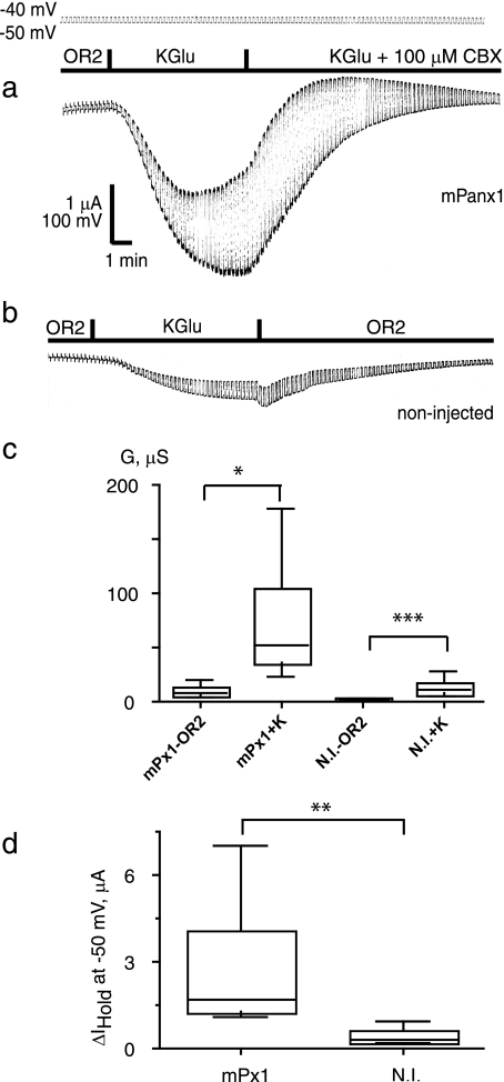
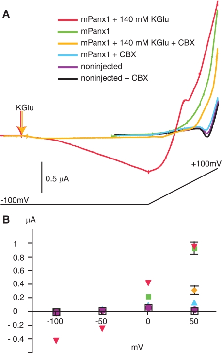
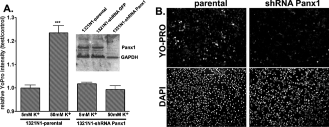
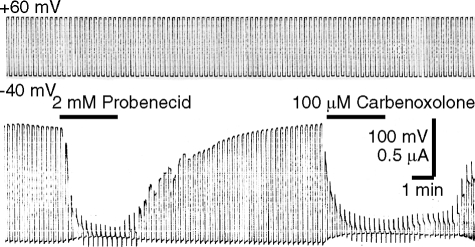
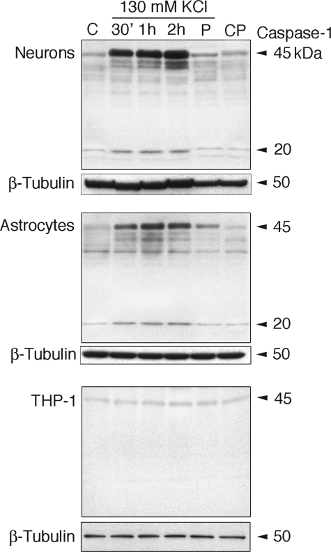
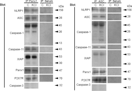
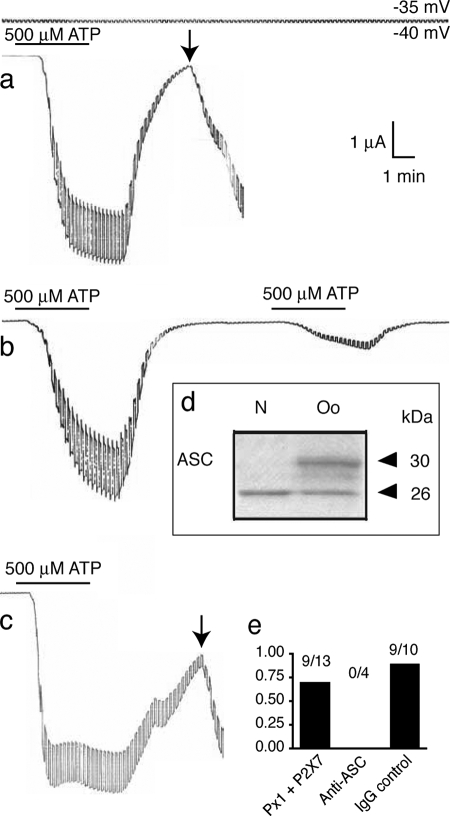
The inflammasome is a multiprotein complex involved in innate immunity. Activation of the inflammasome causes the processing and release of the cytokines interleukins 1beta and 18. In primary macrophages, potassium ion flux and the membrane channel pannexin 1 have been suggested to play roles in inflammasome activation. However, the molecular mechanism(s) governing inflammasome signaling remains poorly defined, and it is undetermined whether these mechanisms apply to the central nervous system. Here we show that high extracellular potassium opens pannexin channels leading to caspase-1 activation in primary neurons and astrocytes. The effect of K(+) on pannexin 1 channels was independent of membrane potential, suggesting that stimulation of inflammasome signaling was mediated by an allosteric effect. The activation of the inflammasome by K(+) was inhibited by the pannexin 1 channel blocker probenecid, supporting a role of pannexin 1 in inflammasome activation. Co-immunoprecipitation of neuronal lysates indicates that pannexin 1 associates with components of the multiprotein inflammasome complex, including the P2X7 receptor and caspase-1. Moreover antibody neutralization of the adaptor protein ASC (apoptosis-associated speck-like protein containing a CARD) blocked ATP-induced cell death in oocytes co-expressing P2X7 receptor and pannexin 1. Thus, in contrast to macrophages and monocytes in which low intracellular K(+) has been suggested to trigger inflammasome activation, in neural cells, high extracellular K(+) activates caspase-1 probably through pannexin 1.


Figures

FIGURE 1.
FIGURE 1.
Activation of pannexin 1 channels by extracellular K+. Oocytes were voltage-clamped at a holding potential of −50 mV, and 10-mV depolarizing pulses at a rate of five/min were applied. Perfusion with a high K+ (130 mm) solution resulted in a large inward current and increase in membrane conductance that was sensitive to carbenoxolone (CBX) (a). High K+ induced a smaller response in uninjected oocytes (b). Box plots of changes in membrane conductance (c) and holding currents (d) induced by high K+ in oocytes expressing mouse pannexin 1 (mPx1) and in noninjected oocytes (N.I.) are shown. The box plots are used conventionally (smallest observation, lower quartile, median, upper quartile, and largest observation). Two-tailed t tests (paired (c) and unpaired (d)) yielded significance levels of <0.05 (*), 0.01 (**), or 0.001 (***) as shown. n = 8 (Panx1) and 10 (noninjected). KGlu, potassium gluconate; G, conductance; μS, microsiemens.
FIGURE 2.
FIGURE 2.
Voltage dependence of activation of pannexin 1 channels by high extracellular K+. A, currents from oocytes held in OR2 or perfused with 140 mm potassium gluconate (KGlu). Cells were held at −100 mV and subjected to a voltage ramp lasting 1 min from −100 mV to + 100 mV as indicated below the current traces. In OR2, cells expressing mPanx1 showed much larger current than noninjected cells, and addition of carbenoxolone (CBX) (100 μm) reduced mPanx1 currents to noninjected levels. Carbenoxolone had no effect on noninjected cells. Addition of potassium gluconate to mPanx1-expressing cells resulted in inward currents at −100 mV and increased outward currents at positive potentials. Carbenoxolone dramatically reduced both the inward and outward currents. B, quantification of currents recorded at −100, −50, 0, and +50 mV. Data are mean ± S.E.; n = 4–5. Where not visible, error bars are smaller than symbols.
FIGURE 3.
FIGURE 3.
Pannexin 1-mediated membrane permeabilization. A, relative YoPro fluorescence intensity (test/control, mean ± S.E., n = 6, two fields from three independent experiments) measured from parental and shRNA-Panx1 1321N1 cells treated for 30 min at 37 °C with normal (5 mm) and high (50 mm) K+ solutions (osmolalities adjusted by reducing equimolar amounts of NaCl). After treatments, cells were fixed and counterstained with 4′,6-diamidino-2-phenylindole. Fluorescence intensity was measured from the whole field of view (10× objective) and normalized to that obtained under control (5 mm K+) conditions. Inset, Western blot showing expression of Panx1 in parental 1321N1 cells and in two stable clones, one expressing an irrelevant shRNA (shRNA-green fluorescent protein (GFP)) and another shRNA-Panx1. B, representative images of YoPro- and 4′,6-diamidino-2-phenylindole (DAPI)-stained parental and shRNA-Panx1 1321N1 cells exposed to 50 mm K+ solution. GAPDH, glyceraldehyde-3-phosphate dehydrogenase.
FIGURE 4.
FIGURE 4.
Probenecid inhibits pannexin 1 currents. Oocytes expressing pannexin 1 were held at −40 mV, and pulses to +60 mV were applied to open pannexin 1 channels. Channel activity was inhibited by probenecid and by carbenoxolone.
FIGURE 5.
FIGURE 5.
Caspase-1 maturation induced by high extracellular K+ in neurons and astrocytes, but not in THP-1 cells, is blocked by probenecid. Primary cortical neurons, astrocytes, and THP-1 cells were maintained in culture and treated for 30 min (30′), 1 h, and 2 h with 130 mm KCl. Other cultures (P) were pretreated with 1 mm probenecid for 10 min, and the medium was removed and replaced with medium containing probenecid and 130 mm KCl for 30 min, whereas controls (CP) received probenecid alone. β-Tubulin was used as an internal standard and control for protein loading. C, media change only.
FIGURE 6.
FIGURE 6.
Caspase-1 activation is attenuated in pannexin 1 knockdown cells. A, representative immunoblot of 1321N1 parental cells and knockdown cells probed for pannexin 1. B, stimulation of both cell types with 130 mm KCl for the indicated times and immunoblot for caspase-1. 30′, 30 min; C, media change only.
FIGURE 7.
FIGURE 7.
The NLRP1 inflammasome in neurons interacts with pannexin 1 and the P2X7 receptor. Left, co-immunoprecipitation (IP) with pannexin 1 of lysates of primary neurons (left lane; C) and neurons treated with 130 mm KCl for 30 min (right lane; KCl). Pannexin 1 immunoprecipitates were blotted for NLRP1, ASC, caspase-1, caspase-11, XIAP, pannexin 1 (Panx1), P2X7 receptor (P2X7R), and caspase-3 (control). Pannexin 1 immunoprecipitated NLRP1, ASC, caspase-1, XIAP, and caspase-11, thus indicating association of these proteins in a multiprotein complex. In reciprocal co-immunoprecipitations (right), anti-ASC immunoprecipitated NLRP1, ASC, caspase-1, XIAP, caspase-11, Panx1, and P2X7R, indicating protein interactions. Preimmune serum did not immunoprecipitate inflammasome proteins, Panx1, or P2X7R and was used as control.
sFIGURE 8.
sFIGURE 8.
Prevention of cell death by anti-ASC antibody. a, stimulation of oocytes co-expressing pannexin 1 and P2X7 receptor with 500 μm ATP for 4 min resulted in membrane breakdown (arrow) at a time when pannexin 1 and P2X7 currents had dissipated. b, preinjection (1 h) of 6 ng of anti-ASC prevented membrane breakdown, and oocytes survived repeated applications of ATP. c, preinjection of an unrelated rabbit IgG (Cx43; 1.5 ng) did not prevent membrane breakdown following ATP application. d, immunoblotting of oocyte lysates (Oo) with ASC antibody reveals the presence of ASC. The identity of the second band reacting with the anti-ASC antibody is not known. Neuronal lysates (N) served as control. e, quantitative analysis of cell death. The fraction of oocytes with membrane breakdown is plotted.

References

    1. Panchin Y., Kelmanson I., Matz M., Lukyanov K., Usman N., Lukyanov S. ( 2000) Curr. Biol. 10, R473– 474 - PubMed
    1. Dahl G., Locovei S. ( 2006) IUBMB Life 58, 409– 419 - PubMed
    1. Huang Y., Grinspan J. B., Abrams C. K., Scherer S. S. ( 2007) Glia 55, 46– 56 - PubMed
    1. Locovei S., Bao L., Dahl G. ( 2006) Proc. Natl. Acad. Sci. U.S.A. 103, 7655– 7659 - PMC - PubMed
    1. Iglesias R., Locovei S., Roque A., Alberto A. P., Dahl G., Spray D. C., Scemes E. ( 2008) Am. J. Physiol. Cell Physiol. 295, C752– 760 - PMC - PubMed

Publication types

MeSH terms